
This article is part of our comprehensive guide to exercise and rehabilitation for hypermobility.
Nearly everyone with hypermobility has been told at some point that their proprioception is poor. It usually turns up as a throwaway line in the last 5 mins of an appointment. Normally, somewhere between something about the Beighton score and a leaflet about pacing. Nobody ever fully explains what it actually means though. Which is a real shame, as it’s probably the most useful single thing anybody could tell you about why your body behaves the way it does, and it’s certainly the thing that decides whether the rehab you’ve been handed has any chance of actually working.
You already know what it feels like, even if nobody has given you the word for it. You put a mug down and it’s an inch from where you thought the worktop was. You misjudge a kerb you’ve walked over a thousand times. You reach behind you for a coat sleeve and your shoulder goes somewhere you didn’t ask it to go. You’re fine in a bright kitchen and genuinely unsafe on the stairs at two in the morning. And every so often you look at your own arm and feel, just for a second, like you’re operating it by remote control from another room.
It’s essentially, a measurement problem, and this article is about exactly what’s being measured, what’s gone slightly wrong with the measuring, and what any of that should change about what you do in the gym.
If your brain can’t figure out where a joint is, then every single thing built on top of that can get worse. How you stand, how you land, which muscles fire, and in what order.
This is the second half of a two part piece. Part 1 made the case that the standard strength model for hypermobility falls a bit short, because how much force a muscle produces depends on the signal and the movement strategy that come before it, and shouting at a wobbly system to push harder doesn’t make it less wobbly. This part is the other end of the same argument, and it’s the end people find most surprising once it’s laid out properly.
I’m going to assume you know nothing whatsoever about neuroanatomy, and a great deal about living in a body that doesn’t report back properly. We’ll build the whole thing from the ground up, and by the end you’ll understand your own symptoms considerably better. However, this will be long. One thing to flag now though, as it’s the claim you’re most likely to have had repeated at you. Chronic pain smudging your brain maps has not survived better brain scanners [3][4]. That isn’t a small correction either, as it’s the justification sitting underneath a good deal of expensive rehab that people have already paid for!
What Proprioception Actually Is
Close your eyes and touch your nose. The fact that you get somewhere near it, rather than just poking yourself in the eye (I have seen this so many times), is proprioception doing its job. It’s the sense that tells you where your body is and what it’s up to, without you having to look at it, and it’s the only sense nobody thinks about until it starts misbehaving, at which point it becomes pretty much the only thing you can think about.
The first problem with the usual explanation is that proprioception isn’t one sense at all. It’s at least three separate abilities that all got bundled under one word, and they can fail independently of each other [5].
The first is joint position sense, the static one. Where exactly is my knee right now, at this angle, with my eyes shut. This is the one nearly all the research measures, mostly because it’s the easiest one to measure in a lab.
The second is kinaesthesia, the moving one. My knee is bending, it’s bending now rather than a second ago, and it’s bending at roughly this speed. That’s movement detection rather than position, and it’s a different skill altogether.
The third is force sense, the effort one. How hard am I pushing right now, and is it anywhere near the amount I meant to do. This one gets ignored almost entirely in hypermobility rehab, which is odd really, as it’s the one that decides whether you can carry a full cup of tea across a room, without throwing it at poor old Aunt Betty.
Now, where does the raw information come from you ask. Your tissues are full of mechanoreceptors, which are nerve endings that fire when the tissue around them physically gets squashed, stretched or pulled out of shape [5]. Muscle spindles sit inside the muscle itself and report how long it currently is, and how fast that length is changing. Golgi tendon organs sit where muscle meets tendon and report how much tension is going through the muscle. There are receptors in the joint capsule and in the ligaments too. And there are mechanoreceptors all through your skin, which matter far more than the textbook diagrams ever let on, as skin stretches in a very particular pattern over a moving joint, and that is information in its own right.
The molecular detail is worth thirty seconds of your time, as it makes the whole thing a lot less abstract. These receptors convert physical deformation into an electrical signal using ion channels, and the main one doing that job for proprioception is called PIEZO2. People born with PIEZO2 not working properly have profound difficulty knowing where their limbs are in space [6]. So proprioception is a physical measurement, taken by a physical sensor, and it depends on tissue actually moving that sensor by a big enough amount.
Which is where the hypermobility story is usually left, and also where it usually goes wrong.
Because muscle spindles are nothing remotely like passive rulers. They have their own motor supply, called gamma motor neurons, and your nervous system uses it to turn a spindle’s sensitivity up and down on the fly, depending on what you’re about to do [7]. So, the spindle isn’t reporting a plain fact about muscle length, it’s reporting muscle length at whatever your brain has currently dialled in. Which means your proprioceptive input has already been shaped by “expectation” before it even leaves the muscle, and that’s a slightly unsettling thought, as well as being the reason that what you’re paying attention to, genuinely changes how well you can feel a joint.
There’s one finding in here that should make everybody a bit more humble about all of this. A very small number of people are born with no working muscle spindles at all, and you would absolutely expect them to have no position sense whatsoever. Their elbow position sense turns out to be perfectly normal [8], which tells you the system is redundant, that skin and joint and tendon information can carry the job when the spindles can’t, and that “your spindles sit in slack tissue so your proprioception is broken” is much too simple a story, even before we get anywhere near hypermobility.
That redundancy is also why measuring any of this is such a mess in the first place. Joint position matching, movement detection thresholds, force matching and a standing balance task all get called proprioception, and they don’t measure the same underlying thing [9]. Test the same group of children four different ways and the four results barely agree with one another [10]. So whenever you read that proprioception is or isn’t impaired in hypermobility, the first honest question is always which proprioception, measured how, at which joint, and with weight through it or not. And of course, symptomatic or not?
What A Noisy Signal Actually Costs You
Before we go up into the brain, it’s worth spending a minute on the ground floor, as this is the part that tends to click the easiest when we work with people in the studios.
Imagine your knee sends your brain a number every few milliseconds, and that number is how bent it currently is. In most people that number is close to the truth, and close to the truth in the same way each time. In symptomatic hypermobility it’s still roughly right on average, it just wobbles about a great deal more from one reading to the next [2], so the problem isn’t that the number is wrong, it’s that it won’t sit still.
Now, think about what a sensible control system does with a wobbly number. It can’t just act on it, as acting on a wobbly number produces wobbly movement. So it does what any decent engineer would do, and it starts hedging. It stiffens things up so there’s less to get wrong, which is why so many of you are gripping through the shoulders, the jaw, and the hips without realising it. It leans harder on the inputs it can still trust, which is nearly always your eyes. And it checks, constantly, which costs attention it would much rather be spending on something else.
That’s the bit almost nobody explains, and it’s the bit that makes sense of your fatigue. Standing up is supposed to be free. Walking across a car park is supposed to be free. When the position information is noisy, none of it is free any more, it’s all being actively managed, and the managing runs on the same attention you were hoping to use for other things. Which is precisely why performance falls apart the moment somebody adds a second task in [66], and why you come home from a supermarket feeling like you’ve done a shift there.
It also explains the pattern of where things go wrong. You’re fine in the bright kitchen and unsafe on the dark landing, as the dark takes away the sense you’d been quietly propping everything else up with [65]. You’re fine doing the exercise in front of the physio, but not doing the same thing while thinking about something else.
What Happens To The Signal On The Way Up
Most explanations treat this next bit like a wire running from your knee up to a little screen in your head. It really isn’t like that at all, and the ways in which it isn’t, are all the bits that actually matter for you.
The signal from your leg travels up the back of your spinal cord in what are called the dorsal columns, and those columns are sorted, both by which bit of the body the signal came from and by what kind of signal it is, so touch and proprioception travel in partly separate lanes rather than as one big mixed cable [11]. Then it splits. One route heads for the cerebellum at the back of your skull, which runs fast, continuous, completely unconscious housekeeping to keep you coordinated from one moment to the next [12]. The other heads up through the brainstem and the thalamus to the cortex, and that’s the one that gives you conscious awareness of where you are.
So, you’ve effectively got two proprioceptions. The one that keeps you upright without ever asking your permission, and the one you can actually feel and describe.
They can come apart though, and It’s why somebody can be dreadful at a formal position matching test and still walk down a corridor perfectly well, and why somebody else can tell you the exact angle of her shoulder and still have it sublux putting a coat on.
When the conscious stream arrives, it lands in primary somatosensory cortex, which everybody shortens to S1 and nearly everybody treats as one uniform thing. It’s actually a strip of neighbouring areas with different jobs and different input preferences [13]. Some of it is weighted heavily towards skin, some towards the deep receptors in muscle and joint, and further back one region encodes whole limb state rather than single joint angles, so it’s carrying something much closer to “where is my arm in space” than “what angle is my elbow at” [14]. The whole organisation runs as a gradient from front to back rather than as a set of tidy boxes [15].
And the different sorts of information mix earlier than the old teaching ever stated. Touch and proprioception were supposed to stay in their own channels until some late, high level integration step, and in fact they’re already interacting inside S1 itself, in the earliest cortical stages [16]. Which is the actual mechanistic reason why a textured surface under your foot, or a strip of tape across your knee, changes what you can feel about a joint’s position.
Two more things, and then we can talk about maps properly.
The first is that moving yourself and being moved are not the same input at all. Move somebody’s arm for them and the cortical response is different from the response you get when they move it themselves [17]. Your own motor command gets copied and sent across to the sensory side, so the system can predict what it’s about to feel and then compare that prediction against what actually happens [18].
What you consciously feel is closer to the gap between the predicted body and the measured body, than it is to the measurement itself. Which is, if you sit with it for a minute, a genuinely strange way for a sense to work.
If that’s hard to picture, think about walking up a staircase in the dark and meeting a step that isn’t there. Your leg goes down, nothing arrives when it should, and you get that horrible lurch through the middle of you. Nothing hurt you, nothing touched you, and no new information arrived at all. The lurch is entirely the size of the gap between what your brain confidently predicted and what actually came back, and it’s a very good demonstration that the prediction is doing most of the heavy lifting in what you consciously feel.
This is also why being moved by somebody else feels so different from moving yourself, and why a physio pushing your arm into a position teaches you a good deal less than you getting it there under your own effort. No motor command, no prediction, no comparison, much less learning.
The second follows straight on from this. If your brain is constantly predicting what it’s about to feel and then checking, then how good the measurement is decides how much the brain can trust the check. A noisy measurement means you can’t resolve small errors, which means the prediction runs with less correction, which means your movement strategy quietly drifts off course over months and years. That’s a completely different problem from weakness, and it’s exactly why it doesn’t respond to load in the same way weakness does.
So, What Is A Brain Map
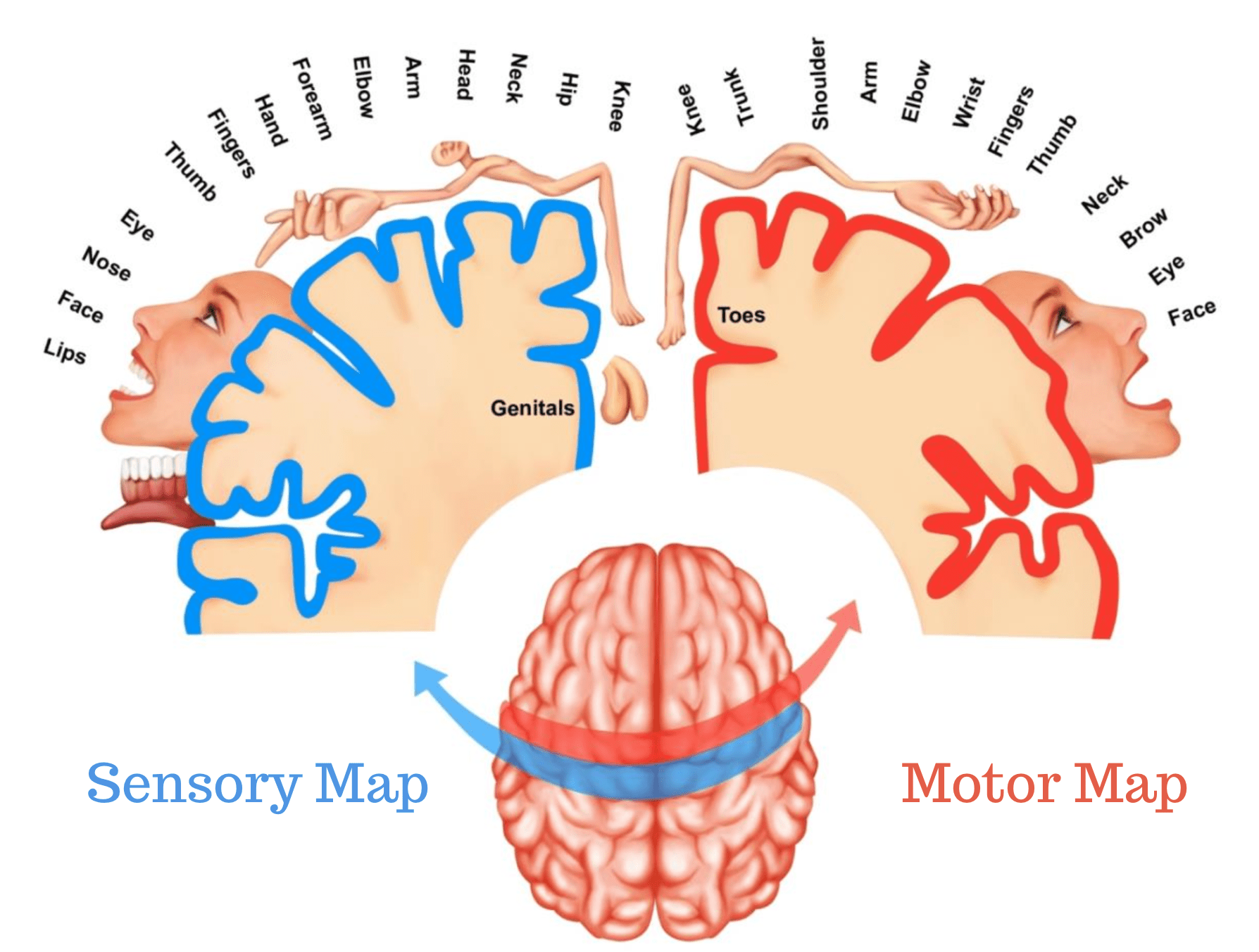
You’ve seen the homunculus. The stretched little man draped over the surface of the brain with the enormous hands and lips (not winning any beauty contests). He’s real enough, in that your body genuinely is laid out in an orderly way across S1 and across the motor cortex sitting next door to it. He’s also badly misleading in about four separate ways, and every single one of them matters for the smudging argument later.
It’s ordered, but it really isn’t neat. Body parts get cortical space in proportion to how much fine detail your brain needs out of them, rather than how big they actually are, which is why your lips and fingertips get an enormous allocation and your back gets almost nothing. The receptive fields overlap far more than the diagram implies, and the boundaries are gradients rather than lines [13]. Ask which patch of cortex belongs to your index finger and the honest answer is that there’s a patch where it dominates, sitting inside a much larger region where it’s present but not in charge.
Information about one body part is also spread right across the whole map, rather than being locked in its own little territory, as you can work out which finger somebody moved from activity well outside that finger’s supposed patch [19]. So, a map drawn by asking “which body part wins here” is only ever a summary of a much richer and much more distributed picture, and losing the winner in one spot doesn’t mean losing the information.
The fine detail is remarkably stable within a person, too. Scan the same individual’s finger layout over and over, weeks apart, and it stays put in its own slightly idiosyncratic shape rather than drifting about [20]. These aren’t things that reshuffle week to week depending on how your body has been behaving lately.
The motor side isn’t a simple strip either. Motor cortex has the classic effector regions, hand and foot and mouth, but interleaved between them are regions that look a lot more like whole body action control, than like individual muscles [21]. And the motor and sensory maps are matched to each other in fine detail across the fold between them, which is presumably rather the point, as you can’t integrate sensation with movement, if the two are filed in different orders [22].
Hold onto that picture, as it’s the exact thing the chronic pain literature has spent twenty years arguing about. A structured, stable, overlapping, distributed layout, in which every part of you is represented in more than one place.

Is Proprioception Actually Worse If You’re Hypermobile
Short answer, yes, often, in symptomatic hypermobility, and mostly not in the way it gets described to you.
Pool the lower limb work together and both position sense and movement detection come out poorer in benign joint hypermobility syndrome, the finger findings are fairly clear, and the shoulder findings don’t line up anywhere near as cleanly [1]. That pattern, strong at the knee and patchy at the shoulder, turns up over and over again.
At the knee specifically, women with the hypermobile type of EDS repositioned the joint less accurately than controls, even after accounting for how much sport they did, which matters, as how active somebody is would otherwise muddy the whole thing. In that same group, shoulder position sense wasn’t significantly different, and vibration sense wasn’t either [23]. That last detail is the genuinely the most interesting one. Vibration is carried by exactly the same class of large sensory fibres, so if hypermobility caused some general failure of large fibre sensation, vibration would have gone with it, and it didn’t. Whatever this is, it’s selective rather than global.
Older and more specific work fills the picture in nicely. Detecting that the knee has started to move takes a bigger movement in hypermobility syndrome before it registers at all [24]. At the finger, position matching errors are larger across the whole range, with a bias towards underestimating how far a joint has actually travelled at the extremes [25]. Which is worth us actually thinking about for a second, as the error isn’t random. It runs in the direction of thinking you’re less bent than you really are, precisely at the point where the tissue has the least to say.
Then there’s the finding that reframes everything. Ask someone with EDS to say where her unseen hand is and, averaged out, she’s about as accurate as anybody else. The scatter around that average is roughly twice as wide [2].
So, the aim is fine and the grouping is poor, which is a completely different problem from not knowing where your hand is. And that imprecision didn’t track how bad her chronic pain was, which is a genuine inconvenience for anybody wanting to make pain the cause of the sensory problem.
Think about throwing darts, badly, which in my case is the only way I throw them. Bad accuracy means every dart lands in the same wrong place, so your sense of where your knee is could be reliably out by ten degrees, which the brain can learn to correct for and largely does. Bad precision means the darts are scattered all over the board around roughly the right spot, so there’s no consistent error sitting there to correct, and the brain gets handed a slightly different answer to the same question every time it asks it.
That second one is the version you’ve got, and unfortunately it’s the harder one, as you can’t calibrate out noise. You can only give the system more to work with, or get better at telling small differences apart, which is exactly what the training evidence further down points at.
Two more pieces from the same line of work, and they’re both quietly hopeful. Give her a task where the visual feedback has been deliberately distorted and she recalibrates her sense of hand position perfectly normally, so the machinery for updating a body estimate is completely intact, even when the estimate arriving is noisy [26]. And when the judgement has to lean purely on proprioception with nothing else to go on, the imprecision gets worse, and how imprecise it is relates to how hypermobile she is [27]. Both of those point at the tissue and the receptors, which is really good news, dressed up as a boring technical detail.
Elsewhere in the body it’s much the same shape. Elbow and knee position sense come out poorer in hypermobile adults while grip strength and functional performance don’t differ at all [28], which is precisely what compensation looks like from the outside. Cervical position error is larger in hypermobile people with neck pain, and, tellingly, how hypermobile you are only relates to how badly you do it if you’re in the pain group [29].
One honest negative belongs here too. Higher level body perception tasks, recognising where a body part is in a more “abstract” sense, or identifying an object by touch alone with your eyes shut, shows no significant relationship with hypermobility [30]. So, this isn’t a story about hypermobile brains processing the body strangely in general. It’s a good deal more specific, and a good deal more peripheral, than that.
Hypermobile Is Not The Same Thing As Symptomatic
This next bit gets missed in nearly every article written on the subject, and it’s the bit most likely to change what you conclude about yourself.
Hypermobile children, tested four different ways in an ordinary community sample, show no proprioceptive differences at all from their normally mobile classmates, and the four tests barely agree with each other anyway [10]. Put young hypermobile children on a loaded standing task, judging heel height with weight going through the foot, and they actually outperform the controls [31]. Across a much larger group of children, the hypermobile ones came out with lower dynamic balance reach and lower isometric force, and yet better functional lower limb strength and no proprioceptive difference whatsoever [32]. None of which looks much like a group with a broken sense.
Now, put symptoms into it and watch what happens. Children with clinically defined hypermobility syndrome, meaning pain and problems, do have poorer knee position sense and poorer movement detection, alongside reduced knee flexor and extensor torque [33]. Those same children also walk differently, with more passive knee range available, less peak knee flexion when the limb is loading and during swing, and more knee extension through mid stance [34]. So, the sensory difference and the movement difference arrive together in the symptomatic group, and largely don’t in the asymptomatic one.
The adult picture matches up pretty well. Pain free hypermobile shoulders show normal average position sense [35], and asymptomatic hypermobile volleyball players don’t come out as the high injury risk group you’d confidently predict [36]. And when balance reactions to a forward shove get measured, it’s the presence of symptoms, rather than the amount of laxity that separates people out [37].
The best current read of all this is a threshold model. Laxity on its own often produces no measurable deficit at all, whereas laxity plus pain, plus recurrent instability, plus fatigue, plus fear of moving, plus years of a slightly different movement history, very often does. Asymptomatic hypermobility looks a lot like compensation that’s working, and symptomatic hypermobility looks like compensation that’s not.
Which is why two people with identical Beighton scores can have completely different bodies to live in, and why “but I’m hypermobile and I’m absolutely fine” and “I’m hypermobile and I can’t keep my shoulder in” are both perfectly true statements about the same trait.
Keep in mind though, that Generalised Hypermobility decreases with age in children, mainly through the elbows and knees losing range, and hypermobile children end up less physically active without being clearly worse off on pain, fitness or quality of life over a couple of years [38]. And in a large group of Nigerian schoolchildren, pain was uncommon and wasn’t linked to how mobile they were [39].
And in children with joint hypermobility syndrome and hypermobile knees, sense into the hyperextended range turned out to be no worse than it was in early flexion. About a tenth of a degree separated the two, tested actively and in weight bearing, which is nothing [40]. Worth sitting with, because it undoes the instruction most people with hypermobile knees get handed, the one where the end of the range is treated as somewhere you’re not allowed to go. The authors’ own conclusion was that this sort of training should be done through the whole range, the hypermobile part of it included.
Why It Happens, And How Confident Anybody Can Honestly Be
There are three plausible mechanisms here, and they aren’t really competing with each other, so much as stacking up on top of one another.
The first is the slack receptor idea, and it’s the one everybody reaches for first. If a mechanoreceptor is sitting in connective tissue that’s more stretchy than average, the deformation it needs in order to fire arrives later and smaller. The amount of movement required to trigger it effectively goes up and the quality of what it sends goes down [41]. Mechanically that’s very coherent, it fits the selectivity we saw at the knee and not the vibration sense, and it fits imprecision tracking how hypermobile somebody is [27].
It also has almost no direct human evidence behind it, as nobody has ever recorded receptor firing in a living symptomatic hypermobile person and compared it against an asymptomatic one [41]. So it’s a plausible mechanism rather than a demonstrated one.
One relevant piece of evidence does narrow it down though. Testing quadriceps control in hypermobile people, the excitability of the pathway running from brain to muscle came out steeper while the spinal reflex measures were unchanged [41], which argues against this being a simple spinal reflex failure and points instead at altered input that the brain is having to work harder to handle. Proprioceptive inaccuracy also partly explains why muscle strength doesn’t map cleanly onto what somebody can actually do in daily life, so strength and function come apart in a way that force production on its own can’t account for [42].
The second mechanism is the tissue itself acting as a sensory organ. Human superficial fascia turns out to be substantially innervated, including with autonomic fibres, which makes it a plausible contributor to nociception and to bodily sensing rather than the inert packing material it always used to be treated as [43]. The work pulling all of this together for hypermobility is exactly that though, a mechanistic synthesis, and nobody has shown that fascial dysfunction causes the proprioceptive deficits in hypermobility [44]. So it’s a reason to suspect something rather than a reason to believe it, and the fascia world does have a bit of a habit of getting ahead of itself.
The third is cumulative damage, and this is where the evidence is strongest, although all of it is borrowed from elsewhere. Ligament injury and chronic instability degrade proprioception, and that much is very well established outside of hypermobility. Position sense is measurably worse in injured limbs after a cruciate ligament rupture [45], and in functionally unstable ankles, the spindle related traffic coming up during ligament stress is altered [46]. So, there’s a solid model in there of what recurrent subluxation, microtrauma and years of joints going where they shouldn’t could plausibly be doing to the quality of your signal. And symptomatic hypermobile women do show more passive forward movement at the tibia than asymptomatic hypermobile women, so the mechanical starting point genuinely differs between those two groups too [47].
Put all of that together and you get slightly compromised sensors, sitting in sensorially active tissue that behaves differently, in a body that’s been in pain for a good long while.
That last one does considerably more than people tend to think.
What Pain Does To The Signal, And What It Doesn’t Do
This is where the two easy stories live. One says the pain is the whole cause of everything above. The other says the pain is irrelevant and it’s all purely mechanical. Neither of them survives more than about five minutes of contact with what’s actually been researched.
Let’s start with the thing that annoys me the most. There’s no such thing as a pain nerve or a pain signal. What you’ve got are called nociceptors, which are sensory endings that respond to things capable of damaging tissue, and what they send up the line is nociceptive input. Pain is what the whole system eventually produces out of that input, along with a great deal else, so the two are genuinely different phenomena, and that’s why the amount of pain somebody is in can’t be read off the amount of traffic coming out of a joint. I’m not being pedantic for the sake of it, and I’m absolutely not hinting that any of this is in your head. I’m saying it because the rest of this section only makes sense once you stop picturing one dial feeding another dial.
Start with the anatomy, as it explains everything that follows. The region of S1 most loaded with deep muscle and joint input also responds to nociceptive input [48]. Proprioception and nociception overlap in the same early cortical territory rather than running as two tidy separate streams that meet up somewhere late on. Which makes it a great deal less surprising that tonic muscle pain changes early cortical processing of touch and position, at the same time as distorting how accurately somebody knows where their finger is [49].
Now, the nuance, because pain does not degrade all of proprioception equally.
Across the experimental pain work, postural control usually gets worse, force sense is often affected, kinaesthesia sometimes suffers, and joint position sense was unchanged in four of the five cases where anybody bothered to look [50]. Induce acute knee pain in healthy people and knee position sense doesn’t change at all [51]. Put pain at a site that genuinely matters “proprioceptively” during a real task though, and it bites hard, as nociceptive input during walking raised the threshold for detecting ankle movement by about a third [52]. In the shoulder, kinaesthesia is moderately supported as impaired by pain, force sense weakly so, and the position sense findings genuinely conflict with each other [53].
So, acute pain isn’t a general proprioception destroyer. It picks its targets, and the targets it picks are the dynamic ones, the ones that matter for standing up and not falling over, rather than the static ones we mostly bother to measure in an assessment.
Chronic pain, is a completely different animal, and this is the part that’ll feel familiar. In long standing back pain, proprioceptive errors turn up widely, including at joints nowhere near the painful area [54]. In fibromyalgia, joint level position errors and postural instability both show up together [55].
And the psychological load is real, which is not a polite way of calling any of this imaginary or that it’s in your head. Limits of stability come out lower in hypermobility syndrome alongside larger lumbar repositioning errors, and both fear of movement and fatigue significantly mediate the relationship between the two [56]. Fear and fatigue aren’t sitting alongside the sensory problem as separate complaints, they’re part of the machinery by which it produces the instability you actually live with. Biology, circumstance and psychology all loading the same system.
In joint hypermobility syndrome and the hypermobile type of EDS, standard testing finds no damage to the somatosensory nerves, while heat and cold pain thresholds come out lower and the wind up from repeated stimulation comes out increased, which is the signature of central sensitisation rather than classic nerve injury [57]. In women with hEDS, pressure pain thresholds are lower, temporal summation is increased, the pain relief you’d normally get from exercise is reduced at the quadriceps but not at the trapezius, and the descending inhibition measure doesn’t differ from controls at all [58]. Pressure pain thresholds come out lower across the whole body, including at sites that aren’t painful! [59].
Which is what generalised hyperalgesia actually means, and it’s why “but it hurts in places where nothing is wrong” is a mechanistic observation.
None of which means one single thing causes all of it. Chronic pain states combine nociceptive, neuropathic and nociplastic mechanisms rather than sorting themselves neatly into one box [60], and in hypermobility the symptom picture splits into distinguishable clusters, with the non musculoskeletal burden being the thing that predicts worse health status and worse pain interference [61]. So the gut, the fatigue and the dizziness aren’t a side show. They’re doing a great deal of the damage to your week, and we’ve been saying that in the studios for years.
There’s a small fibre and autonomic layer sitting underneath all of this as well, at least in the people who end up in specialist care. Skin biopsy in hEDS shows generalised small fibre neuropathy in a majority, a substantial minority meeting criteria for PoTS, and reduced nerve fibre density both proximally and distally [62]. In a larger hEDS group, widespread but mild autonomic failure was very common, along with reduced cerebral blood flow velocity on standing and small fibre neuropathy in most of them [63]. Take that one carefully though please, as these are people who were referred to specialist clinics for exactly these problems, so those rates tell you about a referred group, rather than about everybody with hEDS. It does mean that “your nerves are fine, it’s just your joints” is not a safe assumption in this population.
Balance, Falls, And The Vision Crutch
Balance is where all of this stops being theoretical and starts being your actual life.
Across the adult hypermobility balance work, the broad picture is more sway, different muscle activity, different movement patterns, frequent falls and frequent fear of falling, with the strong caveat that the classification systems used are all over the place , and the overall level of evidence is low [64]. So the direction is consistent and the confidence really should stay moderate.
The detail is where it gets useful for us though. In hEDS, sway area comes out larger than in controls, and the instability gets noticeably worse once vision is removed, particularly side to side. In that same work, compressive garments and insoles partly improved postural stability, most clearly with the eyes closed, although this has only been looked at in a very small group so far, so treat that as a look in the right direction [65].
That eyes closed pattern is more or less the entire story in one observation.
If you lean on vision more than the average person does in order to know where your body is, then you’ll look completely fine in a well lit room on a flat floor, and you’ll be genuinely unsafe in the dark, on a cobbled street, carrying your shopping home, or with your head turned to talk to somebody. And the thing your physio measured you doing was, unfortunately, the easy version.
Which brings in the finding that ought to be pinned to the wall of every hypermobility rehab room in the country. Women with the hypermobile type of EDS show greater sway, slower gait with shorter steps across walking conditions, and a much larger drop in performance when they have to do something else at the same time. Almost everybody asked had fallen in the previous year [66]. That dual task decrement is the real tell, as holding yourself together is costing you attention. It isn’t automatic any more, so anything that steals attention, a conversation, a phone, fatigue, brain fog, a chocolate covered toddler, comes straight out of the budget you were quietly using to stay upright.
In EDS, muscle activation during walking is altered, gait is slower, stride is shorter and the hip and ankle muscles are weaker, while the actual joint angles stay similar to controls [67]. So the shapes look nearly normal and the strategy generating them really doesn’t. Somebody could watch you walk and see nothing much wrong at all, which is more or less the universal experience of being assessed.
The word for what your nervous system is doing here is reweighting, and it’s the system working as designed. Balance gets built out of three streams, what your eyes report, what the balance organs in your inner ear report, and what your body reports from the ground up. Downgrade the reliability of one stream and the others get weighted up to make up for it, which is a perfectly sensible thing for a control system to do. It’s also why asymptomatic hypermobile people can test completely normally, while quietly leaning on vision far more than average [31][36].
The trouble with a strategy that leans heavily on one stream though, is that it’s brittle in exactly the situations that take that stream away, and it costs attention to run, which is what the dual task finding is really measuring [66]. Add pain on top and the reweighting shifts further towards protection and away from efficiency [50].
There’s an uncomfortable corollary to all of that as well. If your compensation is good enough to pass the test, then the test tells you absolutely nothing about how close to the edge you’re living. Which is why “your balance is fine” and “I fall over constantly” are both routinely true of the same person on the same day, and why nobody should be taking a clean assessment as evidence that nothing needs doing.
The Brain Map Story, And What Actually Happened To It
This is the part where something you’ve very probably been told with a great deal of confidence turns out to be, at best, unsettled. If you’ve been in this world for any length of time you’ll have met the brain map story in one form or another, and it usually arrives wearing a lab coat.
It goes like this. Your brain holds a map of your body, a patch of brain for each bit of you, and long term pain is supposed to smudge the patch belonging to the sore part, so it goes blurry and starts bleeding into its neighbours. The blurring is then what keeps the pain going. Sort the map out and you sort the pain out, which is why sensory work was said to help, as it was redrawing the thing.
It came out of good research, and it came out of amputation. In people who had lost an arm, the changes measured in the brain afterwards lined up closely with how bad their phantom limb pain was, and the same sort of rearranging seemed to explain why pain got felt in a limb that was no longer there. From there it got generalised into an entire rehab framework built around the map as the thing you treat [68][69][70][71].
There was a version of it you can test without a scanner as well, and you may well have had it done to you. How finely you can tell two points of touch apart on your skin comes out worse in several long term pain conditions, and that got read as the map going blurry, although the studies behind it disagreed with each other quite a lot and many of them were poorly run [72]. On the movement side of the brain, the amount of smudging was reported as tracking how bad somebody’s back pain was [73]. It’s a tidy story, it makes mechanistic sense, and that’s exactly why you’ve heard it about nine times.
Then the scanners got better.
When somebody went back and mapped the fingers properly, in much finer detail, in people who had lived with complex regional pain syndrome for years, the painful hand’s map looked much like the other hand’s and much like everybody else’s. Same size, same place, same shape, and no relationship to how severe the pain or the disability was [3].
Phantom limb pain went the same way, only more so. The old hand area was still sitting there, fully intact, and the people with the worst pain were the ones holding on to it most firmly, which is pretty much the opposite of what the story predicted [74][75]. Step back and look across the wider literature and adult body maps turn out to be remarkably stable even when they lose their input altogether, and a lot of what looked like the brain rearranging itself is better explained by changes lower down, in the nerves and the spinal cord [76]. Even the textbook example, the face taking over the hand’s territory after spinal cord injury, hasn’t held up the way it was first described [77].
And in long term back pain, where the smudging idea has had by far the most influence on what people actually get told in a treatment room, the maps of the back come out normal [4]. So the honest position in 2026 is that a badly distorted body map isn’t a reliable feature of long term pain, the strong version of the claim doesn’t hold up, and the theory the whole thing was built on has had to be rewritten [78].
Now, that doesn’t mean the brain isn’t in this, and that’s the bit that gets lost the second somebody hears that smudging is out. What holds up far better is the wiring rather than the shape. The map is still there and it’s still fine, and what’s changed is who it talks to, and how urgently the rest of the brain treats what it says. In complex regional pain syndrome, the sensory region talks less to its opposite number and more to the areas that deal with threat and with turning pain up and down, and some of that does track how long somebody has had it and how bad it is [79]. Different research centres haven’t all found the same thing though, and the data quality is a genuine concern, so hold that one loosely [80]. In back pain the strength of those connections tracks how intense the pain is, and most of all in people who worry about it more, which is a sentence that gets misread constantly, as it’s a description of wiring rather than an accusation [81][82].
Which is honestly a far more interesting story than smudging ever was.
There’s no published study that has mapped the sensory part of the brain in hEDS or HSD in any detail. Not one. Every confident claim you’ve ever read about hypermobile brain maps being smudged, blurred, degraded or in need of restoring is borrowed from amputation and CRPS research, and that research no longer says the thing it keeps getting quoted for. Nobody has actually looked (surprise, surprise).
What does sit next door to it is thin, and worth knowing about anyway. In teenagers with long term pain, adding hypermobility into the mix made no difference at all to how they did on sensory testing, although the hypermobile ones did have pain in more places, more fatigue and worse sleep. There were some brain differences during a task that mixed the senses together, but they only turned up at one of the two research sites, so they badly need somebody to repeat them [83]. That’s genuinely the state of the art on hypermobile bodies and body sensing at the moment. It’s a start, and it’s nowhere near a mechanism.
If The Maps Aren’t Smudged, Why Does Sensory Work Help
Because a smudged map was never the only explanation going, and because the thing that actually works isn’t the thing the old story said would work.
The better way to think about it is your whole sense of your own body, rather than one strip of brain tissue. Touch, pain, where you feel your limbs to be and whether they feel like yours are all worked out together, which is why an illusion that messes about with one of them can shift the others, pain included [84]. So there’s genuinely something in there to aim at. It just isn’t the map.
The cleanest bit of evidence on this is also the most quietly awkward for the wobble board industry. In people with long term limb pain, touch training where they had to work something out, which spot was being touched, which of two things they could feel, brought their pain down. The same amount of touch with nothing to work out did nothing at all [85]. So the active ingredient is the deciding. Not the sensation, not the kit, and not the novelty of standing on something squashy.
Graded motor imagery, which is the staged programme where you start off judging whether a photo of a hand is a left or a right, then move on to imagining movements, then work with mirrors, does reduce movement pain in long standing CRPS of the arm, and nobody can properly tell you why yet [86]. Which is about the right amount of confidence to have, in our opinion, as it does something and the reason is still an open question.
In hypermobility specifically, the closest thing we have is a fortnight of sensory work on the soles of the feet in young hypermobile adults, which improved light touch, telling two points of contact apart, vibration sense, how accurately they could place a joint, and balance reach in one direction, with no change in strength at all [87]. That’s encouraging, although who it was done in matters a great deal here, as they were pain free eighteen to twenty five year olds rather than people with hEDS and fifteen years of pain behind them. So it’s a reason to try it, and not a reason to promise anybody anything.
Put all of that together and you get a position that needs no smudged maps in it whatsoever. If the information arriving is vague, then making it clearer, or getting better at telling things apart within it, gives the brain something better to work with. That’s a very different claim from redrawing a map inside somebody’s head, it can actually be tested, and it’s the one the evidence supports.
Motor Learning, And Why You Can Practise And Keep Nothing
Everything so far has been about the information going in, this last part is about what your nervous system then does with it, and it’s where rehab either works or quietly eats two years of your life.
First, let’s get rid of the idea that pain switches muscles off, as it doesn’t. What it does is change who does what, from the reflexes in your spinal cord all the way up to the instructions leaving the movement part of your brain, and the whole pattern looks protective rather than broken [88]. Look right down at the level of individual muscle fibres being switched on and it’s a reshuffle, as some go quieter while others pick up the slack [89][90]. Your body isn’t failing to switch things on, it’s choosing differently, on purpose, to protect something.
Which is exactly why you can look completely fine while the strategy underneath has changed entirely. Reach for something with a sore muscle and the movement anybody can see stays much the same, while the way the muscles share the job out underneath it reorganises substantially [91]. Easy tasks look normal precisely because the spare muscles quietly cover for the problem, so watching whether somebody got their hand to the cup tells you very little [92]. Which is why the easy tests keep passing you, and life keeps not passing you.
Make the task harder though, and it shows up straight away. Put somebody with a sore ankle on a balance task where they have to reach in several directions and the distance comes down. Put somebody with a sore back on a task that needs steady force and the steadiness goes, along with how well the brain and the muscle are keeping time with each other [93][94]. The output from the movement part of the brain drops over the first minutes and hours of pain, and by the time you’re into days and weeks it stops being one tidy group effect and becomes far more individual, which is a polite way of saying it depends on the person [95][96].
Now, two things turn up in this research that should change how you train, and I don’t think either of them is widely known outside of it. The first is that expecting pain changes which muscle fibres get used before anything has actually happened [97], so the expectation alone is enough to do it. The second is that when a limb is expected to hurt, the brain damps down the line to that particular limb, and damps it down harder in people who are more anxious generally [98]. So the guarding you’ve been told repeatedly to stop doing is partly a decision made well below the level of choice, in that specific limb, scaled by how threatening the whole situation feels. Telling somebody to relax into it isn’t an intervention, it’s a request.
And then the one I’d put on a poster if anybody let me. In a study on learning a new walking pattern, pain during the training left performance on the day completely intact, and wrecked how much of that performance was still there the next morning [99]. Which is worth reading twice, as in plain terms it means you can practise in pain, do it well, and keep none of it.
So a session that felt genuinely productive can teach you absolutely nothing, and the only way you’d ever find out is by testing tomorrow rather than admiring today. It’s probably the most expensive misunderstanding in this entire field, and almost nobody scores their sessions that way.
It gets self reinforcing if you leave it long enough as well, as a poor signal coming from the joints seems to help drive longer term changes in how movement gets organised, rather than simply being a result of those changes [100]. So a year of practising in pain isn’t a neutral year in which not very much happened. It’s a year of teaching a strategy you’d rather not have, really quite efficiently, to a system that learns whether you meant it to or not.
There’s one more thing worth saying about how movement skills get built, as it decides what a session should actually look like. A skill isn’t a recording you play back. It’s closer to a rule your nervous system works out for solving a type of problem, and you only ever get a rule out of variety. Do the identical rep on the identical surface at the identical speed three hundred times and you’ll get very good at that, and surprisingly bad at the version life hands you, which is never the one you rehearsed. Change the surface, the speed, the direction, the load, the shoes, whether your head is turned, and the system has to work out what all of those attempts had in common. That common thread is the rule, and the rule is the bit that turns up for you in a situation you’ve never practised.
Which matters even more in a body working off a vague signal, as a rule built out of variety is far more forgiving of a bad reading on any given day, than one rehearsed pattern is. It’s also why three sets of ten, same everything, is such a poor fit here, however well meant.
The training evidence in this population is worth being precise about, as it gets overstated in both directions. Exercise and movement training do appear to work in HSD and hEDS, while the evidence for equipment, hands on treatment, being handed instructions and generic gym work is weaker, and every question about how much, how often and how hard is still wide open [101]. So the broad direction is supported, nobody can currently tell you the right sets and reps, and anybody handing you a precise number is handing you their preference.
Worth knowing as well that strength work does improve joint sense at the shoulder [102]. So none of this was ever an argument against getting strong, it’s an argument about the order you do things in.
How We Train It
Part 1 argued this from the strength end, and this is the same argument from the sensory end. It’s our read rather than a settled finding, and it comes down to two things, what you do first and how you decide whether it worked.
– Signal before load. If the information arriving at your brain is vague, then adding weight to a joint nobody can locate accurately mostly adds consequences. Sort out the information and the movement strategy first, then load it [41][42].
– Work the range you actually live in. The far end of a hypermobile range isn’t a sensory dead zone you have to fence off. In children with JHS the knee read it about as accurately as it read early flexion [40], and the researchers took that to mean training should cover the lot. Build into it gradually, the way you would with anything you’ve not trained, rather than avoiding it on principle and then being surprised it feels unfamiliar.
– Give the system something to work out. The training that brought pain down was the version where people had to tell one thing from another, not the version that simply applied touch [85]. So put a judgement in the task. Which surface is this, which way did that move, which foot is further forward. Textures under the feet, tape across a joint, different floors, all fine, as long as you’re deciding something rather than sitting there receiving it.
– Take vision away on purpose, carefully. Balance gets noticeably wobblier without vision, side to side most of all [65]. That’s a weak point, it’s trainable, and it’s also not something to surprise yourself with at the top of a flight of stairs. Do it near a wall, do it early, do it often.
– Train the two things at once version. Performance drops further than average as soon as you add a second job to do [66], which means holding yourself together is eating attention. So practise holding a position while counting backwards, talking, catching something or turning your head, as that’s the version of the skill real life is going to ask you for.
– Judge the session by tomorrow. Practising in pain can leave today intact and cost you the lot by morning [99]. So the honest measure of a session isn’t how it felt or what you lifted, it’s whether the thing is still there the next day. That one change in scoring will reorganise an entire programme, and it’s the first thing we change with a new client.
– Expect the guarding, and work with it. Expecting pain changes which muscles get used before anything hurts, in that limb specifically [97][98]. So making a movement smaller, slower, more predictable or better supported isn’t coddling anybody, it’s taking away something that’s currently sitting in the way.
– Vary the rep, don’t repeat it. Same movement, different surface, speed, direction, head position or shoes. You’re building a rule your nervous system can use in a situation it hasn’t met before, and you can’t build one of those out of three identical sets.
– Don’t abandon strength, resequence it. Strength work does improve joint sense [102], and the evidence in this population backs exercise and movement training over the passive stuff [101]. Load isn’t the enemy here, loading a joint that nobody can accurately locate, is.
What Nobody Knows Yet
A fair amount of this is still missing, and the holes are where the next decade of it goes.
Nobody has mapped the sensory part of the brain properly in hEDS or HSD. Every claim about hypermobile brain maps, including the ones we used to make ourselves, is borrowed from amputation and CRPS work, and that literature has moved a very long way since [3][4][76].
Nobody has measured what the receptors in a living, symptomatic hypermobile person are actually doing and compared it against somebody equally hypermobile who feels fine. The slack receptor idea is worked out from what the tissue is like, repeatedly, by a lot of people, and almost never measured [41].
The change itself has never been followed. Hypermobile people who feel fine haven’t been tracked forwards into becoming symptomatic, so the research is full of snapshots comparing one group against another, and very thin on how somebody actually gets from the first to the second, which is precisely the question that matters if you want to stop it happening [30].
How much to do is completely open. Exercise and movement training look like they work, and the studies behind that are mixed enough that the sets, the reps, the frequency and the progression are all still guesses [101].
And nobody knows which sort you are. Some of the trouble in hypermobility comes from the tissue and the small nerve fibres sitting in it, some from the nervous system turning pain up, some from the autonomic side of things, and the mix clearly differs a great deal from one person to the next [60][61][63]. Working out which of those is running your week is a judgement call rather than a test at the moment, and pretending otherwise is how people end up doing the wrong rehab very diligently for years.
The Fibro Guy

References
[1] Smith, T.O., Jerman, E., Easton, V., Bacon, H., Armon, K., Poland, F. et al. (2013) ‘Do people with benign joint hypermobility syndrome (BJHS) have reduced joint proprioception? A systematic review and meta-analysis’, Rheumatology International. https://doi.org/10.1007/s00296-013-2790-4
[2] Clayton, H.A., Jones, S.A.H. and Henriques, D.Y.P. (2015) ‘Proprioceptive precision is impaired in Ehlers–Danlos syndrome’, SpringerPlus. https://doi.org/10.1186/s40064-015-1089-1
[3] Mancini, F., Wang, A.P., Schira, M.M., Isherwood, Z.J., McAuley, J.H., Iannetti, G.D. et al. (2019) ‘Fine-Grained Mapping of Cortical Somatotopies in Chronic Complex Regional Pain Syndrome’, The Journal of Neuroscience. https://doi.org/10.1523/jneurosci.2005-18.2019
Read More[4] Dörig, M., Cole, D.M., Guekos, A., Stämpfli, P., Schütz, P., Schibli, L. et al. (2026) ‘Stable Mechanoreceptive Somatotopy in Chronic Low Back Pain: Is Cortical Map Reorganization a Myth?’, The Journal of Neuroscience. https://doi.org/10.1523/jneurosci.2069-25.2026
[5] Delhaye, B.P., Long, K.H. and Bensmaia, S.J. (2018) ‘Neural Basis of Touch and Proprioception in Primate Cortex’, Comprehensive Physiology. https://doi.org/10.1002/j.2040-4603.2018.tb00045.x
[6] Nagel, M. and Chesler, A.T. (2022) ‘PIEZO2 ion channels in proprioception’, Current Opinion in Neurobiology. https://doi.org/10.1016/j.conb.2022.102572
[7] Dimitriou, M. (2022) ‘Human muscle spindles are wired to function as controllable signal-processing devices’, eLife. https://doi.org/10.7554/elife.78091
[8] Smith, L., Norcliffe‐Kaufmann, L., Palma, J., Kaufmann, H. and Macefield, V.G. (2020) ‘Elbow proprioception is normal in patients with a congenital absence of functional muscle spindles’, The Journal of Physiology. https://doi.org/10.1113/jp279931
[9] Hillier, S., Immink, M. and Thewlis, D. (2015) ‘Assessing Proprioception’, Neurorehabilitation and Neural Repair. https://doi.org/10.1177/1545968315573055
[10] Ituen, O.A., Smits-Engelsman, B., Ferguson, G. and Duysens, J. (2024) ‘Proprioception and its relationship with range of motion in hypermobile and normal mobile children’, Experimental Brain Research. https://doi.org/10.1007/s00221-024-06937-1
[11] Niu, J., Ding, L., Li, J.J., Kim, H., Liu, J., Li, H. et al. (2013) ‘Modality-Based Organization of Ascending Somatosensory Axons in the Direct Dorsal Column Pathway’, The Journal of Neuroscience. https://doi.org/10.1523/jneurosci.3429-13.2013
[12] Pop, I.V., Espinosa, F., Blevins, C.J., Okafor, P.C., Ogujiofor, O.W., Goyal, M. et al. (2021) ‘Structure of Long-Range Direct and Indirect Spinocerebellar Pathways as Well as Local Spinal Circuits Mediating Proprioception’, The Journal of Neuroscience. https://doi.org/10.1523/jneurosci.2157-20.2021
[13] Harding-Forrester, S. and Feldman, D. (2018). Somatosensory maps. Handbook of clinical neurology, 151, pp. 73-102. doi: 10.1016/b978-0-444-63622-5.00004-8.
[14] Chowdhury, R.H., Glaser, J.I. and Miller, L.E. (2020) ‘Area 2 of primary somatosensory cortex encodes kinematics of the whole arm’, eLife. https://doi.org/10.7554/elife.48198
[15] Ngo, G.N., Haak, K.V., Beckmann, C.F. and Menon, R.S. (2021) ‘Mesoscale hierarchical organization of primary somatosensory cortex captured by resting-state-fMRI in humans’, NeuroImage. https://doi.org/10.1016/j.neuroimage.2021.118031
[16] Kim, S.S., Gomez-Ramirez, M., Thakur, P.H. and Hsiao, S.S. (2015) ‘Multimodal Interactions between Proprioceptive and Cutaneous Signals in Primary Somatosensory Cortex’, Neuron. https://doi.org/10.1016/j.neuron.2015.03.020
[17] London, B.M. and Miller, L.E. (2013) ‘Responses of somatosensory area 2 neurons to actively and passively generated limb movements’, Journal of Neurophysiology. https://doi.org/10.1152/jn.00372.2012
[18] Gandolla, M., Ferrante, S., Molteni, F., Guanziroli, E., Frattini, T., Martegani, A. et al. (2014) ‘Re-thinking the role of motor cortex: Context-sensitive motor outputs?’, NeuroImage. https://doi.org/10.1016/j.neuroimage.2014.01.011
[19] Muret, D., Root, V., Kieliba, P., Clode, D. and Makin, T.R. (2022) ‘Beyond body maps: Information content of specific body parts is distributed across the somatosensory homunculus’, Cell Reports. https://doi.org/10.1016/j.celrep.2022.110523
[20] Kolasinski, J., Makin, T.R., Jbabdi, S., Clare, S., Stagg, C.J. and Johansen-Berg, H. (2016) ‘Investigating the Stability of Fine-Grain Digit Somatotopy in Individual Human Participants’, The Journal of Neuroscience. https://doi.org/10.1523/jneurosci.1742-15.2016
[21] Gordon, E.M., Chauvin, R.J., Van, A.N., Rajesh, A., Nielsen, A., Newbold, D.J. et al. (2023) ‘A somato-cognitive action network alternates with effector regions in motor cortex’, Nature. https://doi.org/10.1038/s41586-023-05964-2
[22] Schellekens, W., Petridou, N. and Ramsey, N.F. (2018) ‘Detailed somatotopy in primary motor and somatosensory cortex revealed by Gaussian population receptive fields’, NeuroImage. https://doi.org/10.1016/j.neuroimage.2018.06.062
[23] Rombaut, L., De Paepe, A., Malfait, F., Cools, A. and Calders, P. (2009) ‘Joint position sense and vibratory perception sense in patients with Ehlers–Danlos syndrome type III (hypermobility type)’, Clinical Rheumatology. https://doi.org/10.1007/s10067-009-1320-y
[24] HALL, M.G., FERRELL, W.R., STURROCK, R.D., HAMBLEN, D.L. and BAXENDALE, R.H. (1995) ‘THE EFFECT OF THE HYPERMOBILITY SYNDROME ON KNEE JOINT PROPRIOCEPTION’, Rheumatology. https://doi.org/10.1093/rheumatology/34.2.121
[25] MALLIK, A.K., FERRELL, W.R., McDONALD, A.G. and STURROCK, R.D. (1994) ‘IMPAIRED PROPRIOCEPTIVE ACUITY AT THE PROXIMAL INTERPHALANGEAL JOINT EN PATIENTS WITH THE HYPERMOBILITY SYNDROME’, Rheumatology. https://doi.org/10.1093/rheumatology/33.7.631
[26] Clayton, H.A., Cressman, E.K. and Henriques, D.Y.P. (2013) ‘Proprioceptive sensitivity in Ehlers–Danlos syndrome patients’, Experimental Brain Research. https://doi.org/10.1007/s00221-013-3656-4
[27] Clayton, H.A., ’t Hart, B.M. and Henriques, D.Y.P. (2021) ‘Sensing hand position in Ehlers-Danlos syndrome’, Somatosensory & Motor Research. https://doi.org/10.1080/08990220.2021.1973403
[28] Akaras, E., Deniz, G., Eymir, M. and Sönmez, M. (2025) ‘The effects of joint hypermobility on strength, proprioception, and functional performance’, Scientific Reports. https://doi.org/10.1038/s41598-025-24199-x
[29] Reddy, R.S., Tedla, J.S., Alshahrani, M.S., Asiri, F. and Kakaraparthi, V.N. (2022) ‘Comparison and correlation of cervical proprioception and muscle endurance in general joint hypermobility participants with and without non-specific neck pain—a cross-sectional study’, PeerJ. https://doi.org/10.7717/peerj.13097
[30] Hanzlíková, I., Ruská, A., Jančíková, K. and Hébert-Losier, K. (2025) ‘No significant links between somatognosia, stereognosia, and hypermobility: sensory processing unlikely to drive common complaints in hypermobile population’, BMC Musculoskeletal Disorders. https://doi.org/10.1186/s12891-025-08307-6
[31] Ituen, O.A., Smits-Engelsman, B., Ferguson, G. and Duysens, J. (2020) ‘Judging heel height: A new test for proprioception while standing reveals that young hypermobile children perform better than controls’, Gait & Posture. https://doi.org/10.1016/j.gaitpost.2019.09.012
[32] Ituen, O.A., Duysens, J., Ferguson, G. and Smits-Engelsman, B. (2024) ‘The strength of balance: Strength and dynamic balance in children with and without hypermobility’, PLOS ONE. https://doi.org/10.1371/journal.pone.0302218
[33] Fatoye, F., Palmer, S., Macmillan, F., Rowe, P. and van der Linden, M. (2008) ‘Proprioception and muscle torque deficits in children with hypermobility syndrome’, Rheumatology. https://doi.org/10.1093/rheumatology/ken435
[34] Fatoye, F.A., Palmer, S., van der Linden, M.L., Rowe, P.J. and Macmillan, F. (2011) ‘Gait kinematics and passive knee joint range of motion in children with hypermobility syndrome’, Gait & Posture. https://doi.org/10.1016/j.gaitpost.2010.12.022
[35] Jeremiah, H.M. and Alexander, C.M. (2010) ‘Do hypermobile subjects without pain have alteration to the feedback mechanisms controlling the shoulder girdle?’, Musculoskeletal Care. https://doi.org/10.1002/msc.178
[36] Hanzlíková, I., Klimešová, K., Lehnert, M., Bizovská, L., Smékal, D. and Hébert-Losier, K. (2025) ‘Decoding injury risk: Exploring the impact of asymptomatic hypermobility on lower limb injury risk factors in young female volleyball players’, Journal of Sports Sciences. https://doi.org/10.1080/02640414.2025.2511358
[37] Bates, A.V., McGregor, A. and Alexander, C.M. (2021) ‘Adaptation of balance reactions following forward perturbations in people with joint hypermobility syndrome’, BMC Musculoskeletal Disorders. https://doi.org/10.1186/s12891-021-03961-y
[38] Ituen, O.A., Duysens, J., Ferguson, G. and Smits-Engelsman, B. (2025) ‘Age- and sex-related changes in children with and without generalized joint hypermobility: a two-year follow-up study’, BMC Musculoskeletal Disorders. https://doi.org/10.1186/s12891-025-08684-y
[39] Ituen, O.A., Anieto, E.M., Ferguson, G., Duysens, J. and Smits-Engelsman, B. (2023) ‘Prevalence and Demographic Distribution of Hypermobility in a Random Group of School-Aged Children in Nigeria’, Healthcare. https://doi.org/10.3390/healthcare11081092
[40] Pacey, V., Adams, R.D., Tofts, L., Munns, C.F. and Nicholson, L.L. (2014) ‘Proprioceptive acuity into knee hypermobile range in children with Joint Hypermobility Syndrome’, Pediatric Rheumatology. https://doi.org/10.1186/1546-0096-12-40
[41] Long, M., Kiru, L., Kassam, J., Strutton, P.H. and Alexander, C.M. (2022) ‘An investigation of the control of quadriceps in people who are hypermobile; a case control design. Do the results impact our choice of exercise for people with symptomatic hypermobility?’, BMC Musculoskeletal Disorders. https://doi.org/10.1186/s12891-022-05540-1
[42] Scheper, M., Rombaut, L., de Vries, J., De Wandele, I., van der Esch, M., Visser, B. et al. (2016) ‘The association between muscle strength and activity limitations in patients with the hypermobility type of Ehlers–Danlos syndrome: the impact of proprioception’, Disability and Rehabilitation. https://doi.org/10.1080/09638288.2016.1196396
[43] Fede, C., Petrelli, L., Pirri, C., Neuhuber, W., Tiengo, C., Biz, C. et al. (2022) ‘Innervation of human superficial fascia’, Frontiers in Neuroanatomy. https://doi.org/10.3389/fnana.2022.981426
[44] Wang, T.J., Stecco, A., Hakim, A.J. and Schleip, R. (2025) ‘Fascial Pathophysiology in Hypermobility Spectrum Disorders and Hypermobile Ehlers–Danlos Syndrome: A Review of Emerging Evidence’, International Journal of Molecular Sciences. https://doi.org/10.3390/ijms26125587
[45] Kim, H.J., Lee, J.H. and Lee, D.H. (2017) ‘Proprioception in Patients With Anterior Cruciate Ligament Tears: A Meta-analysis Comparing Injured and Uninjured Limbs’, The American Journal of Sports Medicine. https://doi.org/10.1177/0363546516682231
[46] Needle, A.R., Swanik, C.B., Farquhar, W.B., Thomas, S.J., Rose, W.C. and Kaminski, T.W. (2013) ‘Muscle Spindle Traffic in Functionally Unstable Ankles During Ligamentous Stress’, Journal of Athletic Training. https://doi.org/10.4085/1062-6050-48.1.09
[47] Stettler, M., Luder, G., Schmid, S., Mueller Mebes, C., Stutz, U., Ziswiler, H. et al. (2016) ‘Passive anterior tibial translation in women with and without joint hypermobility: an exploratory study’, International Journal of Rheumatic Diseases. https://doi.org/10.1111/1756-185x.12917
[48] Whitsel, B.L., Vierck, C.J., Waters, R.S., Tommerdahl, M. and Favorov, O.V. (2019) ‘Contributions of Nociresponsive Area 3a to Normal and Abnormal Somatosensory Perception’, The Journal of Pain. https://doi.org/10.1016/j.jpain.2018.08.009
[49] Rossi, S., della Volpe, R., Ginanneschi, F., Ulivelli, M., Bartalini, S., Spidalieri, R. et al. (2003) ‘Early somatosensory processing during tonic muscle pain in humans: relation to loss of proprioception and motor ‘defensive’ strategies’, Clinical Neurophysiology. https://doi.org/10.1016/s1388-2457(03)00073-7
[50] Efstathiou, M.A., Giannaki, C.D., Roupa, Z., Hadjisavvas, S. and Stefanakis, M. (2022) ‘Evidence of distorted proprioception and postural control in studies of experimentally induced pain: a critical review of the literature’, Scandinavian Journal of Pain. https://doi.org/10.1515/sjpain-2021-0205
[51] Bennell, K., Wee, E., Crossley, K., Stillman, B. and Hodges, P. (2005) ‘Effects of experimentally‐induced anterior knee pain on knee joint position sense in healthy individuals’, Journal of Orthopaedic Research. https://doi.org/10.1016/j.orthres.2004.06.008
[52] Bertrand-Charette, M., Le Quang, M., Roy, J.S. and Bouyer, L.J. (2022) ‘Alteration of ankle proprioceptive threshold during gait in the presence of acute experimental pain’, PLOS ONE. https://doi.org/10.1371/journal.pone.0263161
[53] Ager, A.L., Borms, D., Deschepper, L., Dhooghe, R., Dijkhuis, J., Roy, J.S. et al. (2020) ‘Proprioception: How is it affected by shoulder pain? A systematic review’, Journal of Hand Therapy. https://doi.org/10.1016/j.jht.2019.06.002
[54] Poesl, M., Carvalho, G.F., Adamczyk, W.M., Schüßler, B., Richter, M., Luedtke, K. et al. (2023) ‘Widespread Proprioceptive Acuity Impairment in Chronic Back Pain: A Cross-sectional Study’, Archives of Physical Medicine and Rehabilitation. https://doi.org/10.1016/j.apmr.2023.02.016
[55] Kandakurti, P.K., Reddy, R.S., Alnakhli, H.H., Alkhamis, B.A., Koura, G.M., ALMohiza, M.A. et al. (2025) ‘Joint-level proprioceptive deficits and postural instability in Fibromyalgia: a biomechanical assessment using digital inclinometry and dynamic posturography’, Frontiers in Bioengineering and Biotechnology. https://doi.org/10.3389/fbioe.2025.1622679
[56] ALMohiza, M.A. and Reddy, R.S. (2025) ‘Exploring the dynamics of stability and lumbar proprioception in hypermobility syndrome: a comparative and mediation analysis’, Journal of Orthopaedic Surgery and Research. https://doi.org/10.1186/s13018-025-05683-6
[57] Di Stefano, G., Celletti, C., Baron, R., Castori, M., Di Franco, M., La Cesa, S. et al. (2016) ‘Central sensitization as the mechanism underlying pain in joint hypermobility syndrome/Ehlers–Danlos syndrome, hypermobility type’, European Journal of Pain. https://doi.org/10.1002/ejp.856
[58] De Wandele, I., Colman, M., Hermans, L., Van Oosterwijck, J., Meeus, M., Rombaut, L. et al. (2022) ‘Exploring pain mechanisms in hypermobile Ehlers‐Danlos syndrome: A case–control study’, European Journal of Pain. https://doi.org/10.1002/ejp.1956
[59] Rombaut, L., Scheper, M., De Wandele, I., De Vries, J., Meeus, M., Malfait, F. et al. (2014) ‘Chronic pain in patients with the hypermobility type of Ehlers–Danlos syndrome: evidence for generalized hyperalgesia’, Clinical Rheumatology. https://doi.org/10.1007/s10067-014-2499-0
[60] Kosek, E., Clauw, D., Nijs, J., Baron, R., Gilron, I., Harris, R.E. et al. (2021) ‘Chronic nociplastic pain affecting the musculoskeletal system: clinical criteria and grading system’, Pain. https://doi.org/10.1097/j.pain.0000000000002324
[61] De Wandele, I., Rombaut, L., Malfait, F., De Backer, T., De Paepe, A. and Calders, P. (2013) ‘Clinical heterogeneity in patients with the hypermobility type of Ehlers-Danlos Syndrome’, Research in Developmental Disabilities. https://doi.org/10.1016/j.ridd.2012.11.018
[62] Igharo, D., Thiel, J.C., Rolke, R., Akkaya, M., Weis, J., Katona, I. et al. (2022) ‘Skin biopsy reveals generalized small fibre neuropathy in hypermobile Ehlers–Danlos syndromes’, European Journal of Neurology. https://doi.org/10.1111/ene.15649
[63] Novak, P., Systrom, D.M., Marciano, S.P., Witte, A., Warren, A., Felsenstein, D. et al. (2025) ‘Hypermobile Ehlers-Danlos Syndrome: Cerebrovascular, Autonomic and Neuropathic Features’, American Journal of Medicine Open. https://doi.org/10.1016/j.ajmo.2025.100111
[64] Wang, Y., Strutton, P.H. and Alexander, C.M. (2025) ‘Falls and balance impairment; what and how has this been measured in adults with joint hypermobility? A scoping review’, BMC Musculoskeletal Disorders. https://doi.org/10.1186/s12891-025-08318-3
[65] Dupuy, E.G., Leconte, P., Vlamynck, E., Sultan, A., Chesneau, C., Denise, P. et al. (2017) ‘Ehlers-Danlos Syndrome, Hypermobility Type: Impact of Somatosensory Orthoses on Postural Control (A Pilot Study)’, Frontiers in Human Neuroscience. https://doi.org/10.3389/fnhum.2017.00283
[66] Rombaut, L., Malfait, F., De Wandele, I., Thijs, Y., Palmans, T., De Paepe, A. et al. (2011) ‘Balance, gait, falls, and fear of falling in women with the hypermobility type of Ehlers‐Danlos syndrome’, Arthritis Care & Research. https://doi.org/10.1002/acr.20557
[67] Robbins, S.M., Cossette‐Levasseur, M., Kikuchi, K., Sarjeant, J., Shiu, Y., Azar, C. et al. (2020) ‘Neuromuscular Activation Differences During Gait in Patients With Ehlers‐Danlos Syndrome and Healthy Adults’, Arthritis Care & Research. https://doi.org/10.1002/acr.24067
[68] Flor, H., Elbert, T., Knecht, S., Wienbruch, C., Pantev, C., Birbaumers, N. et al. (1995) ‘Phantom-limb pain as a perceptual correlate of cortical reorganization following arm amputation’, Nature. https://doi.org/10.1038/375482a0
[69] Knecht, S., Henningsen, H., Elbert, T., Flor, H., Höhling, C., Pantev, C. et al. (1995) ‘Cortical reorganization in human amputees and mislocalization of painful stimuli to the phantom limb’, Neuroscience Letters. https://doi.org/10.1016/0304-3940(95)12186-2
[70] Vartiainen, N., Kirveskari, E., Kallio-Laine, K., Kalso, E. and Forss, N. (2009) ‘Cortical Reorganization in Primary Somatosensory Cortex in Patients With Unilateral Chronic Pain’, The Journal of Pain. https://doi.org/10.1016/j.jpain.2009.02.006
[71] Moseley, G.L. and Flor, H. (2012) ‘Targeting Cortical Representations in the Treatment of Chronic Pain’, Neurorehabilitation and Neural Repair. https://doi.org/10.1177/1545968311433209
[72] Catley, M.J., O’Connell, N.E., Berryman, C., Ayhan, F.F. and Moseley, G.L. (2014) ‘Is Tactile Acuity Altered in People With Chronic Pain? A Systematic Review and Meta-analysis’, The Journal of Pain. https://doi.org/10.1016/j.jpain.2014.06.009
[73] Schabrun, S.M., Elgueta-Cancino, E.L. and Hodges, P.W. (2017) ‘Smudging of the Motor Cortex Is Related to the Severity of Low Back Pain’, Spine. https://doi.org/10.1097/brs.0000000000000938
[74] Makin, T.R., Scholz, J., Filippini, N., Henderson Slater, D., Tracey, I. and Johansen-Berg, H. (2013) ‘Phantom pain is associated with preserved structure and function in the former hand area’, Nature Communications. https://doi.org/10.1038/ncomms2571
[75] Kikkert, S., Johansen-Berg, H., Tracey, I. and Makin, T.R. (2018) ‘Reaffirming the link between chronic phantom limb pain and maintained missing hand representation’, Cortex. https://doi.org/10.1016/j.cortex.2018.05.013
[76] Makin, T.R. and Bensmaia, S.J. (2017) ‘Stability of Sensory Topographies in Adult Cortex’, Trends in Cognitive Sciences. https://doi.org/10.1016/j.tics.2017.01.002
[77] Howell, P., Rabe, F., Meneghin, C., Schading-Sassenhausen, S., Schone, H., Sonar, H.A. et al. (2025) ‘Revisiting face-to-hand area remapping in the human primary somatosensory cortex after a cervical spinal cord injury’. https://doi.org/10.1101/2025.03.11.642631
[78] Vittersø, A.D., Halicka, M., Buckingham, G., Proulx, M.J. and Bultitude, J.H. (2022) ‘The sensorimotor theory of pathological pain revisited’, Neuroscience & Biobehavioral Reviews. https://doi.org/10.1016/j.neubiorev.2022.104735
[79] Hotta, J., Saari, J., Harno, H., Kalso, E., Forss, N. and Hari, R. (2023) ‘Somatotopic disruption of the functional connectivity of the primary sensorimotor cortex in complex regional pain syndrome type 1’, Human Brain Mapping. https://doi.org/10.1002/hbm.26513
[80] Hok, P., Strauss, S., McAuley, J., Domin, M., Wang, A.P., Rae, C. et al. (2024) ‘Functional connectivity in complex regional pain syndrome: A bicentric study’, NeuroImage. https://doi.org/10.1016/j.neuroimage.2024.120886
[81] Kim, J., Mawla, I., Kong, J., Lee, J., Gerber, J., Ortiz, A. et al. (2019) ‘Somatotopically specific primary somatosensory connectivity to salience and default mode networks encodes clinical pain’, Pain. https://doi.org/10.1097/j.pain.0000000000001541
[82] Parker, R.S., Lewis, G.N., Rice, D.A. and McNair, P.J. (2016) ‘Is Motor Cortical Excitability Altered in People with Chronic Pain? A Systematic Review and Meta-Analysis’, Brain Stimulation. https://doi.org/10.1016/j.brs.2016.03.020
[83] Huft, K., Gaydos, E.F., White, J.N., Pascual-Diaz, S., Schubert-Hjalmarsson, E., Harrison, N. et al. (2026) ‘Hypermobility and chronic pain in adolescents: diverging functional and neural profiles without sensory differences’, Pain. https://doi.org/10.1097/j.pain.0000000000003999
[84] Moseley, L. (2011) ‘Pain while you are out of your body—A new approach to pain relief? Commentary on a paper by Hänsel et al. (2011)’, European Journal of Pain. https://doi.org/10.1016/j.ejpain.2011.07.001
[85] Moseley, L.G., Zalucki, N.M. and Wiech, K. (2008) ‘Tactile discrimination, but not tactile stimulation alone, reduces chronic limb pain’, Pain. https://doi.org/10.1016/j.pain.2007.10.021
[86] Strauss, S., Barby, S., Härtner, J., Pfannmöller, J.P., Neumann, N., Moseley, G.L. et al. (2021) ‘Graded motor imagery modifies movement pain, cortical excitability and sensorimotor function in complex regional pain syndrome’, Brain Communications. https://doi.org/10.1093/braincomms/fcab216
[87] Yildiz, R., Yildiz, A., Camli, O., Akkaya, H., Aydin, M. and Basaran, Z. (2025) ‘Effect of Plantar Sensory Stimulation on Sensorimotor Organization in General Joint Hypermobility: A Randomized Controlled Study’, Healthcare. https://doi.org/10.3390/healthcare13202572
[88] Bank, P.J.M., Peper, C.E., Marinus, J., Beek, P.J. and van Hilten, J.J. (2012) ‘Motor consequences of experimentally induced limb pain: A systematic review’, European Journal of Pain. https://doi.org/10.1002/j.1532-2149.2012.00186.x
[89] Sanderson, A., Wang, S.F., Elgueta‐Cancino, E., Martinez‐Valdes, E., Sanchis‐Sanchez, E., Liew, B. et al. (2021) ‘The effect of experimental and clinical musculoskeletal pain on spinal and supraspinal projections to motoneurons and motor unit properties in humans: A systematic review’, European Journal of Pain. https://doi.org/10.1002/ejp.1789
[90] Hodges, P.W., Butler, J., Tucker, K., MacDonell, C.W., Poortvliet, P., Schabrun, S. et al. (2021) ‘Non-uniform Effects of Nociceptive Stimulation to Motoneurones during Experimental Muscle Pain’, Neuroscience. https://doi.org/10.1016/j.neuroscience.2021.03.024
[91] Muceli, S., Falla, D. and Farina, D. (2014) ‘Reorganization of muscle synergies during multidirectional reaching in the horizontal plane with experimental muscle pain’, Journal of Neurophysiology. https://doi.org/10.1152/jn.00147.2013
[92] Struyf, F., Lluch, E., Falla, D., Meeus, M., Noten, S. and Nijs, J. (2014) ‘Influence of shoulder pain on muscle function: implications for the assessment and therapy of shoulder disorders’, European Journal of Applied Physiology. https://doi.org/10.1007/s00421-014-3059-7
[93] Bertrand-Charette, M., Roy, J.S. and Bouyer, L.J. (2023) ‘Effect of acute ankle experimental pain on lower limb motor control assessed by the modified star excursion balance test’, Frontiers in Sports and Active Living. https://doi.org/10.3389/fspor.2023.1082240
[94] Parolini, F., Becker, K., Ervilha, U.F., Santos, R., Vilas-Boas, J.P. and Goethel, M.F. (2025) ‘Disrupted Corticomuscular Coherence and Force Steadiness During Acute Low Back Pain’, Bioengineering. https://doi.org/10.3390/bioengineering12111269
[95] Chowdhury, N.S., Chang, W.J., Millard, S.K., Skippen, P., Bilska, K., Seminowicz, D.A. et al. (2022) ‘The Effect of Acute and Sustained Pain on Corticomotor Excitability: A Systematic Review and Meta-Analysis of Group and Individual Level Data’, The Journal of Pain. https://doi.org/10.1016/j.jpain.2022.04.012
[96] Murray, G.M. and Sessle, B.J. (2024) ‘Pain-sensorimotor interactions: New perspectives and a new model’, Neurobiology of Pain. https://doi.org/10.1016/j.ynpai.2024.100150
[97] Tucker, K., Larsson, A.K., Oknelid, S. and Hodges, P. (2012) ‘Similar alteration of motor unit recruitment strategies during the anticipation and experience of pain’, Pain. https://doi.org/10.1016/j.pain.2011.11.024
[98] Betti, S., Badioli, M., Dalbagno, D., Garofalo, S., di Pellegrino, G. and Starita, F. (2024) ‘Topographically selective motor inhibition under threat of pain’, Pain. https://doi.org/10.1097/j.pain.0000000000003301
[99] Bouffard, J., Bouyer, L.J., Roy, J.S. and Mercier, C. (2014) ‘Tonic Pain Experienced during Locomotor Training Impairs Retention Despite Normal Performance during Acquisition’, Journal of Neuroscience. https://doi.org/10.1523/jneurosci.5303-13.2014
[100] Meier, M.L., Vrana, A. and Schweinhardt, P. (2018) ‘Low Back Pain: The Potential Contribution of Supraspinal Motor Control and Proprioception’, The Neuroscientist. https://doi.org/10.1177/1073858418809074
[101] Garreth Brittain, M., Flanagan, S., Foreman, L. and Teran-Wodzinski, P. (2023) ‘Physical therapy interventions in generalized hypermobility spectrum disorder and hypermobile Ehlers-Danlos syndrome: a scoping review’, Disability and Rehabilitation. https://doi.org/10.1080/09638288.2023.2216028
[102] Salles, J.I., Velasques, B., Cossich, V., Nicoliche, E., Ribeiro, P., Amaral, M.V. et al. (2015) ‘Strength Training and Shoulder Proprioception’, Journal of Athletic Training. https://doi.org/10.4085/1062-6050-49.3.84