- Ivabradine for POTS: What the Evidence Actually Says - 19 July 2026
- Hypermobility and Throat Problems: Why hEDS Affects Swallowing, Voice and Reflux - 8 July 2026
- Headaches and Migraines in Hypermobility and EDS - 8 July 2026
If you’ve got hypermobility, you’ve been handed the same advice on a loop. Build muscle around the joint. Lift heavier. Do more exercise. Tighten the core. Pull the navel to the spine. Hold the brace. Do the same exercise the same way every single session, because consistency is what builds the pattern. And so you train. You stick at it. And the dislocations carry on. The pain carries on. The fatigue carries on. The sense that your body just isn’t reading the room carries on.
So either you’re not trying hard enough, or the advice is wrong. We’re going to argue here that the advice is wrong. Not because muscle doesn’t matter. Of course it matters. But because muscle is the wrong place to start when the actual bottleneck sits one layer further in, at the nervous system. And once you frame the problem that way, the answer to “why doesn’t traditional strength training fix this” stops being a mystery.
This piece sits alongside a second piece on how your brain actually maps your joints, what the sensory inputs are, and how tactile cues plug into all of that. This one, the one you’re on, is about why the old model fails and what motor learning has to say about it, including a fairly blunt look at why Pilates style isolation keeps people stuck.
The Old Model: Build Muscle, Lift Heavier, Carry On
The mainstream advice for hypermobility, when it isn’t just “stop doing yoga”, boils down to a hypertrophy programme. The thinking is intuitive. Joints are loose. Muscle holds joints together. So build the muscle and the joint will be held. It sounds tidy. It’s also incomplete in a way that matters.
When the systematic reviews are read in full, the picture isn’t one of strong, well controlled exercise programmes that produce reliable change. It’s one of small studies, inconsistent protocols, and modest effects, with the more recent reviews flagging that we still don’t have clear answers on what works best [1, 2, 3, 4]. The work coming out of Scheper and colleagues makes the point in another way. In adolescents and adults diagnosed with hypermobility related disorders, disability is the rule rather than the exception [5]. And in a separate piece of work from the same group, muscle strength does track with activity limitations in EDS hypermobility type, but the relationship is confounded by proprioception. Controlling strength on the basis of proprioceptive input, not strength on its own, is what tracks with reduced activity limitations [6]. Strength matters. It just doesn’t carry the whole load on its own.
The simplest way to see why is to think about what a strong muscle actually does in real life. A strong muscle on its own is just a strong muscle. To stabilise a joint, that muscle has to fire at the right time, in the right sequence, with the right amount of force, in the right direction, in response to information about where the joint is and where it’s going. That last bit, the information, is the bit that hypermobility tends to mess with. And no amount of bigger biceps fixes that.
When It Comes To Hypermobility, Strength Is Downstream Of The Signal
Here’s the order that actually matches the research. Sensory input first. Pattern second. Load third. We’ve laid this out in more depth in our piece on motor learning for hypermobility and in the muscle tone article, so we won’t repeat the full case here. The short version is that your nervous system can only drive a muscle to the extent that it can predict and sense what that muscle is doing. If the prediction is fuzzy and the sensing is patchy, the output is going to be either too much, too little, or out of time. Often all three.
Now, you might be hypermobile and reading this and thinking, my proprioception feels fine. Studies on healthy children with generalised joint hypermobility have at times shown no clear difference on standard joint position matching tests [7]. But the more careful work, including studies that look at hand position when only afferent information is available, shows that those with EDS are reliably less precise than controls, with imprecision tracking the Beighton score [8]. Add in the studies showing reduced foot sole sensation and ankle proprioception in children with generalised joint hypermobility [9], and the picture starts looking less like “no problem” and more like “the problem only shows up when you stress test it”.
So when it comes to building stability, the question isn’t really how much muscle can we add. It’s how good is the signal feeding the system that drives the muscle. If you skip the signal, you can lift twice your bodyweight and still find your shoulder slipping out when you reach for a glass on a top shelf, because the act of reaching at full range is exactly where the signal goes thinnest.
Motor Learning, In Plain English
Motor learning is the field that asks how the nervous system goes from “can sort of do this with a lot of effort and a lot of attention” to “does it automatically, well, every time, even tired, even distracted”. It’s the science of how a skill gets baked in. And every model in motor learning, from Bernstein’s coordination framework (originally 1930s to 1940s, translated into English in 1967) to Fitts and Posner’s three stages of skill acquisition [10] to Schmidt’s schema theory [11], lands on the same broad conclusion. Skills are built by sensing what’s happening, predicting what should happen next, comparing the two, and updating the plan. Then doing it again. And again. And again.
Now, here’s the bit that breaks most exercise advice for hypermobility. If you only ever do the same exercise, in the same way, on the same surface, with the same cue, the only thing your nervous system learns is “this exact context”. It doesn’t generalise. Schmidt called this out in 1975. His schema theory said, in essence, that the brain builds a flexible rule, not a fixed copy. To build that rule, the system has to encounter variation. Different speeds. Different ranges. Different surfaces. Different angles. Different starting positions. Variation isn’t a distraction from learning. It is the learning [11].
Wulf’s review of attentional focus brought another piece in [12]. When you tell someone to focus internally, “feel your TVA contract”, “draw the navel in”, “engage your core”, the brain narrows down on muscle activation in a way that, paradoxically, makes the movement worse. External focus, paying attention to the outcome or to an object in the environment, consistently produces better movement learning. Across fifteen years of studies. Across multiple populations. It’s one of the most boringly solid findings in the field.
Now put those two things together. Variability is required. External focus beats internal focus. And then look at what most rehab for hypermobility actually looks like. Lying on a mat. Doing the same exercise. With an internal cue. Pull the navel in. Hold for ten. Twenty reps. Same thing tomorrow. The method violates two of the cleanest principles in motor learning. So when it doesn’t fix things, the surprise isn’t that it didn’t. The surprise is that anyone expected it to.
The Pilates Problem, Through A Motor Learning Lens
We need to be careful here. We’re not saying every Pilates instructor is wrong about every cue, and we’re not saying Pilates can’t be made useful. Plenty of people use Pilates and feel better for it, especially in early stages where any movement is better than no movement. What we’re going to argue is more specific. When it comes to building lasting joint control in a hypermobile body, the core operating principles of classical Pilates pull in the opposite direction to what motor learning research tells us we need.
Start with isolation. The original transversus abdominis story comes from Hodges and Richardson in the 1990s [13]. They showed delayed onset of the deep abdominal wall in people with low back pain. From that, the rehab world built a whole programme around isolating the TVA, drawing it in, hollowing the belly, holding the brace. It became dogma. The trouble is that the follow up work, including from Hodges’ own lab and others, has shown the TVA doesn’t actually fire that way. It fires asymmetrically. It fires in response to the task. It fires as part of a system, not as a single trigger to be flicked on before everything else [14]. Lederman’s piece, bluntly titled The Myth of Core Stability, walked the field through what the evidence actually said, and the answer was “the core stability story you’ve been sold is not what the data shows” [15].
That alone would be enough to question isolation. But the deeper problem is what isolation does to learning. When you tell a hypermobile body to isolate one muscle and hold it on, you’re asking the nervous system to do the exact opposite of what coordinated movement requires. The brain doesn’t organise by muscle. It organises by action. Reach. Grasp. Step. Catch. The action is the unit. Inside that unit, the brain coordinates dozens of muscles in patterns that change with every micro variation in the task. Asking it to pin one muscle on and then layer movement on top is a bit like asking the wifi router to do the dishes. It’s not what the system is built for.
Then there’s the same way every time problem. Classical Pilates is built around the idea that there’s a correct form for each exercise, drilled until it’s clean, with the same cue, same tempo, same setup, same teacher demonstration. The whole pedagogy prizes reproducibility. And reproducibility feels like progress, because the movement gets prettier. But the scoping review of movement variability in Pilates makes the point plainly. Across the literature, there’s almost nothing examining how variability is used in Pilates as a learning principle, and the one study that did look at it found Pilates work was associated with maintaining variability in joint angle, which the authors framed as a benefit [17]. The honest read is that the method, as taught, doesn’t have variability built in as a deliberate ingredient. It’s an outcome at best.
For someone in the average gym class, doing the same Pilates flow each week probably won’t do much harm and might do a bit of good. For a hypermobile body trying to build genuine joint control, doing the same thing the same way every week is the part that keeps you stuck. The nervous system isn’t being asked to predict, adapt, or generalise. It’s being asked to repeat a context. And so when the context changes, when you reach a bit further, when the surface is uneven, when you’re tired, the pattern doesn’t hold. Because the pattern was never abstracted in the first place.
Layer on the internal cueing, “feel your powerhouse”, “scoop the abs”, “lengthen through the crown of the head”, and you’ve got a method that’s pulling against external focus, pulling against variability, and pulling against action level coordination. Three of the most replicated findings in motor learning, all neatly walked over in the name of precision.
If your Pilates class makes you feel calmer, more aware of your body, and less stuck, that’s a real benefit and you should keep doing it. But if you’ve done years of Pilates and you’re still subluxing your shoulder when you reach overhead, the lesson isn’t that you need to do more Pilates harder. The lesson is that the method, as it’s structured, isn’t built to fix the bit that’s broken.
Ready to work through this with structure and support?
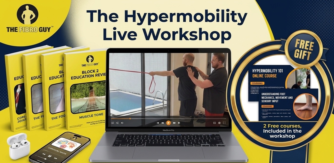
The Hypermobility Live Workshop is four weeks of breaking down the frameworks we use in our studios, with live Q&A so you can ask about your specific situation. Two free courses are included.
The Core Matters. “Engage Your Core” Often Makes It Worse
We need to be careful with this one. We’ve taken a longer swing at it in our piece on hypermobility core exercises, but the headline matters too much to skip here. The core does matter in hypermobility. It matters more than in most other groups. The mistake is in what people are usually taught to do about it.
Forget the six pack for a second. The core, when it comes to actual function, is a pressure system. Diaphragm at the top. Pelvic floor at the bottom. Transversus abdominis, the obliques, and multifidus around the sides and back. It’s a canister, and it works by managing pressure as you breathe and move. Inhale and the diaphragm drops, the pelvic floor gives a touch, the abdominals lengthen, the system makes room. Exhale and the whole thing recoils. It’s a constant, quiet rhythm running through the trunk every breath you take. It only works as a system. The components don’t get to be hired and fired independently.
Now drop a hypermobile nervous system into that canister. The joints aren’t giving the brain clean information about where they are. So the brain hedges. The working model, consistent with the proprioception data and what we see in studios every week, is that the system co contracts. It braces. It holds on to multiple muscle groups at once, substituting mechanical rigidity for the sensory clarity it can’t get. The pelvic floor very often gets recruited into that bracing. It tightens up. It stays on. It loses its ability to switch between contraction and release the way a healthy pelvic floor does. The diaphragm, meanwhile, can’t drop properly into an already braced abdomen, so the breath drifts up into the chest. Accessory neck and shoulder muscles do more work. The canister stops moving like a canister and starts moving like a pressurised box. This is what people in this space call breathing pattern dysfunction, and it’s far more the norm than the exception in those with hypermobility.
The pelvic floor side of this isn’t hypothetical. In a large international survey study of cisgender women with Ehlers-Danlos syndrome, stress urinary incontinence was reported by sixty per cent, urgency incontinence by fifty four per cent, pelvic organ prolapse by twenty one per cent, and pelvic pain by seventy one per cent [16]. The pelvic floor is, by any measure, deeply affected in this population. The clinical experience, ours and many others’, is that a meaningful chunk of that comes not from a pelvic floor that’s too weak to switch on, but from a pelvic floor that’s been switched on for years and doesn’t know how to switch off. That’s a working model rather than a single trial finding, but it’s consistent with the wider pattern of over recruitment and impaired sensation that runs through the rest of the picture.
Hormonal conditions add a layer to exercise prescription too. PCOS, renamed PMOS in 2026, brings its own metabolic considerations that need to interact with hypermobile programming. We cover the joined-up picture in PCOS to PMOS and hypermobility.
Now put yourself in that body and listen to the standard advice. Engage your core. Pull the navel in. Brace before you move. Kegel for the pelvic floor. What you’re being told to do, in plain terms, is add more tension to a system that’s already over tense. You’re telling someone whose jaw is already clenched to bite down harder. The diaphragm has even less room to descend. The pelvic floor has even less ability to lengthen on the inhale. The breath gets even more shallow. The bracing strategy that was already a problem gets reinforced. Pain often goes up. Performance often gets worse. People walk away thinking they failed at the exercise, when actually the exercise failed them.
The research is supportive here, at least in part. The Hodges 1996 transversus abdominis work is the seed paper most of “engage your core” was grown from [13]. The follow up evidence, including Allison’s 2008 work on directional asymmetric feedforward, and Lederman’s blunt 2010 myth of core stability review, has progressively walked the original story back [14, 15]. Lederman went so far as to flag potential harm from chronic tensing of trunk muscles. The argument isn’t that the core doesn’t exist, or that breathing and trunk control don’t matter. The argument is the opposite. They matter so much that getting them wrong has consequences, and “isolate, brace, hold” is the wrong instruction for a system that needs to be moving, breathing, and responding.
So what does actually work for the hypermobile core? Boringly, the answer doesn’t make a good Instagram post. You teach the diaphragm to descend properly. You let the pelvic floor relax as well as contract. You re establish the seesaw between them so the canister can actually work as a canister. You build trunk control through whole body action, breathing, and integrated movement, not through static bracing held against a stopwatch. The take home for here is that the core matters enormously in hypermobility. It just doesn’t respond to the instruction most people have been given for it.
What Actually Has To Change
If the old model is wrong in the ways we’ve laid out, then the new model has to do a few specific things. We’ve spelled out the full mechanics in the motor learning piece, but the shape of it is this.
First, signal before load. Before you ask a hypermobile shoulder to hold a heavy weight, you need to give the brain a clear, repeatable sense of where the shoulder is and what it’s doing. That means tactile input, that means external focus, that means working in ranges where the system can predict accurately and gradually pushing the edge out. Stick a load on a fuzzy signal and the system braces, co-contracts, and tightens. Which is exactly the readiness tone problem we covered in the muscle tone article.
Second, vary the conditions, not the exercises. This one trips people up. Variability doesn’t mean “do a different exercise every week”. It means take the same fundamental action, the same pattern, and vary the speed, the range, the surface, the focus, the starting position. You’re building a rule the nervous system can apply across contexts, not collecting a folder of one off party tricks.
Third, train actions, not muscles. The brain doesn’t have a folder called “transversus abdominis” with a button to press. It has folders called “reach”, “step”, “stand up from a chair”, “catch yourself when you trip”. Those are the units of training. If your exercise programme is a list of muscles, it’s organised by anatomy, not by how movement is built. That doesn’t make it useless. It makes it inefficient, and for hypermobile bodies, inefficient means a lot of work for not much carry over.
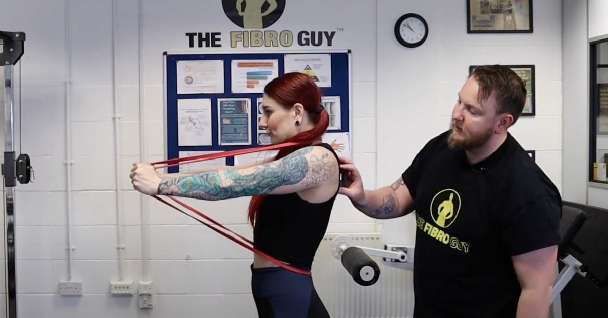
Fourth, use the body’s own information channels. Tactile cues, where a band, a strap, a hand on the skin gives the nervous system a clearer read on where a joint is, are some of the most useful tools we’ve got. We’ll cover them in detail in Part 2, but the principle is simple. Where the signal is thin, you can thicken it from the outside. Once the brain has cleaner information, the motor output gets cleaner too. There’s case level work showing neuromuscular taping applied to the lumbar spine and knees was associated with improved ankle position at initial contact during walking in an EDS case study [18], and pilot work showing somatosensory orthoses improve postural control in hEDS [19]. These aren’t magic tricks. They’re sensory cleaners.
Fifth, accept the timeline. Motor learning works on weeks and months, not days. The first two months of a properly designed programme can look like nothing’s happening on the outside, because what’s happening is happening inside the system. That’s normal. It’s not a sign the programme is wrong. It’s the period where the maps and predictions are being rebuilt. After that, the changes start showing up in real life rather than in the exercise itself, which is the whole point.
Where This Leaves Pilates, Yoga, And Everything Else
People are going to keep doing Pilates. People are going to keep doing yoga. People are going to keep going to the gym. None of that is the enemy. The frame we’re pushing here isn’t anti exercise. It’s anti misdiagnosis. If your exercise method is built on a wrong story about how the body works, it doesn’t matter how hard you grind, the outcome won’t match the effort. The pattern won’t generalise. The joints won’t get more reliable. And after a few years, you’ll have lovely glutes and the same dislocating shoulder.
So use whatever movement you enjoy. Keep moving. But if you want the dislocations, the subluxations, the bracing, and the constant low grade tension to actually change, you’ll need to step outside the “build the muscle and brace the core” frame.
Where The Companion Piece Goes Next
If you want to go deeper on the brain side of all this, our companion piece on how your brain maps your joints picks up where this one finishes. It covers the sensory inputs, the mechanoreceptors, the muscle spindles and Golgi tendon organs, the cutaneous and vestibular and visual systems, and how all of that lands as cortical maps. It also gets into why those maps are plastic, why they smudge in chronic pain, and how tactile cues plug into them as a deliberate training tool. The cortical map work is where most of the new neuroscience lands, and it’s the bit that’s been under represented in the hypermobility space.
A Few Loose Ends
Three things worth saying before we close. First, the people who run our studios see the result of the “build more muscle” approach every week. People who’ve done physiotherapy, who’ve done Pilates, who’ve done gym programmes, who’re stronger than they were five years ago and still subluxing. Strong, not stable. That isn’t a freak occurrence. It’s the predictable outcome of training a body using a model that doesn’t match how the body actually controls itself.
Second, this isn’t an argument against effort. The work we ask people to do is harder, in some ways, than a gym programme. It’s harder because it asks for attention, precision, and patience, not just sweat. When it comes to building a body that doesn’t fall apart on you, the work is real. It just isn’t the work you’ve been told to do.
Third, you don’t need to give up your gym, your yoga, your weights, or your Pilates class. You need to add a sensorimotor layer underneath whatever you already enjoy, so the joints get a chance to learn how to talk to the brain again. That’s the part the old model has been missing.
If you’ve found this useful, the companion piece on cortical maps and proprioception picks the thread up. And if you want the whole thing applied to your body, with feedback, that’s what our work at the studios and online is for.
Frequently Asked Questions
Is strength training useless for hypermobility?
No. Strength training matters. The argument here is about order. If the sensory and motor learning layer underneath isn’t built, strength training delivers nowhere near what it should. Get the signal right first, then load it.
Should I quit Pilates?
Not unless you want to. The point isn’t that Pilates is harmful. It’s that, when it comes to building genuine joint control in a hypermobile body, the operating principles of classical Pilates aren’t aligned with what motor learning research tells us is needed. Use it if you enjoy it. Add a sensorimotor programme underneath if joint instability is your main issue.
What about yoga?
Same answer with a small caveat. Yoga can build awareness and confidence with movement, and the breath work is genuinely useful. The risk for hypermobile people is the deep stretching side, where end range positions get held passively and the joint capsule absorbs load it shouldn’t. We’ve covered this in detail in our piece on stretching and hypermobility.
Do I need a coach or can I do this on my own?
Honestly, the bit that’s hardest to self coach is the early stage of getting the cues right and noticing when a movement is “clean” versus “looks clean but isn’t”. A coach who understands sensorimotor work shortens that bit. People do make progress on their own, but it tends to be slower and patchier. The principles in this article are the principles either way.
References
1. Smith TO, Bacon H, Jerman E, Easton V, Armon K, Poland F, Macgregor AJ. Physiotherapy and occupational therapy interventions for people with benign joint hypermobility syndrome: a systematic review of clinical trials. Disability and Rehabilitation. 2014;36(10):797-803. doi:10.3109/09638288.2013.819388
2. Palmer S, Bailey S, Barker L, Barney L, Elliott A. The effectiveness of therapeutic exercise for joint hypermobility syndrome: a systematic review. Physiotherapy. 2014;100(3):220-227. doi:10.1016/j.physio.2013.09.002
3. Palmer S, Cramp F, Clark E, Lewis R, Brookes S, Hollingworth W, et al. The effectiveness of conservative interventions for the management of syndromic hypermobility: a systematic literature review. Clinical Rheumatology. 2020;39(10):2965-2976. doi:10.1007/s10067-020-05284-0
4. Buryk-Iggers S, Mittal N, Santa Mina D, Adams SC, Englesakis M, Rachinsky M, et al. Exercise and Rehabilitation in People With Ehlers-Danlos Syndrome: A Systematic Review. Archives of Rehabilitation Research and Clinical Translation. 2022;4(2):100189. doi:10.1016/j.arrct.2022.100189
5. Scheper MC, de Vries JE, Verbunt J, Engelbert RH. Chronic pain in hypermobility syndrome and Ehlers-Danlos syndrome (hypermobility type): it is a challenge. Journal of Pain Research. 2015;8:591-601. doi:10.2147/JPR.S64251
6. Scheper MC, Juul-Kristensen B, Rombaut L, Rasmussen EE, Verbunt J, Engelbert RH. Disability in Adolescents and Adults Diagnosed With Hypermobility-Related Disorders: A Meta-Analysis. Archives of Physical Medicine and Rehabilitation. 2016;97(12):2174-2187. doi:10.1016/j.apmr.2016.02.015
7. Pacey V, Tofts L, Adams RD, Munns CF, Nicholson LL. Proprioceptive acuity into knee hypermobile range in children with Joint Hypermobility Syndrome. Pediatric Rheumatology. 2014;12:40. doi:10.1186/1546-0096-12-40
8. Clayton HA, Jones SAH, Henriques DYP. Proprioceptive precision is impaired in Ehlers-Danlos syndrome. SpringerPlus. 2015;4:323. doi:10.1186/s40064-015-1089-1
9. Akkaya KU, Burak M, Yildiz R, Yildiz A, Elbasan B. Examination of foot sensations in children with generalized joint hypermobility. Early Human Development. 2023;180:105755. doi:10.1016/j.earlhumdev.2023.105755
10. Fitts PM, Posner MI. Human Performance. Belmont, CA: Brooks/Cole; 1967.
11. Schmidt RA. A schema theory of discrete motor skill learning. Psychological Review. 1975;82(4):225-260. doi:10.1037/h0076770
12. Wulf G. Attentional focus and motor learning: a review of 15 years. International Review of Sport and Exercise Psychology. 2013;6(1):77-104. doi:10.1080/1750984X.2012.723728
13. Hodges PW, Richardson CA. Inefficient muscular stabilization of the lumbar spine associated with low back pain. A motor control evaluation of transversus abdominis. Spine. 1996;21(22):2640-2650. doi:10.1097/00007632-199611150-00014
14. Allison GT, Morris SL, Lay B. Feedforward responses of transversus abdominis are directionally specific and act asymmetrically: implications for core stability theories. Journal of Orthopaedic and Sports Physical Therapy. 2008;38(5):228-237. doi:10.2519/jospt.2008.2703
15. Lederman E. The myth of core stability. Journal of Bodywork and Movement Therapies. 2010;14(1):84-98. doi:10.1016/j.jbmt.2009.08.001
16. Kciuk O, Li Q, Huszti E, McDermott CD. Pelvic floor symptoms in cisgender women with Ehlers-Danlos syndrome: an international survey study. International Urogynecology Journal. 2022;34(2):473-484. doi:10.1007/s00192-022-05273-8
17. Pereira NS, Bobbio TG, Davis E, Hernandez SSS. Movement variability in Pilates: a scoping review. Frontiers in Psychology. 2023;14:1195055. doi:10.3389/fpsyg.2023.1195055
18. Camerota F, Galli M, Cimolin V, Celletti C, Ancillao A, Blow D, Albertini G. The effects of neuromuscular taping on gait walking strategy in a patient with joint hypermobility syndrome/Ehlers-Danlos syndrome hypermobility type. Therapeutic Advances in Musculoskeletal Disease. 2015;7(1):3-10. doi:10.1177/1759720X14564561
19. Dupuy EG, Leconte P, Vlamynck E, Sultan A, Chesneau C, Denise P, Besnard S, Bienvenu B, Decker LM. Ehlers-Danlos Syndrome, Hypermobility Type: Impact of Somatosensory Orthoses on Postural Control (A Pilot Study). Frontiers in Human Neuroscience. 2017;11:283. doi:10.3389/fnhum.2017.00283
The Hypermobility Fundamentals Bundle
The Hypermobility Fundamentals Bundle is a structured education and movement pathway that starts at the bottom and builds upwards, grounded in motor learning and sensory motor science. Designed for people with hypermobility and persistent pain, it focuses on how strength and stability are organised and learned by the nervous system, beginning with the feet and progressing through sensory input, coordination, and motor learning. The courses are intended to be completed in sequence, with each one establishing the concepts and skills needed for the next.