If you’ve been diagnosed with Parsonage-Turner syndrome and you’re wondering what exercises you should be doing, you’ve probably already discovered that the standard advice is frustratingly vague. “Strengthen your shoulder.” “Do some stretches.” “Work on your range of motion.” And whilst none of that is wrong exactly, it misses something fundamental about what PTS actually does to your body. This isn’t a rotator cuff tear. It isn’t a frozen shoulder. The brachial plexus has been attacked, and the consequences of that go far beyond the muscles themselves.
This article is part of our comprehensive guide to Parsonage-Turner syndrome.
So, this article is about exercise and rehabilitation for Parsonage-Turner syndrome, but probably not in the way you’d expect. We’re not going to hand you a list of 10 shoulder exercises and send you on your way. What we’re going to do is explain why the standard “strengthen and stretch” approach often falls short for those with PTS, what a neurology-first programme actually looks like, and why tactile cues are one of the most underused and underappreciated tools in the whole rehabilitation process. We’ve worked with people who spent months doing conventional physiotherapy with very little progress, and in almost every case the missing piece was the same: nobody was addressing what had happened in the brain.
We’ve covered the full overview of PTS in our comprehensive hub page, and the specific phases of recovery in another article. This one is about the practical side, what to actually do, when to do it, and why the sensory component of rehab matters just as much as the physical one.
Why Traditional Physiotherapy Falls Short
Let’s be really clear about something. Traditional physiotherapy isn’t bad. The exercises your physio gives you for shoulder stability, range of motion and strengthening are all necessary parts of PTS recovery. But they’re not sufficient on their own, and here’s why.
When the brachial plexus is damaged by neuralgic amyotrophy, it doesn’t just affect the muscles. It disrupts the entire communication system between your brain and your arm. The signals that tell your brain where your arm is in space, what your hand is touching, how it’s moving, all of those get scrambled or go silent. And your brain responds to that silence by reorganising itself.
A 2022 fMRI study of 39 patients with neuralgic amyotrophy found decreased cerebral activity in visuomotor areas of the brain, and crucially, a correlation between this decreased activity and persistent pain [1]. That’s not a peripheral problem. That’s a central nervous system problem. The technical term is maladaptive plasticity, your brain’s maps of the affected limb become distorted because the input they rely on has been disrupted.
So what happens when you try to strengthen a shoulder whose cortical representation is fuzzy? You get compensatory movement patterns. The brain can’t properly plan the movement it’s supposed to be doing, so it recruits other muscles to help. You end up hiking your shoulder, rotating your trunk, doing anything to complete the movement except using the muscles you’re trying to target. Sound familiar?
Cup and colleagues found in a 2013 study that standard physiotherapy approaches were ineffective or actually worsened symptoms in more than 50% of PTS patients [8]. That’s not a small number. And it reflects something we see in practice regularly: people who’ve been doing months of conventional shoulder rehab with no meaningful progress, because the cortical component was never addressed. This is exactly why, when it comes to PTS rehabilitation, we take a neurology-first approach. It’s the same approach we use with those with hypermobility and other conditions where the nervous system is the primary driver.
The Neurology-First Approach
The idea behind neurology-first rehabilitation is straightforward, even if the application takes more thought. Before you can effectively strengthen muscles, you need to make sure the brain knows those muscles exist and can communicate with them properly. Before you ask for force production, you need accurate sensory feedback. Before you load a movement pattern, that movement pattern needs to actually be planned correctly by the motor cortex.
A scoping review of cortical plasticity interventions in peripheral nerve injury rehabilitation identified seven distinct intervention categories targeting brain-level reorganisation [2]. These include sensory re-education, mirror therapy, mental motor imagery, cross-modal sensory substitution, action observation with simultaneous peripheral nerve stimulation, and activity-based sensory approaches. What they all have in common is that they’re working on the brain’s representation of the limb, not just the muscles.
Research on mirror therapy after peripheral nerve repair has shown greater cortical activation in multimodal association cortices compared to classical sensory relearning alone, though it’s worth noting that this particular study involved just six participants, so we should treat it as preliminary evidence rather than settled science [3]. Animal research has also shown that enhancing plasticity in central networks improves both motor and sensory recovery after nerve damage, though obviously the translation from rat models to human rehabilitation isn’t direct [9]. A 2025 review of modern rehabilitation methods for peripheral nerve and brachial plexus injuries specifically noted that targeted physical activity enhances intracellular regenerative mechanisms and helps ensure appropriate cortical representation during recovery [11]. The broader picture is consistent: targeting the brain’s organisation alongside the peripheral recovery makes a genuine difference to outcomes.
Now, I want to be honest here. The evidence base for PTS-specific exercise protocols is still thin. Most of the research comes from peripheral nerve injury rehabilitation more broadly, or from brachial plexus studies that aren’t exclusively PTS. But the underlying neurology is the same and the principles transfer directly. We’ve seen this work with our PTS clients, and the research supports the mechanism even if we’re waiting for PTS-specific randomised trials.
What Are Tactile Cues?
Before we get into the phase-by-phase exercise guidance, it’s worth spending some time on tactile cues, because they’re central to how we work with PTS clients and they’re probably the thing that most distinguishes our approach from what you’ll find elsewhere. You might’ve heard the term if you’ve spent any time looking into hypermobility rehab. It gets thrown around quite a lot in our world, and for good reason.
At their simplest, tactile cues are pieces of physical sensory input, things like touch, pressure, resistance, or texture, that give your brain clearer information about where a joint is and how it’s moving. But that description doesn’t quite capture it. This isn’t just about “touching” something. It’s strategic sensory feedback designed to change how your nervous system processes movement.
Think of it this way. Your brain builds maps of your body based on the sensory information it receives. These maps, stored in the somatosensory cortex, determine how accurately your brain knows where each body part is in space, from your elbows to your wrists to your shoulder blades. When the sensory input is clear and consistent, the maps are detailed and precise. When it’s not, the maps become blurry. And blurry maps mean imprecise movement control [1].
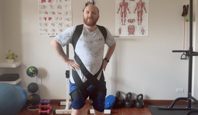
Tactile cues work by adding extra sensory information to sharpen those maps. A resistance band wrapped around the trunk during a shoulder exercise isn’t there for resistance in the traditional sense, it’s there because the pressure gives your brain additional data about where your ribcage is relative to your shoulder blade. A strip of KT tape isn’t just “support”, it’s a constant stream of sensory input telling the brain where that joint is. We’ve written more about the mechanisms behind taping if you want to dig into that specifically.
We’ve been using tactile cues with those with hypermobility for years, and the results speak for themselves. But the science behind why they work is genuinely fascinating, and it applies to far more than just hypermobility. For PTS specifically, they become one of the most important tools in the entire rehabilitation toolbox.
The Science Behind Tactile Cues
So why does adding external sensory input actually change how the brain controls movement? The answer lies in something called cortical mapping, and it’s one of the most important concepts when it comes to understanding rehabilitation after any kind of nerve injury.
Your somatosensory cortex contains a map of your entire body. You might have seen those odd-looking diagrams of a “homunculus”, a distorted human figure where the hands and face are enormous because they have the most sensory nerve endings. The size of each body part on that map corresponds to how much sensory input the brain receives from it. More input means a bigger, more detailed map. Less input means a smaller, fuzzier one.
Now here’s where it gets interesting. These maps aren’t fixed. They change based on experience, injury, and the quality of sensory information coming in. This is neuroplasticity in action. And it cuts both ways. If you lose sensory input from an area, say through nerve damage or through the poor proprioception that comes with hypermobility, the cortical map for that area shrinks and becomes less defined [2]. The brain essentially starts to “forget” the detailed layout of that body part.
However, the really encouraging research shows that this process is reversible. A landmark primate study by Florence and colleagues in 2001 demonstrated that sensory enrichment, providing enhanced tactile stimulation to an area, can actually restore cortical maps after nerve injury [4]. They showed that the brain’s representation of a body part could be rebuilt by giving it richer sensory input. That’s measurable cortical change. Now, it was a macaque study rather than a human trial, so we should be appropriately cautious about direct translation. But the mechanism it demonstrated has been replicated and supported in the wider cortical plasticity literature since.
More recent work has reinforced this. Zink and Philip’s 2020 scoping review of cortical plasticity interventions for peripheral nerve injury identified several rehabilitation approaches based on this principle, including traditional sensory re-education, activity-based sensory re-education, cross-modal sensory substitution, mirror therapy, mental motor imagery, selective deafferentation, and action observation with simultaneous peripheral nerve stimulation [2]. All of these share a common thread: they work by providing the brain with enhanced sensory information to rebuild its internal maps. Tactile cues are our practical tool for delivering that enhanced input. They’re the bridge between the laboratory research on cortical plasticity and what actually happens in a rehab session.
Why Tactile Cues Matter for Hypermobility
If you’ve got hypermobility or EDS, there’s a good chance your proprioception isn’t great. This is well documented in the research. Proprioception, your body’s ability to sense where it is in space without looking, relies on receptors in your joints, muscles and tendons. When those joints move further than expected, or when the connective tissue is laxer than typical, the signals those receptors send become less reliable [5].
We often describe this to our clients using a map analogy. Imagine trying to navigate a city using a really low-resolution map. You can see the rough layout, maybe the main roads, but you can’t see the side streets, the landmarks, or the details that would help you move around accurately. That’s what proprioception is like for a lot of those with hypermobility. The brain has a general idea of where the joint is, but the resolution is too low for precise control.
And what happens when the brain can’t precisely control a joint? It compensates. You get muscle guarding, co-contraction, fear of movement, and those compensatory patterns that so many of our clients recognise, things like rib subluxations from altered shoulder mechanics, or postural adaptations that develop over time. The anxiety that often accompanies hypermobility isn’t separate from the proprioceptive problem, it’s partly driven by it. When your brain doesn’t trust the information it’s getting about where your body is, it ramps up the threat response.
So tactile cues increase the resolution of that map. By adding external sensory input, pressure from a band, feedback from tape, contact with a textured surface, you’re giving the brain more data points. It’s like upgrading from a blurry map to a detailed one. And when the brain has a clearer picture of where a joint is, it can control it more effectively.
This is why we use tactile cues for everything from core stability work to shoulder blade rehabilitation to lower limb motor control. The principle is the same regardless of the body part: give the brain better information and it produces better movement output. We’ve linked to our full exercise guide for those with hypermobility if you want to see how these principles apply more broadly.
Side note, and this is something I find genuinely interesting. We’ve noticed over the years that tactile cues seem to work almost immediately for some people. Not in terms of structural change obviously, that takes time, but in terms of movement quality. Someone who’s been struggling with a particular movement will put a band on and suddenly the movement cleans up. It’s as if the brain was always capable of producing the movement, it just didn’t have enough information to do it confidently. Whether that’s a proprioceptive thing or a confidence thing, or both, I’m not entirely sure. But it’s something we see again and again.
Tactile Cues for Nerve Injury Recovery
Now, this is where things get particularly relevant for anyone recovering from PTS. The reason is that nerve injury creates an even more dramatic version of the same cortical mapping problem we just discussed.
The Full Body Fibro Tool Box
The tool box brings together education on pain and movement alongside a wide range of practical movement, stretching and sensory based resources. Rather than offering a single plan or set of rules, it gives you options you can return to and use as needed.
When the brachial plexus is damaged, the sensory pathways between the arm and the brain are disrupted. The brain stops receiving reliable signals from the affected area. And just like with hypermobility, the cortical maps become disordered. But with nerve injury, it’s often more severe. The 2022 fMRI study by Lustenhouwer and colleagues found decreased cerebral activity in visuomotor areas of the brain in patients with neuralgic amyotrophy, and this directly correlated with persistent pain and dysfunction [1]. The worse the cortical changes, the more pain people experienced.
This is what’s called maladaptive plasticity. The brain adapts to the loss of input, but it adapts in ways that actually make things worse. The cortical map doesn’t just become blurry, it becomes distorted. And this is why some people with PTS still have functional problems even after the nerve itself has regenerated. The peripheral hardware has been repaired, but the central software hasn’t been updated [6]. Our article on brain changes after nerve injury covers this in much more detail if you want the full picture.
However, and this is the encouraging part: if tactile input can restore cortical maps after nerve injury, which Florence et al. demonstrated in that 2001 primate study, then there’s a clear rationale for using tactile cues during PTS recovery [4]. You’re not just waiting for the nerve to regrow and hoping the brain figures it out. You’re actively feeding the brain sensory information to help it rebuild those maps as reinnervation happens.
The same principles we use for those with hypermobility apply directly to PTS rehabilitation, but arguably they matter even more here. When it comes to nerve injuries, the cortical mapping problem is often the thing that determines long-term outcomes. Research on brachial plexus rehabilitation has specifically identified tactile interaction with different textures and temperatures as effective for sensory re-education [7]. Mirror visual feedback has also been shown, in that small 6-participant preliminary study, to create stronger motor-sensory connections than classical rehab alone [3]. It’s a promising finding, but the sample size means we should treat it as a signpost rather than a conclusion.
For those with hypermobility who are also recovering from PTS, this matters even more. You’re dealing with two layers of proprioceptive deficit: the baseline hypermobility-related reduction in sensory clarity, plus the nerve injury disruption on top. When it comes to PTS rehabilitation in this population, tactile cues aren’t optional, they’re essential. The recovery phases article explains exactly when tactile cues become most relevant in the rehabilitation timeline.
How to Use Tactile Cues in Practice
Right, so the science makes sense. But what does this actually look like in a rehab session? There are several approaches and the specific technique depends on what you’re trying to achieve and which body part you’re working with. Here are the main methods we use.
Resistance Bands for Proprioceptive Feedback
This is probably the most common way we use tactile cues. A light resistance band wrapped around the trunk or placed around a joint creates constant pressure feedback. For shoulder blade work, a band around the ribcage gives the brain a clear reference point for where the thorax is, making it much easier to control scapular movement. The band isn’t there to make the exercise harder. It’s there to make the movement clearer.
Taping for Sustained Input
When it comes to providing a constant tactile cue throughout the day, KT tape and similar taping methods are really useful. The tape sits on the skin and provides ongoing sensory feedback about joint position. It’s not providing structural support in any meaningful way, the adhesion simply isn’t strong enough for that, but it doesn’t need to. The sensory input is the point. For those recovering from PTS, taping the shoulder girdle area can provide that constant stream of proprioceptive information while the nerves are recovering. Compression garments work on a similar principle, providing broad area tactile feedback across a larger surface.
Unstable Surface Training
Varying the surface your hands or feet are in contact with during exercises adds an additional layer of sensory input. Something as simple as performing an exercise on a slightly textured mat rather than a smooth floor gives the brain more to work with. For upper limb work during PTS recovery, placing the hand on a varied surface during weight-bearing exercises provides that richer sensory environment we’re looking for. It doesn’t need to be fancy. Different textures, different temperatures, and different surface densities all serve the same purpose: giving the brain more data about where the body is in space.
Manual Pressure and Guided Touch
In clinic, we use manual tactile cues a lot. Placing a hand on a specific muscle or joint while someone performs a movement helps the brain “tune in” to that area. It’s a bit like pointing at something on a map, you’re directing the brain’s attention to a specific body part. This is particularly useful in the early stages of PTS recovery when motor control is significantly compromised. We talk about pain management approaches in the acute phase, but once someone moves into rehabilitation, manual tactile cues become a core part of the work.
Progressive Complexity
The key thing with tactile cues is progression. You start simple, a band around the trunk, some tape on the shoulder, basic movements with manual guidance. Over time you increase the complexity. The tactile cues become less overt as the brain’s maps improve, the movements become more challenging, and you gradually reduce the external feedback as the internal systems take over. It’s not a replacement for exercise, it’s an enhancement that makes the exercise more effective.
For those with hypermobility who are also recovering from PTS, this progressive approach is especially important. You’re dealing with two layers of proprioceptive deficit, and the pace of progression needs to reflect that. Don’t rush the removal of the external cues. The brain will tell you when it’s ready, and that shows up as consistent movement quality even without the band or tape in place.
Breathing and the Brachial Plexus
Something that gets overlooked in most PTS exercise guidance is the impact on breathing. The muscles of the shoulder girdle, the scalenes, upper trapezius, and serratus anterior, they all contribute to breathing mechanics as well as shoulder movement [10]. When these muscles are compromised by PTS, breathing patterns can be affected, and altered breathing patterns contribute to fatigue, which then affects your capacity for rehabilitation.
The scalene muscles in particular deserve attention here. They’re responsible for elevating the upper ribs during inhalation, but they also sit in close anatomical proximity to the brachial plexus. The trunks of the brachial plexus emerge between the anterior and middle scalenes, and the phrenic nerve, which drives diaphragm movement, runs alongside this structure. This means that scalene involvement in PTS isn’t just a shoulder and neck problem. It can have knock-on effects for breathing mechanics that most standard protocols never consider [10].
Likewise, the phrenic nerve itself is derived from the C3, C4, and C5 nerve roots. In cases of PTS affecting the upper brachial plexus, there can occasionally be involvement that contributes to diaphragmatic dysfunction, which is part of why some people with PTS report unexpected fatigue and breathlessness that doesn’t quite fit with their level of physical activity.
If you’re finding that you fatigue much faster than expected during exercises, or if you’re getting breathless with upper body work, this is worth raising with your neurologist or physiotherapist. It’s not just deconditioning. There may be a genuine mechanical reason for it, and addressing breathing patterns should be part of the rehabilitation programme rather than an afterthought.
What Tactile Cues Don’t Do
We should be honest here because there’s a risk of overselling this. Tactile cues are not a miracle fix. They don’t heal damaged nerves. They don’t replace the time that nerve regeneration takes after PTS, and they don’t substitute for a properly structured rehabilitation programme.
What they do is optimise the conditions for recovery. They give the brain the best possible information during the rehabilitation process. Think of it like this: if nerve regeneration is the road being rebuilt, tactile cues are making sure the GPS is updated as the new roads come online. Without that updating process, you can have a perfectly regenerated nerve but still struggle with function because the brain’s maps haven’t caught up [6].
It’s also worth saying that the research on tactile cues specifically for PTS is still emerging. Most of the evidence comes from broader peripheral nerve injury research and from cortical plasticity studies. The principles are sound and the mechanisms are well understood, but we need more PTS-specific trials. We’re honest about that because we think transparency about the evidence matters more than making bold claims. If you’ve been dealing with persistent PTS pain for a while, you’ll probably appreciate that honesty.
Likewise, tactile cues work best as part of a broader approach. They complement motor learning, graded exposure, pacing strategies, and addressing the psychological components of recovery. The brain’s role in pain and recovery goes well beyond just cortical mapping, and for those dealing with the emotional toll of a PTS diagnosis, that side of things needs to be addressed alongside the physical rehabilitation.
Phase-Based Exercise Progression
One of the biggest mistakes people make with PTS exercise is doing the right thing at the wrong time. Rehabilitation for Parsonage-Turner syndrome needs to match the phase you’re in, and pushing ahead too quickly is one of the fastest ways to set yourself back. If you haven’t read our article on the three phases of PTS recovery, it’s worth doing that alongside this one.
The Acute Pain Phase: Less Is More
During the acute phase, when pain is at its worst (and we’re talking 8 to 10 out of 10 for many people), the priority is pain management, not exercise. This is not the time to be doing shoulder strengthening work. Your brachial plexus is inflamed and the nerves are under attack. Forcing movement through that level of pain isn’t brave, it’s counterproductive.
However, that doesn’t mean you do nothing. Gentle range of motion work, if tolerable, can help prevent the shoulder from stiffening up completely. And very gentle nerve glides, not aggressive stretching, can help maintain neural mobility without provoking more inflammation. We cover pain management strategies in more detail in our acute pain management article.
The key is listening to your body. If a movement increases your pain significantly, stop. There’s a difference between mild discomfort during movement and the kind of sharp, burning neuropathic pain that tells you something is being aggravated. And here’s something I think about quite a lot. The acute phase of PTS is genuinely one of the most painful experiences people go through. I’ve worked with clients who’ve been through childbirth and said the PTS pain was worse. So when we say “rest,” we’re not being passive, we’re respecting the biology. That nerve inflammation needs time to settle before rehabilitation can begin in earnest.
Early Recovery: Sensory Re-Education and Motor Control
This is where things start to get interesting, and where the neurology-first approach really comes into its own. As the acute pain begins to settle and weakness becomes the dominant feature, your first instinct might be to start strengthening. But that instinct is wrong, or at least premature.
The first thing we focus on is sensory re-education. Before the brain can plan movements effectively, it needs to know what it’s working with. Sensory re-education involves providing the brain with rich, varied input from the affected area, different textures, temperatures, pressures, and movements that help rebuild the somatosensory map [2]. This is where tactile cues become the centrepiece of the programme rather than just an add-on.
It’s a bit like tuning an old radio, you’re adjusting the signal until the brain can hear it clearly again. Every time you provide a clear, consistent sensory input to the affected arm, you’re helping the cortical map sharpen up. And as that map improves, the quality of motor output improves alongside it. The two are not separate processes, they’re the same process.
Alongside sensory work, we introduce very gentle motor control exercises. These aren’t about strength, they’re about quality of movement. Can you activate the right muscles in the right sequence? Can you move the scapula without compensating through the trunk? The exercises might look easy from the outside, but when your neural pathways have been disrupted they’re genuinely challenging. We use a lot of tactile cues during this phase, bands, taping, manual guidance, to give the brain every possible advantage.
Shoulder Mapping and Progressive Loading
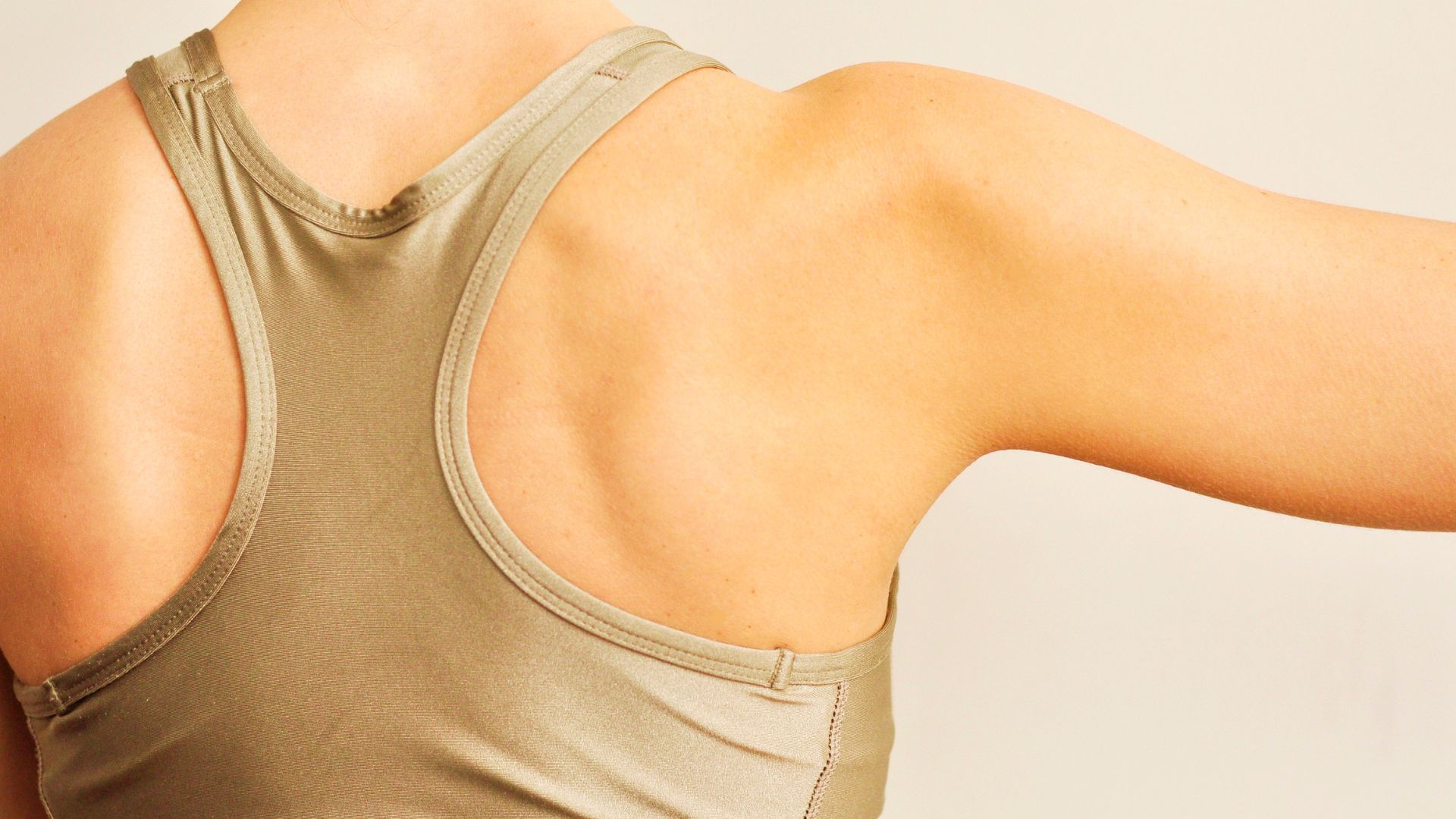
The shoulder is one of the most complex joints in the body, and when PTS hits, it’s usually the shoulder and scapular muscles that take the brunt of it. The suprascapular and long thoracic nerves are commonly affected, which means muscles like the supraspinatus, infraspinatus, and serratus anterior can be weakened or paralysed. Scapular winging, where the shoulder blade sticks out prominently, is one of the most visible signs of PTS.
However, just telling someone to “stabilise their scapula” when their brain can’t properly communicate with the muscles that do that job is a bit like telling someone to drive a car when the steering wheel is disconnected. The intention is there but the mechanism isn’t working.
What we do instead is shoulder mapping. This involves systematically reintroducing the brain to the muscles around the shoulder girdle using a combination of tactile cues, visual feedback, and carefully graded movements. We start with the basics: can you feel this muscle working when I tap it? Can you move the scapula in this direction with a band providing feedback? And we build from there.
The progression looks something like this: isolated scapular awareness with tactile input, then scapular control in supported positions, then control against gravity, then control during arm movement, then control under load. Each stage only progresses when the movement quality is there, not when the client feels like they can push through. This is motor learning in action, and it’s very different from the “3 sets of 10” approach that most shoulder rehab programmes use.
When it comes to PTS rehabilitation, there’s a critical distinction between strengthening and motor learning, and it matters more than most people realise. Strengthening assumes the muscle already knows what to do and just needs more capacity. Motor learning acknowledges that the communication pathway has been disrupted and the brain needs to relearn how to plan, execute, and refine movements. We progress based on movement competency, not just tolerance.
Side note, and this is a bit of a tangent but it connects to something important. I’ve always found it interesting that the rehabilitation world has fully accepted motor learning principles for stroke recovery, where cortical reorganisation is a central target, but hasn’t widely applied the same thinking to peripheral nerve injuries like PTS. The cortical changes are well documented in both conditions [1]. The mechanisms overlap significantly. And yet PTS rehab is still largely stuck in the “strengthen the weak muscles” thinking of 20 years ago. It’s changing slowly, but not fast enough.
Nerve Flossing and Neural Mobilisation
Now, we need to talk about nerve flossing because it comes up a lot in PTS discussions and there’s a right way and a wrong way to approach it. Neural mobilisation, sometimes called nerve gliding or nerve flossing, involves gentle movements designed to help nerves slide through their surrounding tissues. For those with PTS, this can be helpful for maintaining neural mobility and reducing the buildup of adhesions around the damaged nerves.
But here’s where people get into trouble. Aggressive nerve stretching during acute or early recovery can actually make things worse. Remember, the brachial plexus is inflamed and irritated. Pulling aggressively on irritated nerves is not going to help. What we want are gentle, controlled gliding movements that encourage the nerve to move without provoking a pain response.
When it comes to nerve flossing for PTS, the principle is “move the nerve, don’t stretch it.” The distinction matters. A glide involves tensioning one end of the nerve while releasing the other, so the nerve slides through the tissue without being placed under excessive stretch. It’s a much gentler approach and it’s far more appropriate for an inflamed brachial plexus.
We typically introduce nerve glides once the acute inflammation has started to settle, and we progress them very gradually. The fear of movement that develops after PTS pain can make people very anxious about nerve mobilisation, which is completely understandable. Starting gently and building confidence is more important than any specific exercise prescription.
Return to Function
The final phase is about bridging the gap between “rehabilitation exercises” and actual life. Can you reach a shelf? Carry a bag? Get back to your sport or hobby? This requires activity-specific retraining that builds on the motor learning foundation you’ve laid.
For those with hypermobility who are also dealing with PTS, this phase has an extra layer of complexity. The shoulder might have better neural control than before, but the underlying joint laxity is still there. We often find that PTS clients with hypermobility actually end up with better shoulder function than before the episode, because the rehabilitation process forces them to address the neurological component that was always an issue but never treated. That’s not a silver lining worth seeking out. But it is something we see.
Pacing remains essential throughout this phase. Recovery from Parsonage-Turner syndrome isn’t linear and pushing too hard on a good day often leads to a setback. The same principles apply here as with any neurological recovery: consistency over intensity, always.
Common Mistakes in PTS Rehabilitation
Having worked with a fair number of PTS clients, we see the same mistakes coming up repeatedly. And frustratingly, many of these are driven by well-meaning but outdated advice.
Strengthening too early. This is the most common one. Someone’s pain starts to settle, they feel a bit better, and they jump straight into resistance exercises. But the neural pathways haven’t been re-established yet. The result is compensatory movement patterns that can become entrenched and create their own problems, things like rib subluxations from altered shoulder mechanics or postural adaptations that stick around long after the PTS has improved.
Ignoring the sensory component entirely. This is the big one when it comes to why conventional physiotherapy falls short. If nobody addresses the cortical map changes, you can do all the strengthening in the world and still have functional limitations. The brain needs sensory input to rebuild its representation of the arm. Without that, motor planning stays disordered.
Pushing through pain. There’s a difference between working through mild discomfort and forcing movement through neuropathic pain. With PTS, the latter can sensitise the nervous system further and actually slow recovery. Those with experience of coat hanger pain or other neuropathic pain will understand why this matters.
Treating it like a simple shoulder injury. PTS is a neurological condition that happens to affect the shoulder. The rehabilitation needs to reflect that. A frozen shoulder protocol or a rotator cuff programme isn’t going to address the brachial plexus damage, the cortical changes, or the neuropathic pain component. Our diagnosis article covers why this misunderstanding is so common and what to do if you’ve been on the wrong track.
Neglecting the psychological impact. The anxiety and frustration that come with PTS are real barriers to rehabilitation. Months of severe pain followed by visible muscle wasting takes a significant psychological toll. The impact of medical trauma from the diagnostic process itself can compound this further. Addressing the emotional side isn’t optional, it’s a necessary part of getting better.
Frequently Asked Questions
During the acute pain phase, avoid structured exercise. Gentle range of motion work can begin as pain allows, but formal rehabilitation typically starts once the severe pain has settled and weakness becomes the dominant issue. This varies from person to person: for some that’s weeks, for others it’s months. Your physiotherapist or neurologist should guide this decision. We outline the full timeline in our recovery phases article.
The wrong exercise at the wrong time can absolutely slow your recovery. Aggressive strengthening during the acute phase, forceful nerve stretching, or pushing through significant neuropathic pain are all things that can be counterproductive. Cup and colleagues found that standard physiotherapy approaches were ineffective or worsened symptoms in more than 50% of PTS patients [8], which is exactly why phase-matched, neurologically-informed rehabilitation matters far more than generic strengthening protocols. However, appropriate exercise is one of the most important factors in PTS recovery. It’s the mismatch between exercise type and recovery phase that causes problems, not exercise itself.
The cortical mapping changes are measurable on fMRI, so there is a genuine neurological mechanism at work [1]. That said, there’s almost certainly a confidence component as well, people often feel more secure with a band or tape in place, and that reduction in threat perception probably contributes to improved movement. But we’d argue that’s not “just” placebo, it’s the nervous system responding to better information. Both mechanisms are real and both are useful.
Ideally, yes. PTS is uncommon enough that many physiotherapists won’t have seen it before, and as we’ve discussed, treating it like a standard shoulder injury misses the neurological component. Look for someone who understands nerve injury rehabilitation and cortical plasticity, or who has experience with brachial plexus conditions. The diagnosis article has more guidance on navigating the healthcare system with PTS.
Many tactile cue techniques can be used at home. Resistance bands, tape, and textured surfaces are all accessible without specialist equipment. However, the tricky part is knowing which cue to use, where to place it, and how to progress over time. Getting the initial setup right with someone who understands the approach makes a big difference. After that, most of the day-to-day work can be done independently.
The relationship between cortical mapping and pain is well established. The Lustenhouwer study showed that decreased visuomotor cortical activity correlated directly with increased persistent pain in neuralgic amyotrophy patients [1]. So interventions that improve cortical mapping may also help with pain. However, acute PTS pain is a different matter and needs to be managed with appropriate medical support. Tactile cues are more relevant during the rehabilitation and recovery phases than during the initial acute pain period. For the acute phase, our pain management article is the better starting point.
This is more common than you’d think. If you’ve been doing rotator cuff or frozen shoulder exercises that haven’t helped, don’t panic. The good news is that the neurology-first approach can still make a difference even if there’s been a delay. Cortical plasticity works in both directions, the brain can reorganise positively at any stage. Read our diagnosis article and speak to your doctor about getting the right investigations. Likewise, if you’ve been dealing with hypermobile elbow or wrist instability alongside your PTS, these all need to be considered together.
Yes. There’s growing evidence of a link between PTS and both COVID-19 infection and vaccination. We’ve covered this in detail in our article on PTS and COVID. The rehabilitation approach is the same regardless of the trigger, but it’s worth knowing about if your PTS developed in a post-COVID or post-vaccination context.
Some people notice immediate improvements in movement quality during a session once tactile cues are introduced. Lasting cortical changes take longer, weeks to months of consistent practice. For those recovering from PTS, the timeline is also influenced by the rate of nerve regeneration. When it comes to expectations, think of it as months not days for meaningful structural change. Nerve regeneration is estimated to progress at roughly 1 millimetre per day, and cortical reorganisation follows that pace. Patience and consistency matter far more than intensity.
If you’re in the early stages of PTS and feeling overwhelmed, start with our Parsonage-Turner syndrome hub page for the full picture. If you’re past the acute phase and ready to think about rehabilitation, the key takeaway from this article is that effective PTS exercise isn’t just about the muscles. It’s about the brain. The cortical maps need rebuilding. The sensory feedback needs restoring. And the movement patterns need relearning from the ground up.
Tactile cues are one piece of a much larger picture. From there, the articles on brain changes and sensory mapping, getting the right diagnosis, and the COVID-19 connection will give you the full context. For those with hypermobility who want to understand how the proprioceptive principles apply more broadly, our articles on proprioception and core stability cover the same framework applied to everyday hypermobility rehab.
We’ve seen this work with clients who’d been stuck for months, and whilst we can’t promise specific outcomes, the research on cortical plasticity in nerve injury is genuinely encouraging [2][6]. The brain wants to reorganise. You just need to give it the right input.
And if you’d like to work with us directly, whether you’re local or remote, our approach to PTS rehabilitation is built on everything described here: sensory mapping, tactile cues, progressive motor learning, and an understanding of how the brain adapts to nerve injury. You can explore what we offer through the academy.
Remember this: your brain can change. The maps can be rebuilt. And giving your nervous system better information is one of the most powerful things you can do for your recovery.
– The Fibro Guy Team –
Where to Go From Here
If you’re working through recovery from Parsonage-Turner syndrome, the principles we use in our studios (neurology-first rehab, sensory mapping, graded loading) are the same ones that underpin our Hypermobility 101 course. It covers the frameworks for rebuilding joint control and proprioception that are directly relevant to PTS recovery. Have a look through the full course library for everything we offer.