
- Hypermobile Neck Exercises - 8 November 2026
- Swollen legs and hypermobility: pooling, lymphoedema and lipoedema - 4 November 2026
- Walking with fibromyalgia - 2 November 2026
This article is part of our comprehensive guide to living with fibromyalgia.
Burning, gritty, aching eyes come up constantly in fibromyalgia, and the sight test nearly always comes back clean. A sight test checks two things: whether the optics work, and whether the structures look healthy. Almost nothing that makes eyes hurt in fibromyalgia lives in either of them. So a normal result is a genuinely useful piece of information, and what it tells you is where the problem isn’t.
There are four strands worth chasing, and you can have more than one of them at the same time. The nerves in the surface of the eye, which is where the dryness lives too. The neck, sending pain forwards into the socket. The way light gets handled once the signal is inside your head. And the tablets you were given for the pain, which is the one nobody brings up, and the one most likely to be fixable this month.
One thing before any of it, though. Eyes aren’t the organ to gamble with, and “it’s probably the fibro” is a guess dressed up as a diagnosis. There’s a list further down of symptoms that need somebody to look at them the same day, and it’s worth reading that bit even if you skip the rest.
What the eye test actually looked at
A standard sight test covers the optics, meaning the shape of your eye, the lens, the focus and how small a letter you can read, and then the structures, meaning the pressure inside the eye, the front surface under a slit lamp, which is a microscope on a stand with a bright slot of light in it, and the back of the eye through a dilated pupil. It’s a structural and optical survey, and it’s a good one.
What it doesn’t do is measure how your nervous system is handling what your eye is sending it. There’s no part of the test for that, and no number on the printout for it either. Which is why the results read the way they do. Visual acuity usually comes back normal in fibromyalgia even when there are plenty of other visual complaints alongside it [1][2].
That mismatch has been measured properly, and it’s the most consistent finding in this whole area. In dry eye clinics, people who also have a chronic pain condition report worse symptoms while their signs on examination, meaning what the clinician can actually see and measure, come back similar or milder than everybody else’s [3]. Look instead at what predicts the gap between how bad somebody says their eyes are and how bad they look under a lamp, and the same thing falls out [4]. Pool the studies and you get the same answer again [5].
Underneath that sits a smaller and messier thread, and it matches what people describe as it comes and goes. The letter chart stays normal while the finer stuff shifts in some studies: contrast sensitivity, meaning how well you pick out grey on grey rather than black on white, colour discrimination, depth perception, and reading letters in low contrast [6][7]. A five year cohort found that decline happening with no thinning of the retinal cell layer [6], and a later follow up reported both the decline and some thinning [7]. The structural side of it hasn’t settled [8], so keep that in mind.
Where the pain is coming from
The nerves in the surface of your eye
The front of your eye is packed with nerve endings, far more densely than your skin is, which is why a single eyelash under the lid is irritating out of all proportion to its size. Those endings do two jobs. They report damage and irritation, and they run the feedback loop that tells your tear glands to make tears and your lids to blink.
They can also be photographed. There’s a microscope used in research that images the nerve fibres in the cornea, the clear dome over the front of the eye, without touching it. So these small fibres can be counted in a living person rather than inferred from a sample taken somewhere else entirely.
Point it at fibromyalgia and the fibres come back thinner on average, with lower density and shorter total length than in people without it [9][10]. That’s been repeated across several groups. Pool the studies that went looking for small fibre involvement, whether by corneal imaging or by taking a small sample of skin, and it lands at around half of people [11][12], and sorting people by their corneal pictures gives you exactly that split, some abnormal and some not [13].
Half is the number to hold on to: It makes this a real finding and not the explanation for everybody, and it means a normal result wouldn’t rule your symptoms out either.
Where it stops being a tidy story is the correlation. If thinner corneal nerves were the whole story, then the thinner the nerves, the worse the pain would be, and that relationship keeps failing to appear: corneal measurements track pain intensity and sensory testing weakly or not at all [13][14], and the evidence on that link is mixed, with plenty of negative correlation studies and positive associations only in subgroups [15]. The nerve change is real and it isn’t doing the whole job on its own.
It also isn’t the same thing as classical small fibre neuropathy, which gets suggested a lot. Small fibre neuropathy is damage to the thinnest nerves in the body, the ones carrying pain and temperature and running the automatic jobs like sweating, and it has its own workup and its own diagnosis. Compared head to head, corneal nerve density and length came out similar between fibromyalgia and small fibre neuropathy, while branch density was more reduced in small fibre neuropathy [16].
What that adds up to in practice: If your eyes burn, sting and feel raw while the surface looks healthy, there’s a measurable reason that can be true, and it sits in the wiring of the surface, rather than the surface itself.
Your neck
This is the one our readers describe best, usually as pain running from the base of the skull straight into the eyes.
The nerve supplying your face, your forehead and the area around your eye is the trigeminal nerve. The nerves supplying the top of your neck arrive separately, through the upper cervical roots. Those two systems don’t stay separate for long: they land in the same pool of cells in the brainstem, and once two inputs share a relay, the brain has no reliable way of telling which one sent the signal.
So, a problem in the upper neck can refer pain forwards into the forehead, the temple and the orbit, the bony socket the eye sits in, and it can arrive with light sensitivity attached, close enough to migraine to get mistaken for it [17]. Cervicogenic is the word for it, meaning generated in the neck and felt somewhere else. That work comes out of the cervicogenic headache literature rather than out of fibromyalgia studies [18], which is worth knowing, because it means you’re borrowing a well described mechanism rather than reading your own population’s data.
It’s also the most workable of the three, and the one we can usually do something about in a studio. For cervicogenic pain the evidence sits with a combination of exercise, manual therapy and the rest of a proper physiotherapy programme, with moderate to low certainty for short term reductions in how often the headaches come and how bad they are [19][20][21]. Short term and moderate certainty is a long way from a cure. It’s still a considerably better bet than a fourth bottle of drops aimed at a surface that was never the problem.
If your eye pain sits behind the eye rather than on it, comes with a stiff neck, gets worse after a day at a desk and eases when you lie down with your head supported, chase this strand first.
The light
Photophobia is the medical word and almost nobody uses it. People say the sun is unbearable, or that the big light has been off for weeks. Take the word anyway, because it’s the one that gets you taken seriously in a clinic.
For light to hurt you, two separate systems have to meet somewhere, because light hitting the retina is a visual signal and pain is its own system. They do meet. Light evoked pain recruits trigeminal pain networks in the brainstem and the cortex, rather than only the visual pathway [22][23]. The route runs from the light signal in the retina, through the thalamus and hypothalamus, two relay stations deep in the brain, and on into trigeminal circuitry, with CGRP among the chemical messengers involved [24][25]. That’s the same convergence that lets your neck refer pain into your eye, which is a decent reason why the two turn up together as often as they do.
In fibromyalgia, the point at which light becomes uncomfortable sits lower than it does in people without pain, and sharper vision doesn’t explain it [26].

Dry eyes, and when to ask about Sjogren’s
What dry eye actually means
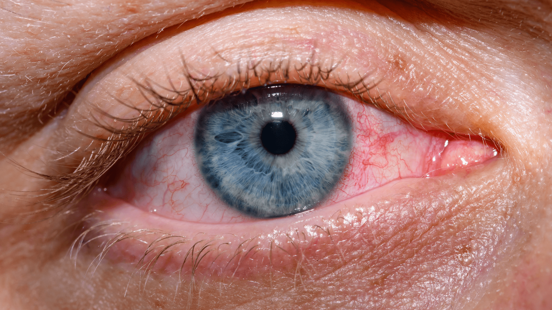
Your tear film is three layers. A watery layer from the lacrimal gland, the tear gland tucked above and behind your upper lid. A thin oily layer over the top of it from the meibomian glands, a row of tiny oil glands along the edge of each lid, and that oil is what stops the water evaporating. And a mucus layer underneath that makes the whole lot stick to the eye. Dry eye can mean a problem with any of the three, they feel broadly the same from the inside, and they need completely different treatment. Which is how people end up working through four brands of drops and getting nowhere.
The association with fibromyalgia is real. Dry eye symptom scores come out higher, and tear breakup time, the measure of how fast the film falls apart between blinks, comes out shorter [10][27]. Set against a control group, the symptom score, the breakup time, the amount of tear fluid produced and the saltiness of the tears have all come out worse together, and the more severe the fibromyalgia, the worse the tear numbers ran [28]. More recently, average tear production came out at 8.2 mm against 17.5 mm in controls, and breakup time at 5.0 seconds against 9.9 [8]. Those millimetres are a paper strip tucked inside the lower lid, and the number is how far the wetting creeps along it.
Population data back that up as well as the clinic data does. A Taiwanese cohort found roughly a 1.4 times higher long term risk of being diagnosed with dry eye [29], and in a large Dutch population study fibromyalgia came out as an independent correlate of dry eye rather than something age and sex explained away [30].
Then the other half of it, which is the half that explains the optician. Plenty of people report genuine eye discomfort and pain with tear production and breakup time both coming back normal, and with corneal sensitivity measuring higher rather than lower [1]. Earlier work found the opposite pattern, lower corneal sensitivity alongside lower tear production [31]. Both are real results in real groups, and together they tell you there isn’t one dry eye in fibromyalgia, there are several, and the whole job is telling them apart.
Get the lids looked at while you’re there. Meibomian gland dysfunction, where those oil glands in the lid margin block up, is one of the things that can contribute alongside the surface disease itself [2], and it’s treated with warmth and lid hygiene rather than with drops.
It doesn’t all point one way, though. Going back further, and following people over time rather than measuring them once, there was no clear overall increase in the damaging kind of dry eye at all, with lid inflammation and tricyclic use picked out as the contributors instead [32]. Tricyclic use, which is where amitriptyline comes in, and we’ll come back to that shortly.
Some of the symptom burden may also come from mood, sleep, medication exposure or the chronic pain itself rather than from tear failure [33][15]. That’s several things loading the same system at once, which is a good deal less tidy than anybody wants it to be. None of it makes the grit in your eyes any less real, and none of it is a polite way of telling you you’re imagining it.
When to ask about Sjogren’s
Sjogren’s syndrome is an autoimmune condition in which the immune system attacks the glands that make tears and saliva. Dry eyes and a dry mouth are its calling card, it’s far more common in women, and it arrives with fatigue and joint pain alongside. From a distance, in other words, it looks a great deal like the thing you already have a label for.
That overlap isn’t hypothetical. In a large group of people being worked up for dryness, a proportion of those carrying a fibromyalgia label came back classified as Sjogren’s instead [34].
The symptom itself won’t sort it out, though. Dry eyes and a dry mouth on their own don’t separate the two conditions, which is exactly why the classification criteria don’t lean on them: the weight sits on an anti-SSA/Ro antibody in the blood and on a lip gland biopsy showing a particular pattern of immune cells, while tear production and saliva flow carry much less discriminating power by themselves [35]. So “my eyes are dry and the tear test was low” doesn’t answer the question in either direction.
If the antibody comes back negative and the dryness won’t settle, the minor salivary gland biopsy is the test with the yield. It’s a small sample taken from inside the lower lip under local anaesthetic, and the validation work puts it and the antibody above the tear and saliva measurements [36][37].
What should prompt you to raise it at all: swelling of the salivary glands, particularly at the angle of the jaw. A genuinely dry mouth alongside the eyes, and not eyes on their own. Abnormal blood counts, or raised immunoglobulins, meaning the antibody proteins, on a routine panel. Or a neuropathy, meaning numbness, burning or pins and needles in a pattern somebody can map, because nerve involvement is a recognised part of Sjogren’s and not a coincidence [38][39].
None of that means walking into your next appointment asking for a biopsy. It means that if the dryness is severe, persistent, and sitting alongside any of the above, “it’s the fibromyalgia” is a reasonable first guess and a poor final answer. Asking your GP about an anti-SSA is a normal and cheap thing to do.
Could it be the tablets?
Nearly everything prescribed for fibromyalgia pain comes with a list of eye side effects attached, and it tends not to get mentioned, because dry eyes and blurry vision get filed as minor and the prescriber is thinking about your pain. If your eye symptoms started or stepped up within a few weeks of starting or increasing something, that timing is worth taking back to whoever prescribed it.
Don’t stop anything on the strength of a blog post, mine included. Some of these are genuinely difficult to come off, and a drug that’s working on your pain may well be worth a bit of dryness. The point is to know the question exists so you can ask it.
Amitriptyline
The clearest one by a distance. Amitriptyline is a tricyclic, an older class of antidepressant given at low doses for pain, and tricyclics block acetylcholine, one of the chemical messengers your nerves use to tell glands and muscles what to do. Your tear production runs on acetylcholine. So does the small muscle that focuses your lens for close work. Reduced tears and blurred vision up close are the predictable consequences of how the drug works [40]. If your eyes went dry and your reading vision went soft after you started amitriptyline, that’s the drug behaving exactly as described on the tin.
Duloxetine
Duloxetine is an SNRI, a different class of antidepressant also used for pain. Its reported eye effects appear to sit more with blurred vision and pupil dilation than with a strong dry eye one [41], so if your complaint is blurring and not grittiness, that fits better. Tear production does come out lower in people taking newer antidepressants, duloxetine among them, although that’s a single snapshot and not people followed over time [42]. Dryness isn’t off the table, though.
Pregabalin and gabapentin
Both are gabapentinoids, a class built for epilepsy and now handed out widely for nerve pain. Pregabalin’s clearest eye effect is blurred vision and double vision, and it shows up both in pooled trial safety data and in the large adverse event reporting databases, which are the systems where side effects get logged once a drug is out in the world [43][44][45]. The dry eye side is much weaker: trials around eye surgery found pregabalin neither increased nor reduced dry eye symptoms afterwards [46][47]. Blurring yes, grittiness probably not.
Gabapentin has reported visual effects too, blurred vision, double vision and wobbling eye movements among them, on a thinner evidence base than pregabalin’s and with no good numbers attached for dryness or focusing problems [48][49]. So it’s worth raising at an appointment, and it’s a thin basis for pinning your symptoms on.
What actually helps
Start with the surface, and don’t stop there
Artificial tears are a reasonable first move and the right place to begin. In this population they’re also, very often, only part of the answer, and that’s a well described pattern and not you using them wrong [50].
The clearest demonstration of it is a dry eye cohort in which the worse somebody’s eye pain, the worse their response to surface treatment across the board: drops, ointments, ciclosporin, serum tears, punctal plugs and lid treatments alike. Fibromyalgia was more common in the group with the high pain scores [51]. Ciclosporin is an anti-inflammatory drop, serum tears are drops made up from your own blood, and punctal plugs are tiny stoppers put into the drainage holes in your lid so the tears you do make hang around longer. None of that is an argument against trying them. It’s an argument for treating the surface properly and then escalating when the symptoms are out of proportion to the signs, rather than cycling the same thing round again in a different box [52].
Treating the surface properly means more than drops, though. If the lid margins are involved, warm compresses and lid hygiene go after the oil layer, which drops don’t touch. If the surface is inflamed, ciclosporin reliably improves the signs more than artificial tears do, while the symptom relief is more variable [53][54]. Those trials were run in general dry eye rather than in fibromyalgia, so take the direction and keep the expectations modest.
Then the cheap end, and none of it costs anything: a humidifier in the room you sit in, the screen moved below eye level so your lids cover more of the surface, deliberately blinking all the way shut when you read, and the fan in the car pointed somewhere other than your face.
Take the dates to your prescriber
The only thing on this list that might resolve the symptom rather than managing it. Put the date your eye symptoms started and the date your medication started or changed side by side, and take both of them to the same appointment.
If it’s your neck
Pain behind the eye with a stiff, sore upper neck in the picture is a physiotherapy question before it’s an ophthalmology one. Exercise, manual therapy and a structured programme is where the evidence sits, and the realistic gain is less often and less intense [19][20][21]. It’s borrowed from cervicogenic headache, so that’s a population sharing the mechanism with you and not the diagnosis.
Tinted lenses
FL-41 is a rose tinted lens originally developed for migraine, and in a small group with chronic eye pain it reduced how unpleasant light felt and damped the pain related activity visible on brain imaging [55]. Early evidence in a small group, cheap, harmless, and worth a go if light is the thing wrecking your day.
What’s worth avoiding is living in sunglasses indoors. Tints have a use, and permanent darkness makes the next bright room worse.
When the surface work has run out
If you’ve done the surface properly, reviewed the medication, and the burning and light sensitivity are still running your week, there are nerve targeted options, and it’s worth knowing they exist, because getting near them usually means asking for them by name.
Botulinum toxin and trigeminal nerve stimulation have both reduced light sensitivity in small groups with chronic eye pain [56][57]. Small groups and early work, and a reason to ask for an ocular pain or neuro-ophthalmology opinion rather than accepting a shrug.
One more thing if you’re already on an SNRI. People with fibromyalgia taking one have been followed over time, with improvement reported in both their symptoms and their corneal nerve measurements [27]. That’s one group followed once, and it’s a fair reason not to assume your pain medication is only ever making your eyes worse.
When eye pain is not something to sit on
A few eye symptoms don’t belong anywhere in the above, and they matter more than usual here, because carrying a chronic pain label is exactly what gets a new symptom waved through as more of the same.
The one to know is pain in the movement itself. If the ache lives in moving your eye, and gets worse as you look further to the side or further up, that presentation belongs to optic neuritis and orbital myositis until somebody has looked and ruled them out [58][59]. Optic neuritis is inflammation of the optic nerve, the cable carrying vision from your eye back to your brain. Orbital myositis is inflammation of the muscles that move the eyeball. Both are treatable, both are time sensitive, and pain on moving the eye is how they classically announce themselves.
Older work in fibromyalgia found changes in how smoothly the eyes track and jump, which points at something central in eye control, and it didn’t describe a syndrome of painful eye movements [60]. Counted directly, eye pain came out at rates comparable to other chronic musculoskeletal pain rather than at anything specific to fibromyalgia, on a single result [61]. So painful eye movement isn’t a fibromyalgia feature you can file and forget about. If the pain is in the movement, that’s the detail to lead with when you ring somebody.
Alongside that, the ordinary list that applies to anybody. Sudden loss or dimming of vision in one eye. A new shower of floaters, flashing lights, or a curtain or shadow coming across part of your sight. A red, painful eye with blurred vision, especially with nausea or haloes round lights. New double vision. A drooping lid, or a pupil that has changed size. Any of those is a same day call to your GP, to an eye casualty, or to the NHS urgent advice line, and not a thing to read another article about.
You’re allowed to go and get it checked without a good reason. Having fibromyalgia doesn’t use up your right to be taken seriously about a new symptom, and nobody in an eye casualty thinks less of you for turning up and being fine.
The short version
Best return: work out which of the four it is. Burning and grit on the surface, ache behind the eye coming from the neck, light sensitivity, or the tablets. They’re treated completely differently and the treatments don’t substitute for each other.
Most likely to be missed: the medication. Amitriptyline for dryness and near blur [40]. Duloxetine appears to sit more with blurring than with dryness [41]. The gabapentinoids for blurring as well [43]. Check the dates against when your symptoms started and take that to your prescriber.
Worth doing properly: the surface. Drops, lid hygiene if the lid margins are involved, and ciclosporin if there’s inflammation, on the understanding that surface treatment works less well the more eye pain somebody has [51].
Worth asking about: Sjogren’s, if the dryness is severe and persistent, the mouth is dry too, or there’s gland swelling or a neuropathy alongside [38][39]. An anti-SSA is a blood test [35].
Reasonable to try: FL-41 tints for light [55], and physiotherapy for the neck if the pain is behind the eye and the neck is stiff [19][20].
- Not fibromyalgia until somebody has looked and ruled the other things out: pain in the eye movement itself, worse towards the extremes of gaze [58][59].
Borrowed evidence: nearly all the treatment evidence here comes from general dry eye, chronic eye pain and cervicogenic headache populations, and it’s applied on the strength of a shared mechanism.
A clean eye test rules out the things it was built to find and narrows what’s left down to a shortlist, and that’s a good place to be starting from rather than a dead end. So go back armed. Ask which layer of the tear film is the problem. Ask whether your medication could be doing it. Ask about your neck. And if the pain is in the movement, say that out loud, early, to somebody who can look.
Adam

References
[1] Aykut, V., Elbay, A., Çigdem Uçar, I., Esen, F., Durmus, E., Karadag, R. et al. (2018) ‘Corneal sensitivity and subjective complaints of ocular pain in patients with fibromyalgia’, Eye. https://doi.org/10.1038/eye.2017.275
[2] Emekli, D.T., Sahutoglu, E., Öztürk, G.Y. and Kocyigit, B.F. (2025) ‘Anterior segment alterations in fibromyalgia syndrome: a cross-sectional study on dry eye disease, meibomian gland dysfunction, and astigmatism’, Rheumatology International. https://doi.org/10.1007/s00296-025-05872-w
[3] Vehof, J., Sillevis Smitt-Kamminga, N., Kozareva, D., Nibourg, S.A. and Hammond, C.J. (2016) ‘Clinical Characteristics of Dry Eye Patients With Chronic Pain Syndromes’, American Journal of Ophthalmology. https://doi.org/10.1016/j.ajo.2015.11.017
Read More[4] Vehof, J., Sillevis Smitt-Kamminga, N., Nibourg, S.A. and Hammond, C.J. (2017) ‘Predictors of Discordance between Symptoms and Signs in Dry Eye Disease’, Ophthalmology. https://doi.org/10.1016/j.ophtha.2016.11.008
[5] Hoffmann, M., Farrell, S., Colorado, L.H. and Edwards, K. (2024) ‘Discordant dry eye disease and chronic pain: A systematic review and meta-analysis’, Contact Lens and Anterior Eye. https://doi.org/10.1016/j.clae.2024.102248
[6] Ordunahospital, E., Viladés Palomar, E., Cordón Ciordía, B., Pérez Velilla, J., José Vicente Altabás, M., Satué Palacián, M. et al. (2019) ‘Progressive impairment of visual function in fibromyalgia’, Acta Ophthalmologica. https://doi.org/10.1111/j.1755-3768.2019.5279
[7] Garcia-Martin, E., Vicente Altabas, M.J., Vilades Palomar, E., Orduna Hospital, E., Cordon Ciordia, B., Garcia-Campayo, J. et al. (2025) ‘Progressive visual dysfunction and retinal neurodegeneration in patients with fibromyalgia’, Archivos de la Sociedad Española de Oftalmología (English Edition). https://doi.org/10.1016/j.oftale.2025.06.008
[8] Yavaş, A.D., Şaipi, A. and Akçay, S. (2026) ‘Eyes of Fibromyalgia’, Biomedicines. https://doi.org/10.3390/biomedicines14081656
[9] Ramírez, M., Martínez-Martínez, L.A., Hernández-Quintela, E., Velazco-Casapía, J., Vargas, A. and Martínez-Lavín, M. (2015) ‘Small fiber neuropathy in women with fibromyalgia. An in vivo assessment using corneal confocal bio-microscopy’, Seminars in Arthritis and Rheumatism. https://doi.org/10.1016/j.semarthrit.2015.03.003
[10] Erkan Turan, K., Kocabeyoglu, S., Unal-Cevik, I., Bezci, F., Akinci, A. and Irkec, M. (2017) ‘Ocular Surface Alterations in the Context of Corneal In Vivo Confocal Microscopic Characteristics in Patients With Fibromyalgia’, Cornea. https://doi.org/10.1097/ico.0000000000001447
[11] Grayston, R., Czanner, G., Elhadd, K., Goebel, A., Frank, B., Üçeyler, N. et al. (2019) ‘A systematic review and meta-analysis of the prevalence of small fiber pathology in fibromyalgia: Implications for a new paradigm in fibromyalgia etiopathogenesis’, Seminars in Arthritis and Rheumatism. https://doi.org/10.1016/j.semarthrit.2018.08.003
[12] Marshall, A., Elshafei, M., Preston, F.G., Burgess, J., Goodson, N., Fallon, N. et al. (2025) ‘Small Fibre Pathology in Fibromyalgia: A review’, Pain and Therapy. https://doi.org/10.1007/s40122-024-00696-1
[13] Oudejans, L., He, X., Niesters, M., Dahan, A., Brines, M. and van Velzen, M. (2016) ‘Cornea nerve fiber quantification and construction of phenotypes in patients with fibromyalgia’, Scientific Reports. https://doi.org/10.1038/srep23573
[14] Marshall, A., Rapteas, L., Burgess, J., Riley, D., Anson, M., Matsumoto, K. et al. (2024) ‘Small fibre pathology, small fibre symptoms and pain in fibromyalgia syndrome’, Scientific Reports. https://doi.org/10.1038/s41598-024-54365-6
[15] Ramírez, M., Guerra-Juárez, A., Miyake, D.Y., Sebastian-Arellano, C., Estrada-Mata, A.G., González-Moyotl, N.J. et al. (2021) ‘Correlation Between Corneal Nerve Density and Symptoms of Small Fiber Neuropathy in Patients With Fibromyalgia: The Confounding Role of Severe Anxiety or Depression’, JCR: Journal of Clinical Rheumatology. https://doi.org/10.1097/rhu.0000000000001592
[16] Jänsch, S., Evdokimov, D., Egenolf, N., Meyer zu Altenschildesche, C., Kreß, L. and Üçeyler, N. (2024) ‘Distinguishing fibromyalgia syndrome from small fiber neuropathy: a clinical guide’, PAIN Reports. https://doi.org/10.1097/pr9.0000000000001136
[17] Shimohata, K., Hasegawa, K., Onodera, O., Nishizawa, M. and Shimohata, T. (2017) ‘The Clinical Features, Risk Factors, and Surgical Treatment of Cervicogenic Headache in Patients With Cervical Spine Disorders Requiring Surgery’, Headache: The Journal of Head and Face Pain. https://doi.org/10.1111/head.13123
[18] Piovesan, E.J., Utiumi, M.A.T. and Grossi, D.B. (2024) ‘Cervicogenic headache – How to recognize and treat’, Best Practice & Research Clinical Rheumatology. https://doi.org/10.1016/j.berh.2024.101931
[19] Jull, G. (2023) ‘Cervicogenic headache’, Musculoskeletal Science and Practice. https://doi.org/10.1016/j.msksp.2023.102787
[20] Bini, P., Hohenschurz-Schmidt, D., Masullo, V., Pitt, D. and Draper-Rodi, J. (2022) ‘The effectiveness of manual and exercise therapy on headache intensity and frequency among patients with cervicogenic headache: a systematic review and meta-analysis’, Chiropractic & Manual Therapies. https://doi.org/10.1186/s12998-022-00459-9
[21] Martins, L., Collet, P., Lafrance, S. and Demont, A. (2026) ‘Efficacy of nonsurgical interventions for the management of adults with cervicogenic headache: a systematic review and meta-analyses’, Annals of Physical and Rehabilitation Medicine. https://doi.org/10.1016/j.rehab.2025.102070
[22] Haddad, E.N., Amin, R.S., Nero, N., Hogue, O., Rao, S., Sayegh, R.R. et al. (2026) ‘A systematic review and narrative synthesis of functional neuroimaging studies for chronic ocular surface pain’, Survey of Ophthalmology. https://doi.org/10.1016/j.survophthal.2026.08.005
[23] Choudhury, A., Reyes, N., Galor, A., Mehra, D., Felix, E. and Moulton, E.A. (2023) ‘Clinical Neuroimaging of Photophobia in Individuals With Chronic Ocular Surface Pain’, American Journal of Ophthalmology. https://doi.org/10.1016/j.ajo.2022.09.020
[24] Diel, R.J., Mehra, D., Kardon, R., Buse, D.C., Moulton, E. and Galor, A. (2020) ‘Photophobia: shared pathophysiology underlying dry eye disease, migraine and traumatic brain injury leading to central neuroplasticity of the trigeminothalamic pathway’, British Journal of Ophthalmology. https://doi.org/10.1136/bjophthalmol-2020-316417
[25] Wang, Y., Wang, S., Qiu, T. and Xiao, Z. (2022) ‘Photophobia in headache disorders: characteristics and potential mechanisms’, Journal of Neurology. https://doi.org/10.1007/s00415-022-11080-4
[26] Martenson, M.E., Halawa, O.I., Tonsfeldt, K.J., Maxwell, C.A., Hammack, N., Mist, S.D. et al. (2016) ‘A possible neural mechanism for photosensitivity in chronic pain’, Pain. https://doi.org/10.1097/j.pain.0000000000000450
[27] Kubat, B., Ozkan, G., Sanal Toprak, C., Demirci, M. and Akkaya Turhan, S. (2025) ‘Corneal confocal microscopy in fibromyalgia syndrome: small fiber neuropathy, and treatment outcomes’, Eye. https://doi.org/10.1038/s41433-025-04132-2
[28] Türkyilmaz, K., Türkyilmaz, A.K., Kurt, E.E., Kurt, A. and Öner, V. (2013) ‘Dry Eye in Patients With Fibromyalgia and Its Relevance to Functional and Emotional Status’, Cornea. https://doi.org/10.1097/ico.0b013e318285c912
[29] Chen, C.H., Yang, T.Y., Lin, C.L., Chen, C.S., Lin, W.M., Kuo, C.N. et al. (2016) ‘Dry Eye Syndrome Risks in Patients With Fibromyalgia’, Medicine. https://doi.org/10.1097/md.0000000000002607
[30] Vehof, J., Snieder, H., Jansonius, N. and Hammond, C.J. (2021) ‘Prevalence and risk factors of dry eye in 79,866 participants of the population-based Lifelines cohort study in the Netherlands’, The Ocular Surface. https://doi.org/10.1016/j.jtos.2020.04.005
[31] Gallar, J., Morales, C., Freire, V., Acosta, M.C., Belmonte, C. and Duran, J.A. (2009) ‘Decreased Corneal Sensitivity and Tear Production in Fibromyalgia’, Investigative Opthalmology & Visual Science. https://doi.org/10.1167/iovs.08-3083
[32] Günaydin, I., Terhorst, T., Eckstein, A., Daikeler, T., Kanz, L. and Kötter, I. (1999) ‘Assessment of keratoconjunctivitis sicca in patients with fibromyalgia: results of a prospective study’, Rheumatology International. https://doi.org/10.1007/s002960050090
[33] Galor, A., Britten-Jones, A.C., Feng, Y., Ferrari, G., Goldblum, D., Gupta, P.K. et al. (2023) ‘TFOS Lifestyle: Impact of lifestyle challenges on the ocular surface’, The Ocular Surface. https://doi.org/10.1016/j.jtos.2023.04.008
[34] Triantafyllias, K., Bach, M., Otto, M. and Schwarting, A. (2023) ‘Diagnostic Value of Labial Minor Salivary Gland Biopsy: Histological Findings of a Large Sicca Cohort and Clinical Associations’, Diagnostics. https://doi.org/10.3390/diagnostics13193117
[35] Shiboski, C.H., Shiboski, S.C., Seror, R., Criswell, L.A., Labetoulle, M., Lietman, T.M. et al. (2017) ‘2016 American College of Rheumatology/European League Against Rheumatism classification criteria for primary Sjögren’s syndrome’, Annals of the Rheumatic Diseases. https://doi.org/10.1136/annrheumdis-2016-210571
[36] Goel, R.R., Jeranko, M., Jones, L., Bishnoi, A. and Meysami, A. (2023) ‘Diagnostic Utility of Minor Salivary Gland Biopsy for Primary Sjögren Syndrome in Patients With Negative Anti-SSA Antibodies’, Cureus. https://doi.org/10.7759/cureus.46207
[37] van Ginkel, M.S., Nakshbandi, U., Arends, S., Haacke, E.A., Liefers, S.C., Verstappen, G.M. et al. (2024) ‘Increased Diagnostic Accuracy of the Labial Gland Biopsy in Primary Sjögren Syndrome When Multiple Histopathological Features Are Included’, Arthritis & Rheumatology. https://doi.org/10.1002/art.42723
[38] Sisó-Almirall, A., Meijer, J.M., Brito-Zerón, P., Conangla, L., Flores-Chavez, A., González de Paz, L. et al. (2021) ‘Practical guidelines for the early diagnosis of Sjögren’s syndrome in primary healthcare’, Clinical and Experimental Rheumatology. https://doi.org/10.55563/clinexprheumatol/pal3z7
[39] Liampas, A., Parperis, K., Erotocritou, M.F., Nteveros, A., Papadopoulou, M., Moschovos, C. et al. (2022) ‘Primary Sjögren syndrome‐related peripheral neuropathy: A systematic review and meta‐analysis’, European Journal of Neurology. https://doi.org/10.1111/ene.15555
[40] Constable, P.A., Al-Dasooqi, D., Bruce, R. and Prem-Senthil, M. (2022) ‘A Review of Ocular Complications Associated with Medications Used for Anxiety, Depression, and Stress’, Clinical Optometry. https://doi.org/10.2147/opto.s355091
[41] Sun, Z., Liu, Y. and Que, L. (2026) ‘Analysis of ocular adverse events associated with SNRIs’, Frontiers in Pharmacology. https://doi.org/10.3389/fphar.2026.1837592
[42] Koçer, E., Koçer, A., Özsütçü, M., Dursun, A.E. and Kirpinar, İ. (2015) ‘Dry Eye Related to Commonly Used New Antidepressants’, Journal of Clinical Psychopharmacology. https://doi.org/10.1097/jcp.0000000000000356
[43] Freynhagen, R., Serpell, M., Emir, B., Whalen, E., Parsons, B., Clair, A. et al. (2013) ‘A Comprehensive Drug Safety Evaluation of Pregabalin in Peripheral Neuropathic Pain’, Pain Practice. https://doi.org/10.1111/papr.12146
[44] Zaccara, G., Gangemi, P., Perucca, P. and Specchio, L. (2011) ‘The adverse event profile of pregabalin: A systematic review and meta‐analysis of randomized controlled trials’, Epilepsia. https://doi.org/10.1111/j.1528-1167.2010.02966.x
[45] Wu, S.N., Chen, X.D., Liu, Q.T., Chen, L., Sun, L., Guan, W.Y. et al. (2025) ‘Drug-related visual blurring: findings from the U.S. Food and Drug Administration Adverse Event Reporting System database’, European Journal of Pharmacology. https://doi.org/10.1016/j.ejphar.2025.177820
[46] Galor, A., Patel, S., Small, L.R., Rodriguez, A., Venincasa, M.J., Valido, S.E. et al. (2019) ‘Pregabalin Failed to Prevent Dry Eye Symptoms after Laser-Assisted in Situ Keratomileusis (LASIK) in a Randomized Pilot Study’, Journal of Clinical Medicine. https://doi.org/10.3390/jcm8091355
[47] Paik, D.W., Lim, D.H. and Chung, T.Y. (2020) ‘Effects of taking pregabalin (Lyrica) on the severity of dry eye, corneal sensitivity and pain after laser epithelial keratomileusis surgery’, British Journal of Ophthalmology. https://doi.org/10.1136/bjophthalmol-2020-317570
[48] ROFF HILTON, E.J., HOSKING, S.L. and BETTS, T. (2004) ‘The effect of antiepileptic drugs on visual performance’, Seizure. https://doi.org/10.1016/s1059-1311(03)00082-7
[49] Murray, M., Guirguis, A., Deslandes, P., Corkery, J.M., Chiappini, S., Parravano, M. et al. (2026) ‘Possible Signals of Ocular Disorders Associated with Gabapentinoids: A Three-Arm Study’, Pharmaceuticals. https://doi.org/10.3390/ph19081175
[50] Kim, M., Lee, Y., Mehra, D., Sabater, A.L. and Galor, A. (2021) ‘Dry eye: why artificial tears are not always the answer’, BMJ Open Ophthalmology. https://doi.org/10.1136/bmjophth-2020-000697
[51] Siedlecki, A.N., Smith, S.D., Siedlecki, A.R., Hayek, S.M. and Sayegh, R.R. (2020) ‘Ocular pain response to treatment in dry eye patients’, The Ocular Surface. https://doi.org/10.1016/j.jtos.2019.12.004
[52] Betz, J. and Galor, A. (2025) ‘Navigating the Dry Eye Therapeutic Puzzle: A Mechanism-Based Overview of Current Treatments’, Pharmaceuticals. https://doi.org/10.3390/ph18070994
[53] Tuan, H.I., Chi, S.C. and Kang, Y.N. (2020) ‘An Updated Systematic Review With Meta-Analysis Of Randomized Trials On Topical Cyclosporin A For Dry-Eye Disease’, Drug Design, Development and Therapy. https://doi.org/10.2147/dddt.s207743
[54] Akpek, E.K., Wirta, D.L., Downing, J.E., Tauber, J., Sheppard, J.D., Ciolino, J.B. et al. (2023) ‘Efficacy and Safety of a Water-Free Topical Cyclosporine, 0.1%, Solution for the Treatment of Moderate to Severe Dry Eye Disease’, JAMA Ophthalmology. https://doi.org/10.1001/jamaophthalmol.2023.0709
[55] Reyes, N., Huang, J.J., Choudhury, A., Pondelis, N., Locatelli, E.V.T., Hollinger, R. et al. (2024) ‘FL-41 Tint Reduces Activation of Neural Pathways of Photophobia in Patients with Chronic Ocular Pain’, American Journal of Ophthalmology. https://doi.org/10.1016/j.ajo.2023.12.004
[56] Reyes, N., Huang, J.J., Choudhury, A., Pondelis, N., Locatelli, E.V., Felix, E.R. et al. (2023) ‘Botulinum toxin A decreases neural activity in pain-related brain regions in individuals with chronic ocular pain and photophobia’, Frontiers in Neuroscience. https://doi.org/10.3389/fnins.2023.1202341
[57] Mehra, D., Mangwani-Mordani, S., Acuna, K., C. Hwang, J., R. Felix, E. and Galor, A. (2021) ‘Long-Term Trigeminal Nerve Stimulation as a Treatment for Ocular Pain’, Neuromodulation: Technology at the Neural Interface. https://doi.org/10.1111/ner.13402
[58] Penkava, J. and Petzold, A. (2025) ‘Diagnosing optic neuritis in the changing landscape of diagnostic criteria’, Practical Neurology. https://doi.org/10.1136/pn-2025-004702
[59] Mombaerts, I. and McNab, A.A. (2022) ‘Idiopathic Orbital Myositis Revisited’, Current Rheumatology Reports. https://doi.org/10.1007/s11926-022-01052-y
[60] Rosenhall, U., Johansson, G. and Orndahl, G. (2020) ‘Eye Motility Dysfunction in Chronic Primary Fibromyalgia With Dysesthesia’, Journal of Rehabilitation Medicine. https://doi.org/10.2340/165019778719139145
[61] Schuster, A.K.G., Wettstein, M., Gerhardt, A., Eich, W., Bieber, C. and Tesarz, J. (2018) ‘Eye Pain and Dry Eye in Patients with Fibromyalgia’, Pain Medicine. https://doi.org/10.1093/pm/pny045