
- Hypermobile Neck Exercises - 8 November 2026
- Swollen legs and hypermobility: pooling, lymphoedema and lipoedema - 4 November 2026
- Walking with fibromyalgia - 2 November 2026
This article is part of our comprehensive guide to exercise and rehabilitation for hypermobility.
Neck pain is close to a standard issue in symptomatic hypermobility. Survey a few hundred people with hEDS or HSD and 76% of them report it [1].
Which means there’s no shortage of advice for it. Spend twenty minutes looking and you’ll be handed a million isometrics, a band round the forehead, a palm pressed into the side of your own head for ten seconds a side, and chin tucks until your jaw aches. It’s all the same instinct wearing different clothes though: the neck feels wobbly, so brace it harder.
The trouble is that nearly all of it gets taken up by one big muscle at the front of your neck that’s already doing more than its fair share, while the small deep ones that are meant to be holding the stack steady from the inside never get a look in. So let’s go a bit deeper than that.
You have probably landed here looking for some exercise videos for your hypermobile neck, so I won’t waffle too much, and I’ll leave all the science bits further down, for those of you wanting to go deeper.
Compare hypermobile people with neck pain against hypermobile people without it, and what separates them is accuracy and stamina. Asked to find their own neutral again with their eyes shut, meaning put the head back to straight ahead after somebody’s moved it, the painful group missed by close to five degrees where the group without pain missed by around two [2]. They also held a gentle neck flexion position for 44.39 seconds against 60.20 seconds [2].
So, the work is about information and control, at small ranges, which is what everybody says, right up to the point where they hand you the exercise.
Where deep neck flexor exercises go wrong
Sternocleidomastoid is the rope that stands out at the front of your neck when you turn your head and look down. It runs from the top of your breastbone and the inner end of your collarbone, up and back, to the bony lump behind your ear.
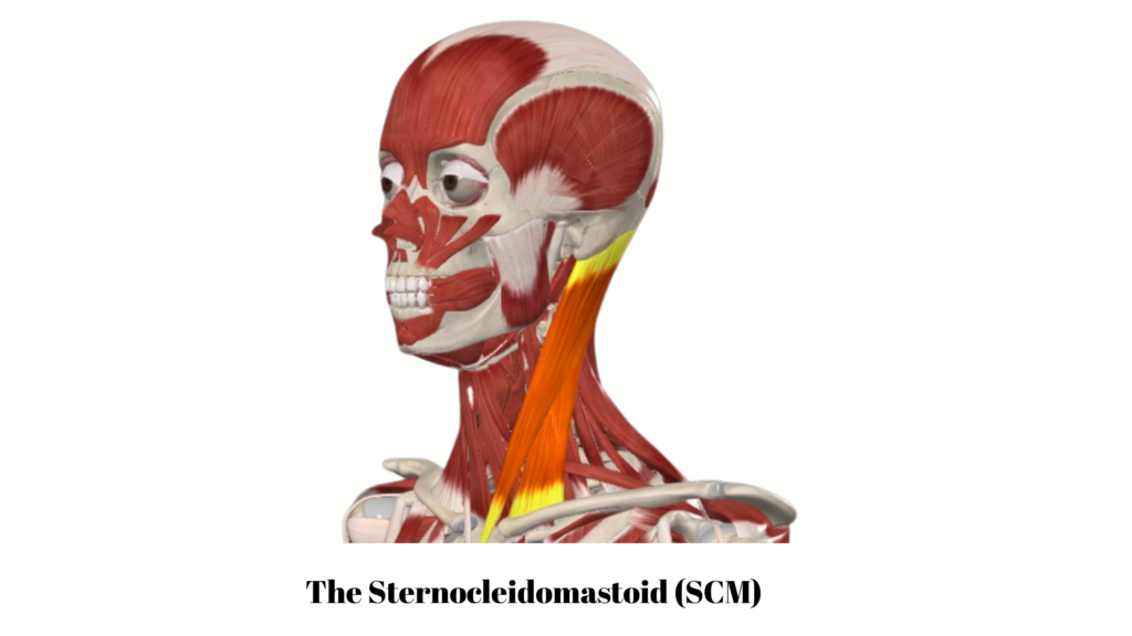
It’s long, it’s right under the skin, and because it attaches so far from the spine it gets a long lever to pull upon. Lift your head off a pillow and it’s the muscle most obviously doing it. It’s also the muscle that so many Instagram coaches keep calling a deep cervical flexor, despite it not being one.
Longus colli runs vertebra to vertebra, and longus capitis runs from the middle of the neck up to the base of the skull, so the most either can produce is a few degrees of tip at the top. They aren’t weaker versions of the sternocleidomastoid, they’re built for a different job, at a different scale, and any exercise big enough to lift your head is a job the big one can do on its own. It will, too.
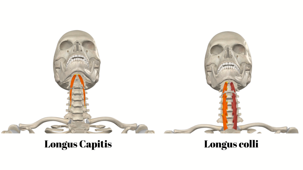
So, the test of anything sold as deep neck flexor work is whether it keeps that one out of it, and the head lift doesn’t. Record the electrical activity and there’s more sternocleidomastoid during an ordinary neck flexion, head coming off the surface, than during a craniocervical nod, the small one where the skull tips forwards on the top of the neck and nothing below it moves [3]. Image it instead and conventional flexion brings up longus colli as well as the sternocleidomastoid, more than the nod does [4].
That second one trips people up. The nod doesn’t produce more deep muscle activity than a head lift, it produces less of nearly everything. Tipping the skull forwards on the top joint is the whole of what longus capitis is built to do, so ask for that movement and nothing else, and longus capitis is the muscle that responds most selectively [5].
Which brings us to the chin tuck, since you’ve probably been given one at some point, along with a few other exercises on A4 paper, and likely left to get on with it. Done as a small tip of the skull it is the nod, and it’s fine. Pulled back hard so the chin jams into the throat it becomes a retraction, sliding the whole neck backwards instead of rotating the skull on it. Different movement, same name on the handout sheet though.
Six weeks of nodding practice raised deep flexor activity and lowered activity in the sternocleidomastoid and the scalenes, the smaller ropes running down the side of the neck behind it, while conventional strength training didn’t raise deep flexor activity at all when the nod was tested [6].
The muscles that actually do this job
Longus colli and longus capitis both sit behind your throat, flat against the front of the vertebrae, about as close to the bone as a muscle gets. Longus colli’s job is holding the stack in line from the inside while something else does the moving. A joint with more available range gives it more positions to hold and less help from the tissue that’s supposed to be holding it.
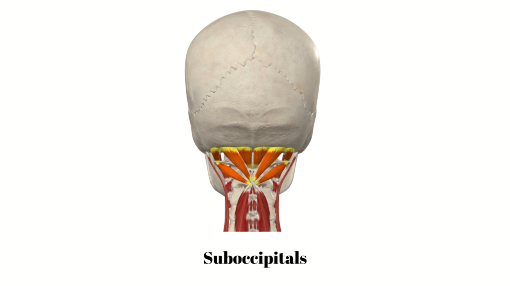
Right at the top, slung between the back of your skull and the top two vertebrae, is a small cluster each side called the suboccipitals. Four muscles a side, none of them long, and between them they tip and turn your head through a few degrees. That’s the whole of their mechanical contribution, and it isn’t much.
What they’re really for is reporting. They’re packed with muscle spindles, the stretch sensors that tell your brain where a joint currently is [7]. A review collecting the anatomical counts puts the two oblique ones at 242 and 190 spindles per gram against 98 for each of the two straight ones, where anything over 50 per gram is generally a muscle built for fine, precise work, and all four come out well above the bigger muscles around them [23]. That’s a reason to take them seriously and not a general case for strengthening them in isolation. Chase force or range through them and you’re using a sensor as a motor.
They’re also rarely underworked, which is the part that gets missed. Recordings of the two straight ones put them at roughly 10 to 18% of maximum effort with the head sitting in neutral, and 34 to 42% once it drifts forwards [23]. For scale, a muscle held at 30% has about one to two and a half minutes in it, at 8% about an hour, and to last a working day it wants to be down at a few percent [23]. So an afternoon with your head slightly forward over a screen is asking a measuring instrument to hold a contraction it was never built for. Which is why the work here starts with taking that guarding tone out of them rather than adding to it, and why the drill further down is an accuracy exercise and not a strengthening one. Those numbers are collected in a narrative review rather than measured in one study, and none of it was done in hypermobile necks.

The exercises
Before you start
If you’re new to any of this, start smaller than what feels worth doing. All three of these are tiny movements, and the version that feels like almost nothing is the version that’s working. There’s nothing here to grind out, and nothing that gets better for being done harder and longer.
Two or three short goes a week is plenty at first.
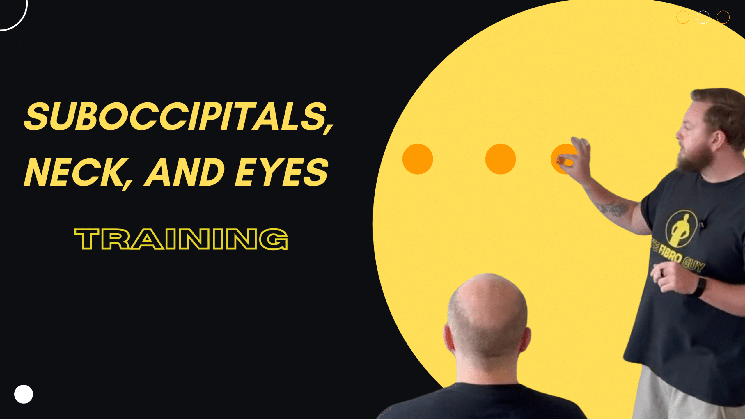
The last one, the eyes and the suboccipitals, is the one to go especially carefully with. It’s the hardest of the 3 to do well and the easiest to overdo, and the thing that limits it isn’t your neck, it’s your eyes. Watch what we say in that video about eye strain, and do the field of vision test before the exercise rather than after it. If your eyes feel strained, your two targets are too far apart. Keep them the same distance from you every session as well, or you’re training something slightly different each time.
Then progress it slowly, and slower than you’ll want to. Widening the field of view, and running the eyes and head together instead of one then the other, are both progressions, and neither of them is earned by one good session. The criteria we use are in the video description..
And if you come away from it queasy or headachy, that’s the signal you went too far too fast. It isn’t a sign that it’s working. Rest up, then go back to a narrower field, fewer repetitions and a proper rest between each one. Worth knowing that the trial we lean on most for the eye work screened out anyone who got dizzy, nauseous or sick while doing the exercise before it even started[24]. So there’s no evidence in there that pushing through that is a good idea, and a fair bit of reason to think it isn’t.
Longus nods
Two fingers resting lightly either side of your windpipe will tell you most of what you need to know. The ropes under them are the sternocleidomastoids, and on a decent repetition they stay quiet. It’s a rough guide and not a readout, because in women with chronic neck pain more superficial activity only loosely goes with less deep activity [8], but it’s the one thing you can feel from the inside. If they harden, if your head lifts, if the chin jams backwards into your throat, or if the movement goes jerky, you’ve gone past the level you can control, and the level you can control is the one to train [9].
Going harder doesn’t improve the share either. Push the effort up and superficial activity comes up with it [10], so the repetition that feels like almost nothing is doing the job better than the one that feels like work. Just play about for a few weeks until you start assigning reps and sets. This isn’t about hammering the poor old tissue with high volume, it’s about connecting to them and getting them functional.
The suboccipital steering wheel
This one is for the top two joints and it’s an accuracy drill. The movement is tiny: the head tilts on the very top of the neck, a few degrees each way, the way you’d correct a steering wheel rather than take a sharp corner. The neck below it doesn’t come along for the ride though, and if it does, you’ve made it too big.
Accuracy is trainable. Six weeks of proprioceptive retraining improved how accurately 64 women with persistent neck pain could find their own neutral again, and six weeks of craniocervical flexion training did much the same [12]. They had ordinary persistent neck pain and not hypermobility, so take it as a principle worth applying and not a promise about your neck.
Adding the eyes
Somebody says your name across a room and your eyes get there first. The head follows a fraction behind. Put fine wires into the neck muscles of a monkey and flash a target off to one side, and 26 of 29 recordings from the suboccipitals showed those muscles responding about 73 milliseconds after the target appeared, well before the animal had shifted its gaze [25]. The neck is being set up for the head turn while the decision to turn is still being made.
Say the species out loud, though, because it matters. The same study ran the same test on 28 people and found that response in 29% of recordings, a bit later at 95 milliseconds, and those recordings came from splenius capitis, a muscle further down the neck, rather than from the suboccipitals themselves [25]. So nobody has measured this directly in a human suboccipital. What there is, is decent evidence of a shared anticipatory system between gaze and neck, and no evidence at all for a tidy reflex where every flick of the eye switches on every muscle at the top of your neck.
Which is where it gets interesting for a hypermobile neck, because a lot of people can’t separate the two. The eyes and the head go at once, or the head swings over first and then corrects itself. That’s the thing the drill below is actually training.
Does bolting it on do anything, though. The closest thing to a direct test is a six week trial in 40 people with chronic neck pain, tension type headache and a forward head position, randomised to manual therapy and biofeedback guided stabilisation work either with eyeball exercise or without it [24]. Both groups got better. The group doing the eye work came out further ahead on neck disability, on headache impact, on quality of life, and on suboccipital muscle tone, which fell more in that group and fell there specifically, since the trapezius change was much the same in both [24]. What it didn’t beat the control group on was pain itself [24].
Adding joint position work, eye movement and balance work on top of manual therapy and exercise improved sensorimotor measures, and helped hold onto the improvements in neck pain and disability out to 6 to 12 months [13]. Narrow it to people with neck pain and visual complaints and the eye work improved gaze stability and the visual symptoms without adding anything to the pain or the disability [14].
One more thing, because it comes up constantly. When what your eyes report, what your inner ear reports and what your neck reports don’t line up, you can feel properly unwell with it. Queasy, headachy, half a second behind the room.
That part isn’t anatomy and inference, somebody has gone and done it to people on purpose. Ten healthy adults sat through up to 40 minutes of passive side to side motion on three separate occasions: once with a small current at the mastoids tuned to make the balance organs disagree with the motion, once tuned to make them agree with it more closely, and once with the electrodes on and nothing running [27]. Sickness followed the disagreement. The mismatch condition came out 56% worse than the no current baseline and the matched condition 26% better, the two active conditions separated reliably from one another, and the trend across all three was significant [27]. Ten people, a motion sled, no necks involved, so hold the numbers loosely. But it’s a manipulation rather than an association, which is rarer in this field than it ought to be.
What that doesn’t license is pinning your own nausea on your own neck. The Bárány Society, who write the diagnostic criteria for this sort of thing, concluded that the evidence for neck pathology causing vertigo is lacking [26], and symptoms that arrive with head position are also what common inner ear problems and vestibular migraine look like. So the mechanism is real, and the neck is one of the three signals feeding it. Which of the three is misbehaving in you, isn’t something an exercise can tell you, so if the room swims when you turn, go and get it looked at either way.
Add this one if finding your own neutral is the part you can’t do, if your eyes and your head refuse to move separately, or if screen days are where your neck goes downhill. Not every neck needs it.
How to train it so something changes
You get what you train. Over ten weeks, endurance training produced endurance gains, coordination training produced coordination gains, and all of the exercise modes improved pain and disability by roughly the same amount [15]. So train the thing you want to change, and don’t expect holding a nod for longer to do anything for how much your neck can carry.
Dose, since that’s the question by now. The trial that put deep flexor work head to head against general neck strengthening used ten repetitions of ten second holds, three times a week, and at four weeks the two groups were level [16]. By eight weeks the deep flexor group had carried on improving on pain, disability and head position while the strengthening group had levelled off [16]. It’s a small trial, so don’t treat ten by ten as scripture. The useful part is the timeline, because four weeks isn’t long enough to judge this on and a lot of people quit at three.
The rehabilitation programmes built for Ehlers-Danlos syndrome are consistent about the how rather than the how much: keep the position supported while you’re learning it, use something that gives you feedback instead of guessing, and don’t hold your breath [17].
Now the ceiling on all of it. Pool forty trials of exercise for chronic neck pain and motor control work, strengthening, yoga and Pilates all beat doing nothing, with no single type coming out better than another [18][19]. So nobody can tell you the nod is the best exercise for a neck, and we won’t either. We use it because it’s the version least likely to hand the job straight back to the muscles already working overtime.
Is this safe if you might have CCI
If you’ve spent an evening reading about craniocervical instability, the idea of deliberately moving the top of your neck is probably why this page is still open, and the towel is still in the cupboard. Fair enough, and nothing here asks you to pretend the question away.
Being hypermobile isn’t itself evidence that your upper neck is unstable, and neck pain and headache don’t establish it either [20]. The international group who wrote the physiotherapy guidance for upper cervical instability say plainly that there’s no validated test and no prediction rule to settle it with [11]. A measurement off a scan isn’t a diagnosis on its own.
Where symptoms are genuinely irritable, that guidance recommends supported positions and control close to the midline, staying away from moderate or large movements, axial loading, meaning pressure down through the top of the spine, and anything beyond minimal force [11]. And small isn’t automatically safe. In three reported cases of highly irritable hypermobility related instability, the clinicians involved judged neck movements, chin tucks and cervical isometrics unsafe to begin with [21].
So, the steer. For a sore, clicky, tired hypermobile neck, the work is information and control, which is everything above. For a smaller number of people it isn’t, and these are the things that mean stop and get assessed rather than train through:
- new or worsening weakness in the arms or legs
- increasing clumsiness of the hands
- changes in walking or balance that are getting worse over weeks and months
- problems with bladder or bowel control
- electric shock sensations down the spine or into the limbs on neck movement
- difficulty swallowing or new persistent voice changes
New weakness, unsteadiness on your feet, trouble swallowing, visual changes or fainting want a clinician looking at them rather than being filed under exercise soreness [22]. How instability actually gets diagnosed, what the imaging shows and doesn’t show, and where surgery comes into it is all in our post on craniocervical instability in hypermobility and EDS.
And the bad days, which exist. A neck that’s clicking at the top, a flare, a morning where your head feels too heavy to hold up: that isn’t the day for more range or more repetitions. Do less of the movement with more attention on it, or do the nods and leave the rest. Stopping altogether is the only choice here that costs you something.
None of this is clever, and that’s rather the point. It’s small, it’s dull, and it’s the one version of neck work that doesn’t start by asking an already overworked muscle to just do more….or has you doing a million isometrics.
Adam

References
[1] Teran-Wodzinski, P. and Kumar, A. (2023) ‘Clinical characteristics of patients with hypermobile type Ehlers–Danlos syndrome (hEDS) and generalized hypermobility spectrum disorders (G-HSD): an online survey’, Rheumatology International. https://doi.org/10.1007/s00296-023-05378-3
[2] Reddy, R.S., Tedla, J.S., Alshahrani, M.S., Asiri, F. and Kakaraparthi, V.N. (2022) ‘Comparison and correlation of cervical proprioception and muscle endurance in general joint hypermobility participants with and without non-specific neck pain—a cross-sectional study’, PeerJ. https://doi.org/10.7717/peerj.13097
[3] O’Leary, S., Falla, D., Jull, G. and Vicenzino, B. (2007) ‘Muscle specificity in tests of cervical flexor muscle performance’, Journal of Electromyography and Kinesiology. https://doi.org/10.1016/j.jelekin.2005.10.006
Read More[4] Cagnie, B., Dickx, N., Peeters, I., Tuytens, J., Achten, E., Cambier, D. et al. (2008) ‘The use of functional MRI to evaluate cervical flexor activity during different cervical flexion exercises’, Journal of Applied Physiology. https://doi.org/10.1152/japplphysiol.00918.2007
[5] Cagnie, B., D’Hooge, R., Achten, E., Cambier, D. and Danneels, L. (2010) ‘A Magnetic Resonance Imaging Investigation Into the Function of the Deep Cervical Flexors During the Performance of Craniocervical Flexion’, Journal of Manipulative and Physiological Therapeutics. https://doi.org/10.1016/j.jmpt.2010.03.010
[6] Jull, G.A., Falla, D., Vicenzino, B. and Hodges, P.W. (2009) ‘The effect of therapeutic exercise on activation of the deep cervical flexor muscles in people with chronic neck pain’, Manual Therapy. https://doi.org/10.1016/j.math.2009.05.004
[7] Peng, B., Yang, L., Li, Y., Liu, T. and Liu, Y. (2021) ‘Cervical Proprioception Impairment in Neck Pain-Pathophysiology, Clinical Evaluation, and Management: A Narrative Review’, Pain and Therapy. https://doi.org/10.1007/s40122-020-00230-z
[8] Jull, G. and Falla, D. (2016) ‘Does increased superficial neck flexor activity in the craniocervical flexion test reflect reduced deep flexor activity in people with neck pain?’, Manual Therapy. https://doi.org/10.1016/j.math.2016.05.336
[9] Izquierdo, T., Pecos-Martin, D., Girbés, E., Plaza-Manzano, G., Caldentey, R., Melús, R. et al. (2016) ‘Comparison of cranio-cervical flexion training versus cervical proprioception training in patients with chronic neck pain: A randomized controlled clinical trial’, Journal of Rehabilitation Medicine. https://doi.org/10.2340/16501977-2034
[10] Park, J., Hur, J.-K. and Ko, T. (2015). Influence of pressure changes on recruitment pattern and neck muscle activities during Cranio-Cervical Flexion Tests (CCFTs). Journal of Back and Musculoskeletal Rehabilitation, 28, pp. 255-259. doi: 10.3233/bmr-140512
[11] Russek, L.N., Block, N.P., Byrne, E., Chalela, S., Chan, C., Comerford, M. et al. (2023) ‘Presentation and physical therapy management of upper cervical instability in patients with symptomatic generalized joint hypermobility: International expert consensus recommendations’, Frontiers in Medicine. https://doi.org/10.3389/fmed.2022.1072764
[12] Jull, G., Falla, D., Treleaven, J., Hodges, P. and Vicenzino, B. (2006) ‘Retraining cervical joint position sense: The effect of two exercise regimes’, Journal of Orthopaedic Research. https://doi.org/10.1002/jor.20220
[13] Sremakaew, M., Jull, G., Treleaven, J. and Uthaikhup, S. (2023) ‘Effectiveness of adding rehabilitation of cervical related sensorimotor control to manual therapy and exercise for neck pain: A randomized controlled trial’, Musculoskeletal Science and Practice. https://doi.org/10.1016/j.msksp.2022.102690
[14] Aggarwal, A., Thakur, J. and Palekar, T.J. (2023) ‘Effect of oculomotor exercises in patients with non-specific chronic neck pain and associated visual complaints’, Hong Kong Physiotherapy Journal. https://doi.org/10.1142/s101370252450001x
[15] O’Leary, S., Jull, G., Kim, M., Uthaikhup, S. and Vicenzino, B. (2012) ‘Training Mode–Dependent Changes in Motor Performance in Neck Pain’, Archives of Physical Medicine and Rehabilitation. https://doi.org/10.1016/j.apmr.2012.02.018
[16] Kim, J.Y. and Kwag, K.I. (2016) ‘Clinical effects of deep cervical flexor muscle activation in patients with chronic neck pain’, Journal of Physical Therapy Science. https://doi.org/10.1589/jpts.28.269
[17] Mittal, N., Santa Mina, D., Buryk-Iggers, S., Lopez-Hernandez, L., Hussey, L., Franzese, A. et al. (2021) ‘The GoodHope Exercise and Rehabilitation (GEAR) Program for People With Ehlers-Danlos Syndromes and Generalized Hypermobility Spectrum Disorders’, Frontiers in Rehabilitation Sciences. https://doi.org/10.3389/fresc.2021.769792
[18] de Zoete, R.M., Armfield, N.R., McAuley, J.H., Chen, K. and Sterling, M. (2020) ‘Comparative effectiveness of physical exercise interventions for chronic non-specific neck pain: a systematic review with network meta-analysis of 40 randomised controlled trials’, British Journal of Sports Medicine. https://doi.org/10.1136/bjsports-2020-102664
[19] Gross, A., Kay, T.M., Paquin, J.P., Blanchette, S., Lalonde, P., Christie, T. et al. (2015) ‘Exercises for mechanical neck disorders’, Cochrane Database of Systematic Reviews. https://doi.org/10.1002/14651858.cd004250.pub5
[20] Mehta, D., Simmonds, L., Hakim, A.J. and Matharu, M. (2024) ‘Headache disorders in patients with Ehlers-Danlos syndromes and hypermobility spectrum disorders’, Frontiers in Neurology. https://doi.org/10.3389/fneur.2024.1460352
[21] Chalela, S. and Russek, L.N. (2024) ‘Presentation and physical therapy management using a neuroplasticity approach for patients with hypermobility-related upper cervical instability: a brief report’, Frontiers in Neurology. https://doi.org/10.3389/fneur.2024.1459115
[22] Gensemer, C., Daylor, V., Nix, J., Norris, R.A. and Patel, S. (2024) ‘Co-occurrence of tethered cord syndrome and cervical spine instability in hypermobile Ehlers-Danlos syndrome’, Frontiers in Neurology. https://doi.org/10.3389/fneur.2024.1441866
[23] Sung, Y.-H. (2022) ‘Suboccipital Muscles, Forward Head Posture, and Cervicogenic Dizziness’, Medicina. https://doi.org/10.3390/medicina58121791
[24] Park, S.-H., Oh, Y.-J. and Lee, M. (2024) ‘Improving Function and Quality of Life in Patients with Chronic Neck Pain, Tension-Type Headache, and Forward Head Posture: The Role of Eyeball Exercise and Cervical Stabilization Programs’, Medical Science Monitor. https://doi.org/10.12659/MSM.944315
[25] Goonetilleke, S.C., Katz, L.N., Wood, D.K., Gu, C., Huk, A.C. and Corneil, B.D. (2015) ‘Cross-species comparison of anticipatory and stimulus-driven neck muscle activity well before saccadic gaze shifts in humans and nonhuman primates’, Journal of Neurophysiology. https://doi.org/10.1152/jn.00230.2015
[26] Seemungal, B., Agrawal, Y., Bisdorff, A., Bronstein, A., Cullen, K., Goadsby, P. et al. (2022) ‘The Bárány Society position on Cervical Dizziness’, Journal of Vestibular Research. https://doi.org/10.3233/VES-220202
[27] Allred, A.R., Gopinath, A. and Clark, T.K. (2025) ‘Validating sensory conflict theory and mitigating motion sickness in humans with galvanic vestibular stimulation’, Communications Engineering. https://doi.org/10.1038/s44172-025-00417-2