

- Teeth, Gums and Jaw Pain in hEDS, and How to Get Through a Dental Appointment - 28 September 2026
- Why ADHD and Hypermobility Keep Turning Up Together - 16 September 2026
- Hypermobility and Early Development: Milestones, Motor Delay, and Predictions - 12 September 2026
This article is part of our comprehensive guide to hypermobility and Ehlers-Danlos syndrome.
Scoliosis and hypermobility get talked about as though one obviously causes the other, and the real picture is a good deal messier than that. They do genuinely turn up together, how often depends entirely on which sort of hypermobility you’re talking about, and nobody has actually shown that the hypermobility produces the curve [1][2]. What’s going on in the muscles and in the nervous system either side of a curve is the part almost nobody bothers to explain, and it’s also where pretty much all of the things you can influence actually live.
So this one covers what scoliosis is, who genuinely gets it alongside hypermobility, what the muscles on each side of a curve are up to, why the nervous system sits right in the middle of the whole thing, and what the exercise evidence does and doesn’t support. That last part is going to be a lot less satisfying than you’d like, as almost nobody has studied people who have both at once.
What Scoliosis Is, and Who Gets It Alongside Hypermobility
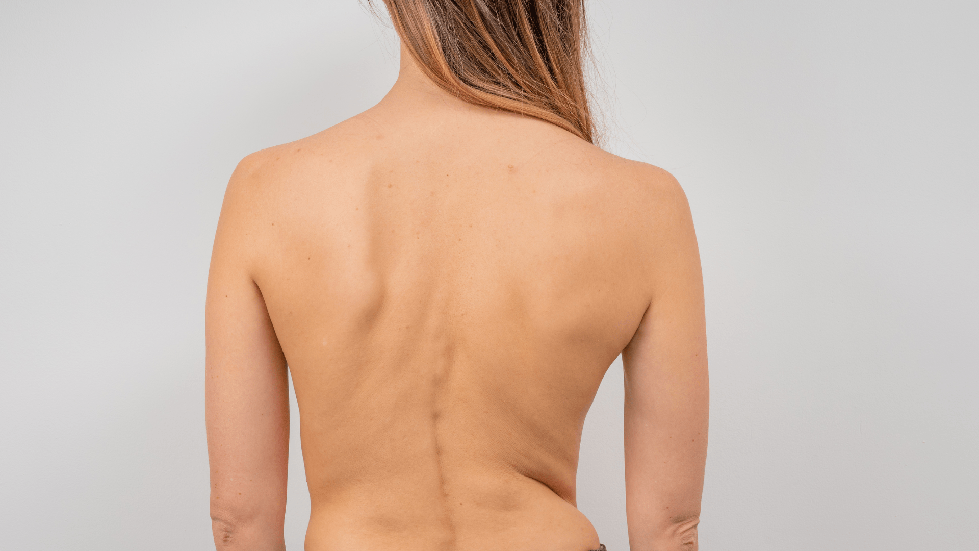
Scoliosis is a sideways curve in the spine, usually shaped like a C or an S, and describing it that way does rather undersell it. The vertebrae rotate as well as tilt, and the front to back curves of the spine change shape at the same time, so what you’ve actually got is a curve happening in three directions at once rather than a bend on a flat drawing [3]. That’s worth knowing, as the rotation is what produces the rib hump, the uneven waist and the shoulder that sits higher than the other one, and it’s also why a curve you can barely make out in a photograph can still leave one side of your back aching all day.
The size of a curve gets measured off a spinal image as an angle (the Cobb angle, if somebody says the words at you in an appointment), and it’s simply the angle between the most tilted vertebra at the top of the curve and the most tilted one at the bottom. Small numbers are common and mostly uneventful, larger ones get monitored, braced, or in a small number of cases operated on.
Now, the link with hypermobility, which is a fair bit messier than either camp tends to make out. In a very large screening of seventeen year olds, being hypermobile went along with having a spinal deformity, and the association was stronger for the moderate and severe deformities than for the mild ones [1]. That reads like a closed case right up until you look at the evidence running the other way. Hypermobile joints on their own, with nothing else going on, haven’t been convincingly linked to adolescent idiopathic scoliosis (the ordinary sort that turns up in teenagers with no known cause) at population level. That’s largely because the work that looked was measuring different things in different people, and couldn’t be pooled sensibly [2]. Looked at again more recently, it’s still sitting unresolved [4].
The distinction that actually holds up isn’t hypermobile versus not hypermobile, it’s between being generally flexible and having a symptomatic connective tissue condition, and then which one of those you’ve got [5]. In adults with hypermobile Ehlers-Danlos syndrome, roughly three in ten had a scoliosis, and most of those curves were mild enough that surgery wasn’t on the table at the point they were diagnosed [6]. In musculocontractural EDS, a rare subtype caused by a specific enzyme deficiency, it’s about two thirds, and the spinal problems there are frequently severe [7]. Same three letters on the diagnosis, and a completely different spinal picture, which is why “EDS causes scoliosis” is much too blunt a sentence to be any real use to anybody.

What the Muscles Either Side of a Curve Are Doing
Two words first, because you’ll meet them constantly and most articles use them as though you were born knowing what they mean. The convex side is the outside of the curve, the side that bulges out, and the concave side is the inside, the short side, where everything is bunched up, so curve to the right and your right side is the concave one.
Those two sides don’t behave the same way at all either. The convex side tends to show more muscle activity, more muscle bulk and more of the slow endurance fibres, while the concave side tends to show more fat mixed in through the muscle and more wasting [8]. Take that as a tendency rather than it being universal though, as the work behind it is small and its quality is mostly rated low, which is a long way from the confidence you’ll usually hear it stated with.
The muscle that keeps turning up here is the multifidus, and it’s worth knowing what that actually is, as it’s a set of short, deep muscles running up the back of the spine, each one spanning only a couple of segments, and its job isn’t really to move you about so much as to hold one vertebra steady on the next while the bigger muscles do the moving [9]. It’s roughly the difference between a mast with guy ropes and a mast without them.
In adolescents with idiopathic curves, scans show more fatty change in the multifidus on the concave side, and it’s at its worst around the apex, which is just the most tilted vertebra in the middle of the curve [10][11]. In severe curves, the muscle tissue itself tells the same story at a much smaller scale, with the concave side carrying smaller slow fibres and fewer of the cells a muscle uses to repair itself [12]. And in adults whose curves developed later through wear rather than in adolescence, the same lopsidedness turns up, again worst at the apex, and it gets more pronounced as the curve gets bigger [13].
That asymmetry does a good deal more than just show up on a scan. In adult curves, the more degenerated the muscles either side of the spine, the worse people rate their own quality of life [14]. It’s worth putting a caveat next to that one though (you know I love them), as the fat and wasting picture in deep back muscle turns up in persistent low back pain generally, with or without a curve, so it’s a fairly common consequence of a spine that’s been guarded for years rather than a signature of scoliosis specifically [15].
Which of those comes first is genuinely not known, as a curve could be holding a muscle in a position that makes it degenerate, or a muscle that’s quietly stopped doing its job could be letting the curve happen, and nobody has untangled it yet [8][10].
Measuring the muscle activity directly muddies things further rather than settling them. In adolescent curves the convex side sometimes comes out working harder during standing and holding tasks [16][17]. Other times what you’re doing at that moment matters a great deal more than which side of the curve it’s on, and we’ll come back to that one [18].
Why the Nervous System Sits in the Middle of This
Scoliosis stopped being a purely structural problem quite a while ago. In adolescents with idiopathic curves, balance is measurably worse, the reliance on vision to work out where the body is goes up, and sensory information gets weighted and combined differently [16][3]. Whether that’s a cause of the curve or a consequence of living with one is still unresolved, and anybody telling you they know which way round it goes is well ahead of the evidence.
The strongest hint that it might be causal doesn’t come from people at all. In mice bred so that the nerves reporting joint and muscle position don’t work properly, spines develop curves that look a lot like scoliosis, and in humans, the genetics points at some of the same position sensing machinery [19][20]. That’s animal work though, and mice are not small people, so it’s a reason to suspect the mechanism rather than a reason to believe it.
Now, your half of it. Hypermobility already comes with a position sense problem, as laxer tissue means the receptors in the joint and the skin report where you are less precisely, and a brain making movement decisions on a noisier signal, makes worse ones [21]. On top of that, adolescents with hypermobility spectrum disorder or hEDS need less pressure before something starts to hurt, across several different muscle groups, and they carry considerably more fatigue than their peers. The usual dip in pain after exercise doesn’t behave the way it should either, which is the pattern you’d expect from a nervous system running its gain too high rather than from something local going wrong in one muscle [22].
Put those two halves next to each other and what you’ve got is a spine that needs unusually good positional feedback to stay organised, sitting on a system that’s supplying unusually poor positional feedback. In our opinion that’s the most sensible way to read it, though it is our read rather than a demonstrated mechanism, as the two sets of research have barely met each other.
Why Moving Costs You More Than It Should
“You just need to strengthen your core” is advice that quietly assumes the exercise itself is free. For a lot of those with hypermobility it really isn’t, and that gets left out of more or less every programme going.
Walking, at the speed you’d naturally pick, costs more energy in hEDS and HSD than it does in those without it, and comes with more leg pain and more fatigue over the same distance [23]. Some of that appears to come from the tissue itself, as lower tendon stiffness and the habit of firing the muscles on both sides of a joint at once to hold it steady both push the energy cost up [24]. So you’re paying a stability tax on every single step, and nobody has ever measured yours, let alone taken it off the exercise prescription.
Then there’s the autonomic side of it, which almost never makes it into a scoliosis conversation, despite being the reason a lot of people can’t tolerate upright exercise at all. Women with HSD and hEDS carry a heavy load of autonomic symptoms, and that burden tracks with worse quality of life [25]. A posture programme that has you holding corrected positions in standing is asking something fairly specific of a system that may already be struggling to keep you upright in the first place.
That’s the real reason scoliosis exercise programmes fall apart in this population, and motivation has very little to do with it. Pain, fatigue, worry about setting something off and low confidence in your own balance are consistently what stops those with persistent pain doing the exercise they were handed [26]. A programme that accounts for none of the four still tends to get blamed on the person doing it.
What Actually Helps
Start with the awkward part. Almost all of the decent exercise evidence for scoliosis comes from adolescents with idiopathic curves and no hypermobility diagnosis, so everything below is a sensible place to start rather than something that’s been shown to work in you [27][28].
Within that population, scoliosis specific exercise does genuinely do something. Programmes built around actively correcting the curve in three dimensions, rather than general fitness work, produce better results on curve size and trunk rotation than non specific exercise, watchful waiting or standard care, and in smaller curves during the growth years they appear to reduce the chance of the curve getting worse [27][28][29][30]. The quality of that evidence is moderate at best and in places low though, so what you’ve got is a real effect that’s been measured imprecisely, which isn’t at all the same thing as no effect.
The principles those programmes are built upon, do a great deal better than any individual exercise does.
– Learning where you actually are: self correction isn’t instinctive. Asked to straighten up without being taught how, most people with a curve can’t do it in a way that improves anything [31]. It has to be coached, with feedback, and given that the position signal in hypermobility is already noisy to start with, this is probably the most transferable thing on the list.
– Body awareness work as an add on: adding structured body awareness training on top of scoliosis specific exercise improves how well people sense where their own trunk is, though whether that shows up as a different curve on an image isn’t established [32]. Useful for how you feel and move, unproven for the shape of the thing.
– Position decides which muscle does the work: prone and bird dog style positions, so face down work and work on hands and knees, get the deep multifidus contributing more relative to the big superficial back muscles under a rotational load. Side lying tends to hand the job over to the superficial muscles instead [18]. If holding one vertebra steady on the next is the thing you’re short of, then the position you do the exercise in isn’t a small detail, it’s most of the exercise.
– Which side to strengthen isn’t settled: you’ll find confident advice to stretch the concave side and strengthen the convex, and equally confident advice to strengthen the concave side precisely because that’s the side that’s wasted [18][17]. Both have a rationale behind them, but neither has been shown to be universally right. Asymmetric exercise genuinely does change which side does the work depending on how you’re positioned, so side specific loading really does depend on your curve and on the task in front of you [29][33]. That’s a much less tidy answer than either side will give you.
– Graded loading rather than blanket strengthening or stretching: exercise and motor control training do help in hypermobility, and the same body of work flags injury caused by the wrong intervention as a genuine risk. So what survives contact with reality is small, individualised, progressive loading rather than a general strength programme applied harder [34][21].
– Doing it regularly is the intervention: the programmes that have been tested run over weeks to months, supervised sessions plus practice at home, and the benefit tracks whether people actually did it rather than whether they were shown it once [30][35]. Nobody has ever got better from being assessed.
One thing to keep in mind though. None of the above validates any particular video, ours included, as a substitute for somebody competent actually looking at your curve. What the research supports is the principles, so three dimensional self correction, stabilising work, breathing, and trained posture awareness [29], and it does not support a stranger on the internet prescribing side specific exercise for a curve they’ve never seen.
What Nobody Has Actually Looked At
The gap here isn’t subtle. The scoliosis research is full of muscle imaging, muscle activity recordings and balance testing, nearly all of it in adolescents with idiopathic curves, while the hypermobility research is full of pain, fatigue and autonomic symptoms, and hardly any of it measures anybody’s spine. Very little at all sits in the overlap where you actually are [2][34][1].
That’s partly a design problem that nobody set out to create. Scoliosis trials recruit idiopathic adolescent samples, which by definition leaves out a lot of the people reading this, and hypermobility rehab work measures general pain and function rather than what a curve is actually doing over time [34]. So until somebody funds the work that sits in the middle, the practical position is to borrow the principles from the scoliosis side, apply them with the loading caution the hypermobility side keeps insisting on, and be clear with yourself that you’re extrapolating.
The Fibro Guy

References
[1] Hershkovich, O., Gordon, B., Derazne, E., Tzur, D., Afek, A. and Lotan, R. (2024) ‘Hypermobility Among Adolescents and the Association With Spinal Deformities: A Large Cross-Sectional Study’, JAAOS: Global Research and Reviews. https://doi.org/10.5435/jaaosglobal-d-24-00047
[2] Shere, C. and Clark, E.M. (2022) ‘Systematic review of the association between isolated musculoskeletal hypermobility and adolescent idiopathic scoliosis’, Archives of Orthopaedic and Trauma Surgery. https://doi.org/10.1007/s00402-022-04508-z
[3] Zhang, T., Zhou, Q., Shen, L. and Gao, S. (2026) ‘The role of proprioception in the management and rehabilitation of adolescent idiopathic scoliosis’, Frontiers in Rehabilitation Sciences. https://doi.org/10.3389/fresc.2026.1806503
Read More[4] Yudistiro, I., Hidayat, A.R., Airlangga, P.A. and Widhiyanto, L. (2026) ‘Association between Generalized Joint Hypermobility and Adolescent Idiopathic Scoliosis: A Systematic Review of Observational Studies’, Spine Surgery and Related Research. https://doi.org/10.22603/ssrr.2026-0245
[5] Hakim, A.J., Tinkle, B.T. and Francomano, C.A. (2021) ‘Ehlers–Danlos syndromes, hypermobility spectrum disorders, and associated co‐morbidities : Reports from EDS ECHO’, American Journal of Medical Genetics Part C: Seminars in Medical Genetics. https://doi.org/10.1002/ajmg.c.31954
[6] Gillas, F., Mekki, A., Foy, M., Carlier, R. and Benistan, K. (2021) ‘Prevalence of Scoliosis in Hypermobile Ehlers-Danlos Syndrome’, European Medical Journal. https://doi.org/10.33590/emj/20-00284
[7] Uehara, M., Kosho, T., Yamamoto, N., Takahashi, H.E., Shimakura, T., Nakayama, J. et al. (2018) ‘Spinal manifestations in 12 patients with musculocontractural Ehlers‐Danlos syndrome caused by CHST14/D4ST1 deficiency (mcEDS‐ CHST14 )’, American Journal of Medical Genetics Part A. https://doi.org/10.1002/ajmg.a.40507
[8] Chan, W.W.Y., Fu, S.N., Chong, T.F., Singh, G., Tsai, D.S.J., Wong, M.C.Y. et al. (2024) ‘Associations between paraspinal muscle characteristics and spinal curvature in conservatively treated adolescent idiopathic scoliosis: a systematic review’, The Spine Journal. https://doi.org/10.1016/j.spinee.2023.11.008
[9] Hodges, P.W. and Danneels, L. (2019) ‘Changes in Structure and Function of the Back Muscles in Low Back Pain: Different Time Points, Observations, and Mechanisms’, Journal of Orthopaedic & Sports Physical Therapy. https://doi.org/10.2519/jospt.2019.8827
[10] Yeung, K.H., Man, G.C.W., Shi, L., Hui, S.C.N., Chiyanika, C., Lam, T.P. et al. (2019) ‘Magnetic Resonance Imaging-Based Morphological Change of Paraspinal Muscles in Girls With Adolescent Idiopathic Scoliosis’, Spine. https://doi.org/10.1097/brs.0000000000003078
[11] Duncombe, P., Dick, T., Ng, P.T.T., Izatt, M.T., Labrom, R.D. and Tucker, K. (2025) ‘Beyond the Curve: The Muscle‐Specific Asymmetries of Adolescent Idiopathic Scoliosis’, JOR SPINE. https://doi.org/10.1002/jsp2.70129
[12] Shao, X., Chen, J., Yang, J., Sui, W., Deng, Y., Huang, Z. et al. (2020) ‘Fiber Type-Specific Morphological and Cellular Changes of Paraspinal Muscles in Patients with Severe Adolescent Idiopathic Scoliosis’, Medical Science Monitor. https://doi.org/10.12659/msm.924415
[13] Schönnagel, L., Pfeiffer, N., Khakzad, T., Muellner, M., Schmidt, H., Pumberger, M. et al. (2026) ‘Asymmetry of muscle changes is most pronounced at the apex of degenerative lumbar scoliosis: a retrospective cross-sectional analysis of older adults’, Spine Deformity. https://doi.org/10.1007/s43390-026-01463-2
[14] Tang, Y., Yang, S., Chen, C., Luo, K., Chen, Y., Wang, D. et al. (2020) ‘Assessment of the association between paraspinal muscle degeneration and quality of life in patients with degenerative lumbar scoliosis’, Experimental and Therapeutic Medicine. https://doi.org/10.3892/etm.2020.8682
[15] Kalli, I., Niglas, M., Naeini, M.K., Freidin, M., Thomas, L., Menni, C. et al. (2025) ‘Paraspinal muscle quality in chronic low back pain: a systematic review and meta-analysis of muscle atrophy and fat infiltration’, European Spine Journal. https://doi.org/10.1007/s00586-025-09454-z
[16] Paramento, M., Passarotto, E., Maccarone, M.C., Agostini, M., Contessa, P., Rubega, M. et al. (2024) ‘Neurophysiological, balance and motion evidence in adolescent idiopathic scoliosis: A systematic review’, PLOS ONE. https://doi.org/10.1371/journal.pone.0303086
[17] Park, Y., Ko, J.Y., Jang, J.Y., Lee, S., Beom, J. and Ryu, J.S. (2021) ‘Asymmetrical activation and asymmetrical weakness as two different mechanisms of adolescent idiopathic scoliosis’, Scientific Reports. https://doi.org/10.1038/s41598-021-96882-8
[18] Kim, S., Suh, J.H. and Ryu, J.S. (2026) ‘Differential Activation of the Multifidus and Erector Spinae During Asymmetric Spinal Stabilizing Exercise in Adolescent Idiopathic Scoliosis’, Annals of Rehabilitation Medicine. https://doi.org/10.5535/arm.250148
[19] Blecher, R., Krief, S., Galili, T., Biton, I.E., Stern, T., Assaraf, E. et al. (2017) ‘The Proprioceptive System Masterminds Spinal Alignment: Insight into the Mechanism of Scoliosis’, Developmental Cell. https://doi.org/10.1016/j.devcel.2017.07.022
[20] Lau, K.K.L., Law, K.K.P., Kwan, K.Y.H., Cheung, J.P.Y. and Cheung, K.M.C. (2023) ‘Proprioception‐related gene mutations in relation to the aetiopathogenesis of idiopathic scoliosis: A scoping review’, Journal of Orthopaedic Research. https://doi.org/10.1002/jor.25626
[21] Russek, L.N., Stott, P. and Simmonds, J. (2019) ‘Recognizing and Effectively Managing Hypermobility-Related Conditions’, Physical Therapy. https://doi.org/10.1093/ptj/pzz078
[22] Schubert‐Hjalmarsson, E., Fasth, A., Ickmans, K., Söderpalm, A. and Lundberg, M. (2024) ‘Exploring signs of central sensitization in adolescents with hypermobility Spectrum disorder or hypermobile Ehlers‐Danlos syndrome’, European Journal of Pain. https://doi.org/10.1002/ejp.4754
[23] Ladell, A.J.M., Golden, D.W., Oliemans, J.P., Walls, K.D., Manocha, R.H.K. and Fletcher, J.R. (2025) ‘Perceived quality of life, fatigue and the metabolic cost of walking in generalized hypermobility spectrum disorder and hypermobile Ehlers-Danlos syndrome’, Frontiers in Rehabilitation Sciences. https://doi.org/10.3389/fresc.2025.1706912
[24] Sheehan, D.S., Oliemans, J.P., Golden, D.W., Walls, K.D., Bennett, E.C., Skaper, S.J. et al. (2026) ‘To what extent do the muscles and tendons influence metabolic cost and exercise tolerance in the hypermobile Ehlers-Danlos Syndrome and Hypermobility Spectrum Disorders?’, Clinical Biomechanics. https://doi.org/10.1016/j.clinbiomech.2025.106695
[25] Collins Hutchinson, M.L., Liang, E., Fuster, E. and Blitshteyn, S. (2025) ‘Autonomic symptom burden, comorbidities and quality of life in women with Hypermobility Spectrum Disorders and hypermobile Ehlers-Danlos syndrome’, Autonomic Neuroscience. https://doi.org/10.1016/j.autneu.2025.103356
[26] Leese, C., Gupte, D., Christogianni, A., Higgins, C., Adair, P., Dall, P. et al. (2024) ‘Barriers and facilitators for physical activity in people living with chronic pain: a systematic review and combined analysis’, Pain. https://doi.org/10.1097/j.pain.0000000000003314
[27] Dong, H., You, M., Li, Y., Wang, B. and Huang, H. (2024) ‘Physiotherapeutic Scoliosis-Specific Exercise for the Treatment of Adolescent Idiopathic Scoliosis’, American Journal of Physical Medicine & Rehabilitation. https://doi.org/10.1097/phm.0000000000002524
[28] You, M.J., Lu, Z.Y., Xu, Q.Y., Chen, P.B., Li, B., Jiang, S.D. et al. (2024) ‘Effectiveness of Physiotherapeutic Scoliosis-Specific Exercises on 3-Dimensional Spinal Deformities in Patients With Adolescent Idiopathic Scoliosis: A Systematic Review and Meta-analysis’, Archives of Physical Medicine and Rehabilitation. https://doi.org/10.1016/j.apmr.2024.04.011
[29] Seleviciene, V., Cesnaviciute, A., Strukcinskiene, B., Marcinowicz, L., Strazdiene, N. and Genowska, A. (2022) ‘Physiotherapeutic Scoliosis-Specific Exercise Methodologies Used for Conservative Treatment of Adolescent Idiopathic Scoliosis, and Their Effectiveness: An Extended Literature Review of Current Research and Practice’, International Journal of Environmental Research and Public Health. https://doi.org/10.3390/ijerph19159240
[30] KARAVIDAS, N., IAKOVIDIS, P., CHATZIPRODROMIDOU, I., LYTRAS, D., KASIMIS, K., KYRKOUSIS, A. et al. (2024) ‘Physiotherapeutic Scoliosis-Specific Exercises (PSSE-Schroth) can reduce the risk for progression during early growth in curves below 25°: prospective control study’, European Journal of Physical and Rehabilitation Medicine. https://doi.org/10.23736/s1973-9087.24.08177-2
[31] Kinel, E., D’Amico, M. and Roncoletta, P. (2021) ‘3D Quantitative Evaluation of Posture and Spine Proprioceptive Perception Through Instinctive Self-Correction Maneuver in Adolescent Idiopathic Scoliosis’, Frontiers in Bioengineering and Biotechnology. https://doi.org/10.3389/fbioe.2021.663394
[32] Yaylacı, S.N., Demirkıran, H.G. and Yağcı, G. (2026) ‘Integrating basic body awareness therapy with scoliosis-specific exercise for trunk proprioception in adolescent idiopathic scoliosis: a randomized controlled trial’, European Spine Journal. https://doi.org/10.1007/s00586-026-10340-5
[33] He, C., Yang, J.T., Zheng, Q., Mei, Z. and Ma, C.Z.H. (2022) ‘How do Paraspinal Muscles Contract during the Schroth Exercise Treatment in Patients with Adolescent Idiopathic Scoliosis (AIS)?’, Bioengineering. https://doi.org/10.3390/bioengineering9060234
[34] Garreth Brittain, M., Flanagan, S., Foreman, L. and Teran-Wodzinski, P. (2023) ‘Physical therapy interventions in generalized hypermobility spectrum disorder and hypermobile Ehlers-Danlos syndrome: a scoping review’, Disability and Rehabilitation. https://doi.org/10.1080/09638288.2023.2216028
[35] Kocaman, H., Bek, N., Kaya, M.H., Büyükturan, B., Yetiş, M. and Büyükturan, Ö. (2021) ‘The effectiveness of two different exercise approaches in adolescent idiopathic scoliosis: A single-blind, randomized-controlled trial’, PLOS ONE. https://doi.org/10.1371/journal.pone.0249492