


- Teeth, Gums and Jaw Pain in hEDS, and How to Get Through a Dental Appointment - 28 September 2026
- Why ADHD and Hypermobility Keep Turning Up Together - 16 September 2026
- Hypermobility and Early Development: Milestones, Motor Delay, and Predictions - 12 September 2026
This article is part of our comprehensive guide to exercise and rehabilitation for hypermobility.
If you have hypermobility or Ehlers-Danlos syndrome and you have ever asked anyone for help with your back, your pelvis, your hips or your dodgy SI joint, there is a very good chance you have been told some version of the same thing. Your core is weak. Build your core. Engage your core. Activate your TVA. Suck your belly button to your spine. Do more planks.
The advice has been so consistent for so long that most people just nod and accept it. And honestly, why wouldn’t you? It comes from physios, from trainers, from doctors, from the bloke at the gym who once read a book. It is the default explanation for almost any back-related complaint, and for the hypermobile crowd it is often the first thing reached for when joints start moving in ways they shouldn’t. The problem is, when you actually sit down with the research, the story falls apart.
It really, really falls apart. And nowhere does it fall apart more dramatically than in the hypermobility population, where the standard “engage your core” advice tends to make things worse, not better. We see this in our studios all the time. People come in having spent two or three years grinding through plank progressions, dead bugs done with breath held and abs locked solid, Pilates classes where they are told to “draw the navel in” and never actually told why, and they cannot figure out why their pain is the same or worse. They have been doing the work. They have been compliant. The work was just wrong for their body.
So this is going to be the proper, deep dive into hypermobility core exercises. What the core actually is. Why “weak core causes pain” doesn’t survive contact with the evidence. What’s genuinely different about the hypermobile core, the hypermobile pelvic floor, and the hypermobile SI joint. And what we actually do in the studios that gets people moving without the bracing, the gripping, and the constant low-grade ache that comes with it.
Bring a cup of tea. This one is long.
What your core actually is (it is not your six-pack)
Ask the average person what their core is and they will point at their abs. Ask someone who’s been to a gym for a year and they will tell you about the transversus abdominis (TVA), the deep wraparound abdominal muscle that the entire core stability industry was built on top of [1]. Both of them are partly right, and both of them are missing the bigger picture by quite a long way.
Your core is not a single muscle. It is not even a sheet of muscles. It is a pressurised cylinder, and it has at least four parts.
– The roof: the diaphragm.
– The floor: the pelvic floor.
– The walls: the transversus abdominis (the deep layer that wraps around like a built-in weightlifting belt), the internal and external obliques, and the rectus abdominis (your six-pack) at the front.
– The back wall: the deep spinal muscles, particularly the multifidus, alongside the erector spinae and the thoracolumbar fascia.
These structures don’t work in isolation. They contract together, in patterns, and what they’re actually doing is managing pressure. Imagine an empty plastic bottle. Squeeze it and it crumples. Now blow some air into it, screw the lid on, and squeeze it again. It barely moves. That is, in very crude terms, what your core does. It pressurises the abdominal cavity and that pressure stabilises the spine and pelvis. The strength of any one muscle matters far less than the coordination of the whole system.
And here is the key bit that almost nobody mentions when they tell you to “engage your core.” The system is supposed to run automatically, on a feedforward basis, based on whatever you’re about to do. In people without back pain, the TVA fires before the deltoid, or near enough at the same moment, when you throw an arm up quickly. The brain is anticipating the load, not reacting to it [2]. That timing, not the strength, is what good core stability looks like.
So when you are told to consciously tense your stomach, hold the brace, suck the belly button in and never let it out, you are doing the exact opposite of what a healthy core does. You are bracing. You are reacting. And in a hypermobile body that has been guarding against perceived instability for years, you are turning the volume up on a system that was already too loud.
The “weak core causes pain” myth (and why it has hung around for thirty years)
The whole modern core stability industry traces back to a small handful of studies in the mid-1990s. Hodges and Richardson published a paper in Spine in 1996 that found a delay, measured in milliseconds, in the activation of the TVA in people with low back pain compared to controls without back pain [2]. The delay turned up in every direction they tested the arm movement in, unlike the more superficial abdominal muscles, which is part of why the TVA got read as a general stiffener rather than as something that fine tunes a particular direction [2]. The TVA still fired. It just fired a little late. That paper became the founding document of the entire “weak core” model, and from a tiny timing difference came a global industry of crunches, planks, abdominal hollowing manoeuvres and “draw your belly button in” cues.
Now, that finding has not been overturned. The delay is real. But what came afterwards is where it gets interesting, and where the standard story gets uncomfortable.
First, follow-up work showed that you can produce the exact same TVA delay in healthy people simply by inducing acute muscle pain. Inject saline into the back muscles of healthy volunteers, give them muscle pain that hadn’t existed five minutes earlier, and the feedforward TVA timing is disrupted [3]. That is a problem for the “weak TVA causes back pain” model. If pain causes the timing change, then strengthening the TVA isn’t the fix, because the TVA was never the cause.
It is also worth knowing how big that delay actually was. In the original core stability studies, the difference in onset time between people without symptoms and people with chronic low back pain was about 20 milliseconds, a fiftieth of a second, and it turned up during fast arm movements but not during slow ones [1]. Lederman’s point about that number is the one that matters: timings on that scale sit well beyond anybody’s conscious control, and beyond a therapist’s ability to test or alter them in a clinic [1]. Nobody is bracing their way to a fiftieth of a second.
Second, when you actually run the trials on whether core stability training works for back pain, the results are underwhelming. Across 29 trials in adults with chronic non-specific low back pain, motor control exercise beat other forms of exercise by a margin too small to matter clinically in the short term, and made no clinically important difference at intermediate or long term follow up [4]. Pool 29 studies of low back pain of any duration and you get the same shape: stabilisation exercise was no better than other forms of exercise for pain or disability in the long term, and its short and medium term edge was too small to matter clinically [7]. A critically appraised summary of five trials came out the same way, ahead of general exercise at three months and no different in the long run [9].
Third, and this is the bit that should really make you raise an eyebrow, when stabilisation exercises do produce clinical improvement, the improvement doesn’t track any measurable improvement in TVA function. You can do all the abdominal hollowing in the world and get someone’s pain down, and how much their TVA changed will not tell you whether they were one of the ones who got better [5]. Pool 15 studies of conservative treatment for non-specific low back pain and the strongest finding is that the change in how much the TVA thickens when it contracts, and the change in its feedforward activation, were unrelated to the change in pain and disability [8]. That same work is more careful about the multifidus. Whether treatment changes in its size and shape track outcomes came out uncertain and conflicting, so that half of it is not settled [8]. Across many different types of exercise therapy for chronic back pain, a good clinical outcome and a change in the thing the exercise was aimed at usually aren’t linked [6]. And on the narrower question of whether core stability exercise resets that onset timing at all, Lederman’s review says flatly that no study has demonstrated it [1].
So what’s actually going on when core training does help? Probably a mix of several things. Reduced fear of movement. The nervous system feeling safer because somebody competent has given the person something structured to do. The general benefits of being more active and less catastrophising. Some genuine improvement in coordination and timing. Endorphins. Education. Confidence.
None of that is core specific. Almost any reasonable form of structured movement, taught with care, would deliver the same gains. The “engage your core” instruction is along for the ride, not the engine.
Even the concept of “spine stability” itself is fuzzier than it sounds. Researchers had been defining and measuring spine stability in completely different ways, so the field was arguing about something it couldn’t actually agree on [10]. Clinical lumbar instability has even been called a “myth”, in the sense that the term gets used clinically without any solid mechanical basis [11]. We are not exactly building from rock-solid foundations.
Then there’s the awkward matter of all the people walking around with weak abdominals and no back pain at all. In one trial, 402 people were identified as having weak abdominal muscles and no back pain. They were given back education alone, or back education plus abdominal strengthening, and across a year there was no significant difference between the groups in episodes of back pain [1]. In 257 collegiate athletes, a core strengthening programme gave no significant advantage in reducing how often low back pain occurred [1]. And after breast reconstruction using the rectus abdominis, where the muscle is surgically moved, no relationship to back pain or to functional impairment showed up when people were followed up to several years later [1].
All three of those are reported in Lederman’s review rather than run by it. It’s a single author critical review with no stated search strategy, so read it as a well argued case against core stability rather than as a systematic one.
And then there’s pregnancy. The abdominal wall stretches enormously and connective tissue becomes more compliant, so by the weak core rule you’d expect back and pelvic pain to hang around for most women after birth. For pelvic girdle pain after delivery, the prognosis is good, with substantial recovery in most women [26]. If a weakened abdominal wall were the cause, that would be hard to explain. And in a trial of 126 women whose pregnancy related low back pain was still there three weeks after delivery, drawn from a cohort of 7,526, a brief self management intervention made a difference to activity limitation that was too small to be clinically relevant over time, and no clear difference to pain [27].
The recruitment figures from one postpartum study make the point more bluntly than any result does. Of 869 pregnant women recruited, 635 were excluded because they recovered spontaneously, with no treatment at all, within a week of delivery [1]. A week. Rectus abdominis takes around four weeks after birth to re-shorten, and around eight weeks for pelvic stability to normalise, so the pain was improving straight through the window of worst abdominal function [1]. Lederman’s question is the obvious one, and nobody has a tidy answer to it. Why doesn’t the spine collapse?
Lederman pulled all of this together back in 2010 in a paper called, brilliantly, “The myth of core stability” [1]. It is over fifteen years old at this point. It is still right.

Why the standard advice is actively unhelpful for hypermobile bodies
If the standard core advice is mediocre for the general population, for those with hypermobility it is often a lot worse. Here is why.
In a typical, non-hypermobile body, the joints provide a steady stream of accurate proprioceptive feedback to the brain. The nervous system knows where everything is. Stability happens automatically because the system has good information.
In a painful hypermobile body, that information stream is a bit degraded. Lax connective tissue means joints can move further than they should, ligaments are more elastic, and the proprioceptors that should be telling the brain “this is where the joint is” are giving fuzzier signals. So, the nervous system, doing its best, compensates. It cranks up muscle tone everywhere. Holds tension as a substitute for the joint stability it can’t get from the connective tissue. Recruits muscle when it can’t trust the ligaments.
It shows up when you measure it. In people with joint hypermobility syndrome and knee pain, responses in the quadriceps to magnetic stimulation of the motor cortex rose more steeply than in people with generalised joint hypermobility and no pain, or people with normal flexibility, and those last two groups didn’t differ from each other [12]. So the difference sat with the pain, not with the bendiness on its own. Children and adults with hypermobility syndrome or hEDS also show generalised hyperalgesia, lower pressure pain thresholds measured across 12 sites of the body, and the authors read that as a possible sign of central nervous system involvement rather than a purely local problem [13].
So now imagine you walk into a physio with widespread hypermobility, dodgy SI joint, low back pain on and off for years, and you’re told to engage your core, hold the brace through every step, every reach, every lift. What you’re being asked to do is add another conscious layer of muscle tension on top of a system that was already gripping. You can guess how that ends. People come out tighter, more painful, with a pelvic floor that won’t switch off and a neck full of lumps because all the secondary muscles got conscripted to keep the chest moving.
Lederman pushes this further than we normally would, and he offers it as a hypothesis rather than a finding, so take it as one. Trunk muscles compress the lumbar spine when they contract, people with chronic back pain already increase co-contraction during movement, and his argument is that instructing them to add more is likely to come at the cost of further compression on joints and discs that are already sensitised [1]. When three strategies were compared directly, abdominal hollowing came out the least effective and didn’t increase stability at all, bracing did improve stability but increased spinal compression, and the group left alone to use their natural strategy got stability without the compression [1]. Tensing the trunk has even been shown to degrade postural control [1]. His conclusion is that anybody trained in complex hollowing and bracing manoeuvres should be discouraged from using them.
This is the bit that gets overlooked. The hypermobile core, in our experience, is almost never under-active. It’s over active. The question isn’t how to make it work harder. The question is how to teach it to coordinate, time properly, and let go when it doesn’t need to be on.
Different problem, different solution.
The breath is the brace (and this is where most people have it backwards)
This is the line we say to clients more than any other:
Core stability is the breath. The brace is the breath.
If you take one thing from this article, take that.
When the cylinder is working properly, the diaphragm does most of the stabilising work. It contracts and domes downwards on the inhale, pressurising the abdomen. The pelvic floor lengthens and descends with it. The deep abdominal wall, including the TVA, responds to the pressure change in a coordinated, low-effort way. The whole cylinder lengthens slightly on the inhale and shortens on the exhale, like a jellyfish opening and closing. There is nothing to consciously squeeze. There is no belly button to suck in.
This isn’t a vibe. The pelvic floor muscles act physiologically as expiratory muscles, working alongside the anterolateral abdominal wall, contracting during expiration and relaxing during inspiration [21]. In a small trial of 33 women with stress urinary incontinence, electrical stimulation training of the pelvic floor increased diaphragm excursion during quiet breathing, forceful breathing and coughing [22]. The two systems aren’t independent. They are mechanically linked, and they need each other to work properly.
The problem is that, in our experience, most people with hypermobility aren’t breathing like this. They default to chest breathing, where the diaphragm hardly moves and the rib cage gets lifted by the accessory muscles in the neck and shoulders, mainly the scalenes, sternocleidomastoid, and upper trapezius. Once those muscles take over the breathing job, they fatigue, they tighten, they protest. A huge amount of the chronic neck and upper back tension we see in hypermobile clients comes from a chest-dominant breathing pattern, not the other way around. And working purely on the neck almost never fixes it long-term.
There’s also a more subtle problem. Adults with hEDS were worse than matched controls at judging how much air they’d breathed in on a small breath, around 30 per cent of their inspiratory capacity, though not on a larger one, and their breathing became more erratic than the controls’ when they were given a mental task at the same time [14]. That was 19 people in each group, measured in one session, and the authors say a larger study is needed to confirm it. Their own hypothesis points at the signal coming from the rib cage, the same kind of less precise signal that affects joint position sense, though they couldn’t rule out the upper airway [14]. The fix is not to “try harder to feel it.” The fix is to clean up the breath pattern so the system has better information to work with.
And then there is the broader picture of respiratory problems in this population, reported across multiple subtypes: breathlessness, decreased respiratory muscle strength, and patterns suggesting dysfunctional breathing [28]. None of this is exotic. It just rarely gets thought about when someone walks in complaining about their back.
There’s also the carbon dioxide piece. Most people think CO2 is a waste gas you breathe out. It isn’t. CO2 is a signal that tells haemoglobin to release oxygen at the tissue level, regulates blood vessel diameter, and is deeply involved in autonomic regulation. When people over-breathe, CO2 drops too low, blood vessels constrict, including in the brain, and the result is the brain fog, dizziness, and air hunger that anyone with POTS or hypermobility will recognise instantly.
Bottom line. Before any of the dead bug, bird dog, plank, brace nonsense, the breath has to be sorted. That is where everything starts.
The hypermobile pelvic floor (where “do your Kegels” can do real harm)
For the general population, the standard pelvic floor advice is mostly fine. Most pelvic floor problems in the wider world come from weakness, particularly after childbirth, or with ageing. Strengthen the muscle, ease the symptoms, job done.
For those with hypermobility, the picture is often different, and the advice can run in the wrong direction. In the pelvic floors we see in the studios, the problem is far more often too much tone than too little. Held in a state of constant low-grade contraction, the way a jaw can be permanently slightly clenched without the person realising. The muscle is not weak in the gym sense. It is exhausted from over-working, and it has lost the ability to fully relax. This pattern, called nonrelaxing pelvic floor dysfunction, is recognised in the wider chronic pelvic pain literature [17]. And hypermobility turns up often there: among women with chronic myofascial pelvic pain at one specialist centre, 24 per cent also met criteria for generalised hypermobility spectrum disorder [16].
The numbers in this group are striking. In an international survey of cisgender women with EDS, self-reported rates of stress urinary incontinence, urinary urgency, prolapse and pelvic pain were high [15]. Across 105 studies of urogenital and pelvic complications in EDS and HSD, urinary problems were the most often reported, although the findings on incontinence were mixed and 4 of 18 studies found no association at all [19]. In an online survey of people with EDS and HSD, rates of painful sex and probable vulvodynia were high, alongside a tangle of co-occurring conditions [20]. And in a prospective cohort following women with joint hypermobility, defined by Beighton score, through the first decade after a first delivery, hypermobility went with a lower rate of operative delivery and was not, in that sample, linked to higher rates of pelvic floor disorders [18]. So the relationship between hypermobility and the pelvis is more nuanced than the simple “laxity equals more problems” story tends to suggest.
Pulling all of that together, this is how we read the shape of the problem. The connective tissue is more compliant, so the passive support of the pelvic organs is reduced. The nervous system compensates by ramping up muscle tone, including in the pelvic floor. The pelvic floor, gripping all day, becomes painful and starts referring symptoms in every direction: incontinence, prolapse sensations, pain on intercourse, constipation, hip pain, back pain. And on top of that, the generalised hyperalgesia mentioned earlier suggests the whole system may be more sensitive, not just the pelvis [13].
There’s one more strand worth putting here, because it points the same way. Tensing the trunk raises intra-abdominal pressure, and it has been estimated that in people with pelvic girdle pain that raised pressure could exert potentially damaging forces on the pelvic ligaments. The study Lederman cites for it recommends teaching people to reduce their intra-abdominal pressure, which is the exact opposite of bracing [1]. That’s an estimate from a model rather than a measurement in anybody, and none of it was done in a hypermobile population. But if your ligaments are already more compliant than average, it’s not a reassuring direction of travel.
This is why blanket Kegel advice can do real harm. If your pelvic floor is hypertonic and you keep adding voluntary contractions on top of it, you are building tension on top of tension. People come in saying their symptoms have got worse since they started doing the Kegels their physio gave them, and in many cases that is exactly what has happened. We’ve seen this pattern dozens of times now. It is not your fault. It is the wrong tool for the job.
What works for hypermobile pelvic floors is the opposite. Lengthening, descending, softening with the inhale. Letting the floor move with the diaphragm rather than overriding it. The cue we use in the studio sounds ridiculous and works really well: imagine you are about to do a pee and a poo at the same time. Not bearing down. Not pushing. Just letting the floor open and drop on the inhale, and rising back up on the exhale.
If pelvic floor symptoms are part of your picture, our full guide to hypermobility, EDS and the pelvic floor goes into this in much more depth than I can here, including how to find a pelvic floor physiotherapist who actually understands hypertonicity in this group rather than assuming the standard weakness model.
The SI joint, force closure, and why nothing fixes it in isolation
If we move from the front of the cylinder to the back, we run into the sacroiliac joint. One on each side, between the sacrum and the ilium. The SI joint relies on a combination of form closure, which is the shape of the joint itself, a wedge with ridges and grooves across its surfaces, plus some very strong ligaments, and force closure, which is compression generated across the joint by muscles, fascia, ligament tension and the ground pushing back at you [23]. That second half is the one that gets mangled in a studio. Force closure is not a stomach held tight. It needs the sacrum to nutate and the compression to be coordinated across the joint, rather than one muscle switched on and kept on [23].
Form closure is the passive bit. The sacrum sits between the two ilia like a keystone in an arch. Strong ligaments hold the assembly together. If everything fits well and the ligaments are tight, you essentially have a self-locking joint that doesn’t need much help. In a typically-stable body, this gives you most of the stability for free.
Force closure is the active bit. Muscle slings cross the SI joint and create compression. The longitudinal sling (erector spinae through the thoracolumbar fascia to biceps femoris). The posterior oblique sling (latissimus dorsi through the fascia to the opposite gluteus maximus). The anterior oblique sling (external oblique to opposite internal oblique to adductors). The transversus abdominis, crossing the joint and clamping the sacrum between the two ilia. The pelvic floor, opposing lateral movement of the pelvic bones. Biomechanical modelling shows that coordinated activation of these muscles can substantially reduce SI joint shear loads [24], and in vivo work using Doppler imaging has confirmed that muscle contraction increases SI joint stiffness in real bodies [25].
Notice that none of those structures are separate from the cylinder we just talked about. The “core” muscles and the “SI joint stabilisers” are the same set of muscles in different combinations. They are not parallel systems. They are the same system seen from a different angle.
In hypermobility, form closure should be reduced, because the ligaments are more compliant. The wedge does not seat as firmly. So force closure has to do more work to keep up. The active system has to work harder, which means more sustained muscle tone around the pelvis, which means more fatigue, more pain, and more grip. That’s our reading of the mechanics. What has been reported is that pelvic girdle pain in pregnancy is more common with hypermobility, 26 per cent against 7 per cent in one comparison, and the authors reviewing it say the research on the link is limited and of varying quality [26].
This is also why fixing the SI joint in isolation almost never works long-term. If your pelvic floor is gripping asymmetrically, if your breath is shallow, if your TVA is firing late, if your nervous system thinks the whole pelvis is unsafe, your SI joint is going to keep getting destabilised regardless of how many specific SI joint exercises or manipulations you do. The pain comes back. The manipulation lasts a few days. The next week you are back at the physio.
The path to a stable SI joint runs through the pelvic floor, which runs through the breath, which runs through the nervous system’s threat level. You cannot pick one piece and skip the rest. Or rather, you can, but you’ll be on a permanent rotation of “feels better for two weeks, gets worse again,” and most of you reading this know that pattern intimately.
This is the bit that’s genuinely different about how we approach this. The exercises themselves are not exotic. The breath work is not exotic. The dead bug and bird dog are not exotic. But the order matters, and the framing matters, and treating the whole cylinder as a system rather than a list of muscles to drill is what actually moves the needle.
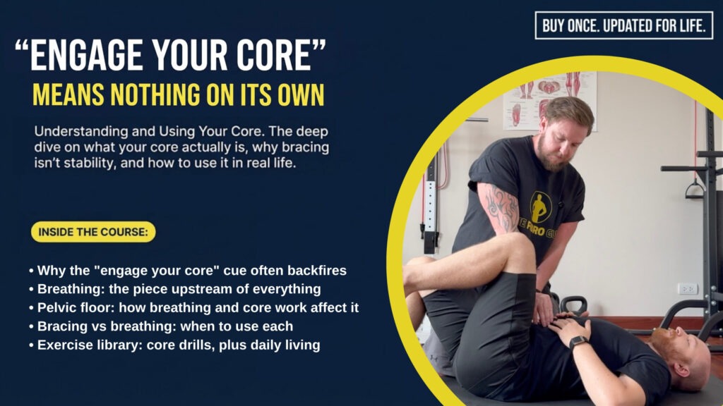
What we actually do in the studios
Right then. Onto the practical stuff.
This is the bit a lot of people skip ahead to, but the reason we spent the first half of this article on the theory is that the practical work makes no sense without it. If you don’t understand why you are doing in-and-down rather than in-and-up, or why we don’t really care about the ten-pack abs you can develop with crunches, the exercises just become another set of moves to grind through.
The video below is the same introduction we use as homework for our Hypermobility Live Workshop and what we have our in-person clients in our studios working through early in their rehab. It walks you through the four breath stages and the entry-level movement patterns. Watch it through once before you try anything. Then read the breakdown underneath so you understand what you are doing and why.
Stage 1: Diaphragmatic breathing on your back
Lie on your back, knees bent, feet flat on the floor. Pillow behind your head if your neck is forward. Comfort first. One hand on the chest, one on the belly.
Breathe through the nose, mouth closed. As you breathe in, the belly should expand outwards and slightly sideways. The chest hand should hardly move. As you breathe out, the belly should soften back down. Slow. Don’t go for big breaths, you will get dizzy. A small weight on the belly, a 1 kg dumbbell or a water bottle, gives you a tactile cue. The job isn’t to push the weight up by force. The job is to let the breath move it.
The single most common error we see is sucking the belly in on the inhale. This is paradoxical breathing, and a lot of chronic chest breathers do it without realising. If you find yourself doing it, slow everything down and use the weight to feel the breath move the abdomen.
Stage 2: 360-degree lateral expansion
Sit up tall. Find the soft space between the bottom of your rib cage and the top of your hips. Wrap the webbing of your hands around your sides at that level, so you can feel the soft tissue under your fingers.
Breathe in and try to push your hands outwards with the breath itself. Most people cannot feel this on the first attempt. Singers and people who play wind instruments often get it straight away because their training already drilled the obliques. Everyone else needs days to weeks. If you can’t feel anything, give yourself three sharp coughs in a row, a “ha, ha, ha”, and feel the obliques punch out into your hands. That’s the muscle group we’re trying to find with the breath.
Stage 3: Back expansion (kneeling lean)
Hips above knees, leaning forward onto your hands or onto a chair seat. Sit-bones underneath, ribs over pelvis, neutral spine. Place your thumbs on either side of the lower spine and breathe into the back. Gravity opens the back of the cylinder rather than the front.
The movement here is small. You are not going to see massive expansion. Most people will see a tiny visible widening of the lower back, and that’s it. Some sensation in the SI joints is normal, because the breath mechanics are pressurising tissues that have been short and compressed for years. If you have had a forward pelvic tilt for decades, this position can feel quite strange the first few times.
Stage 4: In and down (the exhale brace)
This is the one most people get wrong, and it is the most important.
On the exhale, the belly button comes in and down. Not in and up. In and up is hollowing, the old “pull your belly button to your spine” cue, and it’s the wrong pattern. It jacks everything up under the rib cage and the neck gets jammed. In and down flattens the lower abdomen, wraps the obliques inwards like a corset, and softens the ribs over the pelvis.
Just 10 to 20 per cent. You should still be able to hold a conversation while doing it. If you can’t, you are gripping too hard.
One important note on the cue itself, because this catches a lot of people out. “In and down” is the intention, not the visible movement. The belly button doesn’t physically drop towards the floor when you do this properly. What you actually see and feel is the lower abdomen flattening and the front of the pelvis tucking very slightly underneath, because the rectus abdominis attaches from the ribs down into the pubic bone, and when you contract it gently, it pulls those two points a tiny bit closer together. The belly button itself often appears to lift up slightly because the pelvis tips under, not because anything is pushing down. The cue says “in and down” because thinking down stops you from hollowing up under the ribs, which is the wrong pattern. The result on the body is a flatter lower belly, a slight posterior tilt of the pelvis, and the obliques cinching inwards like a corset. Don’t go looking for the belly button to physically descend. Look for the lower abs to flatten and the waist to narrow.
The two movement patterns: dead bug and bird dog
Once the breath is in place, we layer it onto two simple patterns. The modified dead bug (lying on your back, one foot on the floor for stability, the other knee coming towards the chest then the leg slowly extending out, breath-led, return on the inhale). And the bird dog (on hands and knees, neutral spine, sliding one leg back, then progressing to lifting it, then opposite arm and leg).
The rule is the same for both. Inhale to set the cylinder. Exhale on the harder part of the movement, with the belly button moving in and down 10 to 20 per cent. Range only goes as far as the breath and pelvis stay neutral. The moment the pelvis tips forward or the breath gets held, you’ve gone too far. Drop back to the easier version.
Wrist pain in the bird dog? Use forearms instead, or place your hands on a low chair or sofa. The position is adjustable. The pattern is what matters. The same applies if your knees or shoulders object.
That’s the entry point. Five things, in order. Breath one, breath two, breath three, breath four, then the simplest possible movement layered on top. None of it is a hard workout. It is not supposed to leave you sweating. The goal is coordination and timing, not fatigue.

What keeps the nervous system switched on at night in hypermobility, EDS and fibromyalgia, and what actually shifts it. No list about screens and camomile tea.
A note on what NOT to do
This is the bit where I get to be a bit grumpy.
There are still a lot of professionals out there prescribing planks, sit-ups, crunches and traditional ab work to hypermobile clients, often as a fix for back or hip pain. In most cases that is the worst thing you can do. Those exercises hammer the rectus abdominis, which already works fine in most people, encourage breath holding, which collapses the cylinder you are trying to build, and reinforce a “tense everything” pattern that most hypermobile bodies are already stuck in.
If your physio or trainer has you doing planks for a hypermobility related back issue and ignoring your breath, your pelvic floor and your nervous system, you are probably in the wrong place. Not always. There are some genuinely thoughtful practitioners who know how to use those tools well. But the average prescription of “more crunches” for a hypermobile back is, in our experience, almost never the right answer.
The other thing to flag is the constant bracing advice. Some practitioners genuinely tell hypermobile clients to “engage your core every time you walk around” or to keep the belly button pulled in throughout the day. This is, I cannot stress this enough, a fantastic way to wreck your breath. You cannot hold your abdominals hard all day and breathe properly. The two are mechanically incompatible. The brace is meant to be situational, the way you might crank up support to push a heavy shopping trolley around a corner. It is not meant to be on all the time.
Likewise, just doing more rounds of the same Kegel programme that did nothing the first time, especially if you’ve got a hypertonic pelvic floor, isn’t a strategy. It’s repetition.
If you have spent years bracing, the unwinding takes a while. That is normal. Give it time, and don’t expect three weeks of breath and core work to undo a decade of gripping.
If anything in this piece has shifted how you think about core work, or you’re now eyeing up the planks in your existing programme with mild suspicion, that’s the point. The standard advice for hypermobility has been weak for a long time. The evidence supports a different approach, and our experience in the studios has consistently borne it out.
The Fibro Guy

References
[1] Lederman E (2010) ‘The myth of core stability’, Journal of Bodywork and Movement Therapies, 14(1), pp. 84-98. doi: 10.1016/j.jbmt.2009.08.001
[2] Hodges PW, Richardson CA (1996) ‘Inefficient muscular stabilization of the lumbar spine associated with low back pain: a motor control evaluation of transversus abdominis’, Spine, 21(22), pp. 2640-2650. doi: 10.1097/00007632-199611150-00014
[3] Hodges PW, Moseley GL, Gabrielsson A, Gandevia SC (2003) ‘Experimental muscle pain changes feedforward postural responses of the trunk muscles’, Experimental Brain Research, 151(2), pp. 262-271. doi: 10.1007/s00221-003-1457-x
Read More[4] Saragiotto BT, Maher CG, Yamato TP, Costa LOP, Menezes Costa LC, Ostelo RWJG, Macedo LG (2016) ‘Motor control exercise for chronic non-specific low-back pain’, Cochrane Database of Systematic Reviews, (1), CD012004. doi: 10.1002/14651858.CD012004
[5] Mannion AF, Caporaso F, Pulkovski N, Sprott H (2012) ‘Spine stabilisation exercises in the treatment of chronic low back pain: a good clinical outcome is not associated with improved abdominal muscle function’, European Spine Journal, 21(7), pp. 1301-1310. doi: 10.1007/s00586-012-2155-9
[6] Steiger F, Wirth B, de Bruin ED, Mannion AF (2012) ‘Is a positive clinical outcome after exercise therapy for chronic non-specific low back pain contingent upon a corresponding improvement in the targeted aspect(s) of performance? A systematic review’, European Spine Journal, 21(4), pp. 575-598. doi: 10.1007/s00586-011-2045-6
[7] Smith BE, Littlewood C, May S (2014) ‘An update of stabilisation exercises for low back pain: a systematic review with meta-analysis’, BMC Musculoskeletal Disorders, 15(1), p. 416. doi: 10.1186/1471-2474-15-416
[8] Wong AY, Parent EC, Funabashi M, Kawchuk GN (2014) ‘Do changes in transversus abdominis and lumbar multifidus during conservative treatment explain changes in clinical outcomes related to nonspecific low back pain? A systematic review’, The Journal of Pain, 15(4), pp. 377.e1-377.e35. doi: 10.1016/j.jpain.2013.10.008
[9] Coulombe BJ, Games KE, Neil ER, Eberman LE (2017) ‘Core stability exercise versus general exercise for chronic low back pain’, Journal of Athletic Training, 52(1), pp. 71-72. doi: 10.4085/1062-6050-51.11.16
[10] Reeves NP, Narendra KS, Cholewicki J (2007) ‘Spine stability: the six blind men and the elephant’, Clinical Biomechanics, 22(3), pp. 266-274. doi: 10.1016/j.clinbiomech.2006.11.011
[11] Mulholland RC (2008) ‘The myth of lumbar instability: the importance of abnormal loading as a cause of low back pain’, European Spine Journal, 17(5), pp. 619-625. doi: 10.1007/s00586-008-0612-2
[12] Long M, Kiru L, Kassam J, Strutton PH, Alexander CM (2022) ‘An investigation of the control of quadriceps in people who are hypermobile; a case control design’, BMC Musculoskeletal Disorders, 23(1), p. 607. doi: 10.1186/s12891-022-05540-1
[13] Scheper MC, Pacey V, Rombaut L, Adams RD, Tofts L, Calders P (2017) ‘Generalized hyperalgesia in children and adults diagnosed with hypermobility syndrome and Ehlers-Danlos syndrome hypermobility type: a discriminative analysis’, Arthritis Care & Research, 69(3), pp. 421-429. doi: 10.1002/acr.22998
[14] Hakimi A, Bergoin C, De Jesus A, Hermand E, Fabre C, Mucci P (2024) ‘Impairment of lung volume perception and breathing control in hypermobile Ehlers-Danlos syndrome’, Scientific Reports, 14(1). doi: 10.1038/s41598-024-58890-2
[15] Kciuk O, Li Q, Huszti E, McDermott CD (2023) ‘Pelvic floor symptoms in cisgender women with Ehlers-Danlos syndrome: an international survey study’, International Urogynecology Journal, 34(2), pp. 473-483. doi: 10.1007/s00192-022-05273-8
[16] Hastings J, Forster JE, Witzeman K (2019) ‘Joint hypermobility among female patients presenting with chronic myofascial pelvic pain’, PM&R, 11(11), pp. 1193-1199. doi: 10.1002/pmrj.12131
[17] Faubion SS, Shuster LT, Bharucha AE (2012) ‘Recognition and management of nonrelaxing pelvic floor dysfunction’, Mayo Clinic Proceedings, 87(2), pp. 187-193. doi: 10.1016/j.mayocp.2011.09.004
[18] Knoepp LR, McDermott KC, Muñoz A, Blomquist JL, Handa VL (2013) ‘Joint hypermobility, obstetrical outcomes, and pelvic floor disorders’, International Urogynecology Journal, 24(5), pp. 735-740. doi: 10.1007/s00192-012-1913-x
[19] Gilliam E, Hoffman JD, Yeh G (2020) ‘Urogenital and pelvic complications in the Ehlers-Danlos syndromes and associated hypermobility spectrum disorders: a scoping review’, Clinical Genetics, 97(1), pp. 168-178. doi: 10.1111/cge.13624
[20] Glayzer JE, McFarlin BL, Castori M, Suarez ML, Meinel MC, Kobak WH, Steffen AD, Schlaeger JM (2021) ‘High rate of dyspareunia and probable vulvodynia in Ehlers-Danlos syndromes and hypermobility spectrum disorders: an online survey’, American Journal of Medical Genetics Part C: Seminars in Medical Genetics, 187(4), pp. 599-608. doi: 10.1002/ajmg.c.31939
[21] Talasz H, Kremser C, Talasz HJ, Kofler M, Rudisch A (2022) ‘Breathing, (S)Training and the Pelvic Floor: a basic concept’, Healthcare, 10(6), p. 1035. doi: 10.3390/healthcare10061035
[22] Hwang U, Lee M, Jung S, Ahn S, Kwon O (2021) ‘Effect of pelvic floor electrical stimulation on diaphragm excursion and rib cage movement during tidal and forceful breathing and coughing in women with stress urinary incontinence’, Medicine, 100(1), p. e24158. doi: 10.1097/MD.0000000000024158
[23] Vleeming A, Schuenke M (2019) ‘Form and force closure of the sacroiliac joints’, PM&R, 11(S1). doi: 10.1002/pmrj.12205
[24] Pel JJM, Spoor CW, Pool-Goudzwaard AL, Hoek van Dijke GA, Snijders CJ (2008) ‘Biomechanical analysis of reducing sacroiliac joint shear load by optimization of pelvic muscle and ligament forces’, Annals of Biomedical Engineering, 36(3), pp. 415-424. doi: 10.1007/s10439-007-9385-8
[25] van Wingerden JP, Vleeming A, Buyruk HM, Raissadat K (2004) ‘Stabilization of the sacroiliac joint in vivo: verification of muscular contribution to force closure of the pelvis’, European Spine Journal, 13(3), pp. 199-205. doi: 10.1007/s00586-003-0575-2
[26] Ali A, Andrzejowski P, Kanakaris NK, Giannoudis PV (2020) ‘Pelvic girdle pain, hypermobility spectrum disorder and hypermobility-type Ehlers-Danlos syndrome: a narrative literature review’, Journal of Clinical Medicine, 9(12), p. 3992. doi: 10.3390/jcm9123992
[27] Bastiaenen CHG, de Bie RA, Vlaeyen JWS, Goossens MEJB, Leffers P, Wolters PMJC, Bastiaanssen JM, van den Brandt PA, Essed GGM (2008) ‘Long-term effectiveness and costs of a brief self-management intervention in women with pregnancy-related low back pain after delivery’, BMC Pregnancy and Childbirth, 8(1), p. 19. doi: 10.1186/1471-2393-8-19
[28] Chohan K, Mittal N, McGillis L, Lopez-Hernandez L, Camacho E, Rachinsky M, Santa Mina D, Reid WD, Ryan CM, Champagne KA, Orchanian-Cheff A, Clarke H, Rozenberg D (2021) ‘A review of respiratory manifestations and their management in Ehlers-Danlos syndromes and hypermobility spectrum disorders’, Chronic Respiratory Disease, 18. doi: 10.1177/14799731211025313