

- Hypermobility and exercising with osteoarthritis - 18 November 2026
- Hypermobile Neck Exercises - 8 November 2026
- Swollen legs and hypermobility: pooling, lymphoedema and lipoedema - 4 November 2026
This article is part of our comprehensive guide to hypermobility and Ehlers-Danlos syndrome.
Exercise doesn’t wear arthritic joints out faster, despite what the coaches on instagram say. Put people with knee osteoarthritis through months of structured exercise, and then image their joints, and you’ll find that in most of the trials cartilage doesn’t come off any worse [1]. Wear and tear is by far the worst name for this type of thing anyway, because it more accurately describes your car tyres. And there is no end of nocebos out there pushing some fairly normal age related changes as a bad thing.
Now, cartilage is “avascular” meaning it doesn’t have it’s own blood supply, so nothings getting to it through circulation, the way it reaches say your muscle. It’s fed instead, by fluid moving in and out of it, as the joint is squashed and released, meaning a joint you’re not using, is a joint you’re not feeding.
I know protecting it by doing less and less seems like the most sensible instinct, but it’s the one thing in this article that evidence doesn’t support.
The worry underneath all of this for those with hypermobility: that a hypermobile joint was heading for arthritis from the start, and that this was coming regardless of whatever you did anyway. This really depends on which joints we’re talking about though, and for most of them the answer is a straight no.
So, lets get into to the evidence, lets take a look at what helps, and lets give you some videos to try.
Does being hypermobile mean you’re heading for arthritis?
Not as a general rule, and the answer changes joint by joint. The blanket version you’ll have read, loose joints now and arthritic later, falls apart the moment anybody asks which joint.
The base of the thumb
Let’s start with the exception and get the worrying out the way. People with high hand joint mobility carry around 3x the odds of advanced osteoarthritis at the base of the thumb [2]. Among people already being seen for hand osteoarthritis, thumb base involvement is far more common and more severe in the hypermobile ones, and how disabled they are also lines up with how many of hypermobility criteria they meet [3].
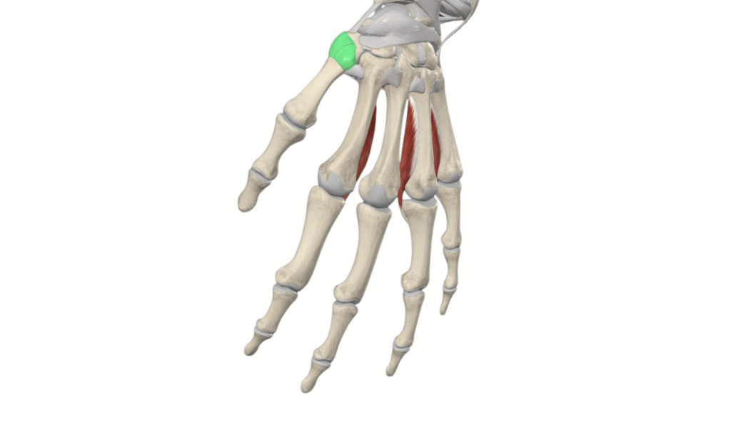
Your thumb meets your wrist at a saddle, two curved surfaces sitting across each other at right angles, one on the end of the thumb bone and one on the trapezium (the small wrist bone sitting underneath it). That shape is what lets the thumb swing around to meet each one of your fingers, but it’s also what leaves the joint without any bony lock. It’s held by ligaments and its capsule, so a joint thats already short on bony stability is now short on ligament stability as well (ouch). When the passive stiffness at that joint drops, the pressure on the trapezium cartilage goes up during a pinch [4].
Pinch isn’t an occasional load either. It’s the jar lid, the key, the zip, the car door, the phone hold in one hand, hundreds of times a day, through a surface not much bigger than a fingernail.
What you may find useful for hypermobile thumb joints, is using KT tape to hold the joint out of a drop thumb position. You may even find that it makes tasks like writing, and general hand use much easier. KT tape isn’t magic, and the evidence is very shaky, but anecdotally least, it appears to help a lot of people.
The knee, the hip and the spine
Move away from the hand and the picture starts to invert.
In community samples, hypermobility came out inversely associated with hand and knee osteoarthritis rather than positively [5], and in a large community cohort in the United States there was no significant link between generalised hypermobility and knee osteoarthritis on X rays in either of the two groups that it compared [6].
The spine is much the same. Pooling three large studies found no significant association between hypermobility, either low back pain or lumbar osteoarthritis, and being able to flex the trunk further went with less symptomatic spine osteoarthritis [7]. At the hip, nothing in the community work turned up an association at all.
And at the middle knuckles it goes past neutral. Hypermobility went with substantially lower odds of osteoarthritis at those joints across more than one cohort [8]. The likeliest explanation is that the two joints are loaded in quite different ways: the middle knuckles are hinges taking force straight down their long axis, the thumb base is a saddle taking force across itself.
What a previous injury does
Now, what reliably predicts arthritic joints is whether or not it’s been hurt before. A previous knee injury raises the odds of knee osteoarthritis by somewhere between roughly 2 and 4 times, and it does it consistently across cohorts [9].
At the ankle it’s much starker: up to 80% of ankle osteoarthritis follows a trauma, rather than turning up out of the blue [10]. In the people that it does happen to, the gap between a lateral ankle sprain and end stage arthritis averaged 34 years [11].
A sprain isn’t only a stretched ligament either, which changes this section a little, as joint surfaces get knocked against each other on the way through, the capsule and ligaments that were reporting position get damaged in the same moment, and the joint comes back working slightly differently from how it previously worked. Do that often enough and the difference stops being slight.
So, does being hypermobile multiply that injury risk once it’s happened? Mostly not.
The interaction between hypermobility and knee injury turned up in 1/3 cohorts and wasn’t there in the population based one [9]. In an ordinary adult population, hypermobility doesn’t reliably add to a risk that’s already large on its own.
Where it does matter is getting injured in the first place, and more importantly…. what happens afterwards.
Hypermobility goes with a higher risk of knee injury in contact sport, though not with more ankle sprains [12]. The ligaments on the outside of hypermobile ankles measure less stiff when imaged [13], which is the structural version of the thing you already know about your ankles. And after surgery, to reconstruct a chronically unstable ankle, people with generalised laxity went on to sprain it again far more often: 15.3% against 3.7% [14].
The useful question here isn’t whether or not your joints are loose, but it’s how many times a particular joint has been properly injured, and whether or not it’s still giving way now. Both of those you can do something about. How your body produced collagen, you can’t.
We are incredibly big on ankle and foot stability here, so if you want a low load exercise to play with the start working on your feet and ankles, you can find it below.
Stiff now, loose then
These studies measured hypermobility and arthritis at the same moment, in people who were already older, and that’s a problem the studies even state themselves. Osteoarthritis reduces how far a joint moves, which can mask hypermobility in older participants [6].
That matters to you specifically, if you’ve ever been told you can’t be hypermobile because you’re stiff now. A knee that could hyperextend easily at 20 years old, can score perfectly normally at 55, and a study run at fifty five records you as not being hypermobile.

What loading actually does to the joint
Long term exercise may “slightly” increase the severity of bone marrow lesions, which are bruise like changes in the bone sitting under your cartilage, although the evidence behind that is pretty low quality [15]. Now, people with knee osteoarthritis who walked for exercise, were less likely to develop new frequent knee pain and they showed less narrowing on the inner side of the joint over time [16]. Narrowing is what an X ray report means by progression. Cartilage doesn’t show up on the picture, so what gets measured is the gap it leaves between the two bones.
Where structural harm does turn up, it tracks with how hard the joint is being hit rather than how often it’s being used. Higher peak forces during walking went with worsening bone marrow lesions and cartilage damage in particular compartments of the knee [17]. Force is something you can change however. How often you move is not something you want to reduce.
It’s been tested from the other side as well. Retrain somebody’s walking pattern to cut the sideways load going through the inner knee, and pain improves while the deterioration in cartilage microstructure slows over a year [18]. So, the joint answers to how you load it, which is a far more hopeful proposition than the tyre it isn’t.
One limit the research names itself: the exercise and cartilage trials didn’t enrol or separate out hypermobile participants [9]. The structural reassurance here is borrowed from people who share the arthritis without sharing the tissue, and it’s the best there is.
What exercise actually does for the pain
Now the size of the benefit, because this gets oversold constantly. Exercise does improve pain in knee osteoarthritis, and how much it does, depends rather heavily on what it’s being compared against. Set against usual care, it’s about 13 points on a 100 point pain scale, which is a bit over one point if you rate your knee out of ten, and against a pretend treatment, it’s about 9, and whether a shift that size is clinically important is a question in itself [19]. Averaged across individual participants rather than across trials, the estimated benefit is smaller yet again, and it’s still there at 12 months [20].
Dont get me wrong, thats a real improvement, but it isn’t a cure.
Harder isn’t better either.
Train at high intensity for 18 months with knee osteoarthritis, and pain comes out no better than training at low intensity, with more non serious adverse events logged along the way, 53 against 30 [21]. Volume behaves the same: across resistance exercise trials, how much was prescribed didn’t predict how much benefit came out [22].
And no single type of exercise has pulled clear of the others [19]. In hip osteoarthritis, progressive resistance training and neuromuscular exercise, which just means exercise aimed at control and coordination rather than at raw strength, produced similar improvements in pain and even in getting out of a chair [23], and bolting aerobic activity on top of the resistance work didn’t improve things [24].
So, there’s no correct protocol you’re failing to find within the depth of the internet. What matters is that you’re loading the joint in a way you can keep doing during the bad weeks.
Actually, I say this great deal of the time. Ive worked with so many hypermobile folks over the years, and the big thing with programming is finding something that people can be consistent with. Thats the sign of getting your exercises right: can you get them done even when things dip a little?
Safety reporting is the thin part in all of this evidence, and only 18% of trials in a large comparison of exercise types reported safety outcomes at all [25]. So when you read that something is well tolerated, part of what you’re reading is that nobody wrote the harms down (happens more than you would think).
Why a lax joint and an arthritic joint want the same thing first
A hypermobile joint and an arthritic joint look like opposite problems. One moves too much, one moves too little and grinds. They meet at the point that decides how a joint behaves in practice, which is how well your nervous system knows where that joint is, and what’s happening to it.
The signal coming out of the joint
Your joints are full of receptors reporting position, movement, speed and load, and they sit in the capsule, in the ligaments and in the muscle itself. Proprioception is the word for what that stream adds up to, and it’s what lets you put a foot on a kerb without looking down at it. It’s also what lets the muscles around a joint fire a fraction before a load arrives instead of a fraction after, and that timing is the difference between a joint that feels solid and a joint that feels like it might give way.
Both conditions degrade that stream, for different reasons obviously.
Knee osteoarthritis commonly comes with poorer proprioceptive accuracy, and often in both knees even when only one of them hurts, with damage to the receptors in and around the joint offered as “plausible” but incompletely established explanation [26]. Both knees, when only one of them hurts, means the problem isn’t simply a consequence of the cartilage in the painful one.
In hypermobility, the trouble starts before the receptor rather than at it. A laxer capsule and ligament deform less predictably for a given amount of movement, so the same movement produces a less reliable report, and the brain ends up predicting from worse information. The measured picture is patchier than that neat explanation suggests though, and it’s joint specific. The clearest deficits in symptomatic hypermobility are in the lower limbs, and upper limb findings are pretty less consistent [27]. In Ehlers-Danlos syndrome, knee position sense came back impaired while shoulder position sense was preserved [28]. For the hand, what came out reduced was the precision of hand position rather than its accuracy [29]. Being imprecise is a different complaint from being wrong: on average you’re in the right place, and you’re off by a different amount each go (it’s what we call scatter). Which is close to what people describe when they say they can do a movement beautifully and then do it badly ten seconds later.
It also matters whether the hypermobility hurts. Painful hypermobility can show larger deficits than hypermobility that doesn’t hurt [30], which is part of why this research reads as contradictory when nobody separates the two groups out.
Control before load
The practical question for you reading is what you do with any of the above information, and the evidence does something useful here. It refuses to treat dedicated stabilisation work as a separate category of training.
Add stabilisation training on top of functional strengthening in people whose knee already gives way, and the stabilisation adds nothing on average. What did improve their reported stability was the strengthening and the functional work both groups were doing [31]. Agility and perturbation training bolted onto exercise therapy produced no additional benefit in a trial of 183 people [32]. And strength training on its own improves knee position sense, not only strength [33], which is the bit that gets missed when strength and control are treated as two separate jobs.
Proprioceptive training does have an effect of its own and it’s a specific one: what it preferentially improves is position sense [34]. In the thumb, proprioceptive exercise improved proprioception and produced no significant between group difference in pain [35].
The reading we take from that, and it’s ours rather than the trials’, is that you don’t need a proprioception programme bolted onto a strength programme. You need strength work done in a way that demands control.
Now, I know what you’re thinking: that phrase gets used a lot and rarely explained!
What it means for you is that the range you can control is the range you can stop in. Take a knee bend. The point where you could pause, hold still without wobbling, and come back up under your own steam is the end of the range you that your own (feel comfortable with). The range you can reach goes well past that if you’re hypermobile, and the last part of it is being held by tissue rather than by muscle, which is exactly the part that hurts afterwards. Working inside the first number, slowly enough that you could stop anywhere in it, with your attention on what the joint is doing rather than on counting the rep, is most of what makes an exercise suitable for a hypermobile joint. It’s where we’d start with an arthritic one too. The gentle, controlled, unglamorous version is the version both conditions want.
Below are two knee exercises you can try, the first one is a regression, so the load is very low, and the second one introduces a little load for you to control through.
Two more things for you: greater quadriceps strength predicted less new and less worsening knee instability over time, although that’s an observational link rather than a trial and it doesn’t crown any particular programme [36]. And the limit on all of it: poorer position sense tracked symptoms getting worse only modestly, and didn’t track the X ray getting worse at all [37]. Better control is a reasonable target for how the joint feels and behaves. It isn’t a way of protecting the cartilage.
The thumb, and what to do about it
Of every joint here the base of the thumb is the one where hypermobility genuinely raises your risk, and it’s also the one where conservative treatment has the best track record (which is a phrase I do not get to say a lot).
Exercise based treatment for thumb base osteoarthritis produces a clinically meaningful reduction in pain in the short term compared with no treatment, along with less disability, on low to moderate certainty evidence, and within that, proprioceptive exercises came out better than standard care alone for pain at the shortest follow ups [38]. Strength work on its own, across hand osteoarthritis generally, has shown little functional benefit [39].
Over a longer run it holds up. Treated without surgery, with exercise and a splint, pain improved by a clinically relevant amount at three months, and at a median follow up of over six years it hadn’t got worse beyond the twelve month mark, with 22% going on to surgery [40], which is to say roughly four in five of them got to six years without one. Narrow that down to people with symptomatic instability at that joint, which is where the hypermobile ones sit, and non surgical treatment improved pain and hand function with 14% needing surgery by just under three years [41].
The splint in those studies isn’t the wrist brace off the shelf at the chemist. It’s a small one that holds the base of the thumb and leaves the tip free to move. Compare a splint plus exercise therapy against a splint on its own and there’s no significant difference in the main pain outcome at three months, but exercise still came out ahead on daily activities, ability to work and satisfaction [42].
And the finding that should change your timing: the combined conservative treatments worked better in people whose thumb joint was less subluxed to start with, meaning the bones had slid less far out of line [43]. So this is a joint to work on while it’s still grumbling, rather than once it’s sitting visibly out of position. If the thumb base is the joint that hurts, that’s where the effort goes, early, with a splint for the jobs that reliably set it off and work that asks the thumb to control a pinch rather than simply to be strong in one.
Telling an ordinary sore day from a real flare
Pain during and after a session and a flare are two different events, and the thing that separates them is mostly how long they last.
The scale the programmes use
The neuromuscular programmes most of this evidence comes from run on a simple scale, and it’s worth knowing because it’s probably the scale your physio is working from. Pain up to 2 out of 10 during the session is labelled safe [44]. Above 2 and up to 5 is labelled acceptable, which means pain doesn’t have to be absent for a session to be a good one [45]. Above 5 is labelled high risk, and you reduce or modify what you’re doing [46]. Then the rule that does the real work: by the following day the joint should be back to its usual level, and if it isn’t, you reduce the loading next time [47].
Those numbers get quoted as though they were biology and they’re nothing of the sort. They’re programme rules rather than measured boundaries where tissue starts being damaged [44]. The foundational study behind them didn’t actually measure 24 hour pain, despite telling participants to monitor it [44]. Use the scale to keep a session sensible. Don’t read it as a gauge on the joint.
For scale, in a programme run with people who had severe hip or knee osteoarthritis, pain was rated acceptable after 93.8% of sessions, while 22% of participants went above 5 out of 10 at least once [44]. Going over it now and then happens, and it doesn’t mean something has gone wrong.
What a flare actually looks like
The pain you get during and just after a session tends to shrink as you keep going. Over eight weeks of neuromuscular exercise, average pain fell from 3.6 to 2.6 out of 10 and the immediate spike after each session got progressively smaller [46]. In another programme, rated effort went up by 2.6 points on the exertion scale while maximum exercise pain came down by about a point [48]. The exercise got harder and hurt less, and that’s a trajectory you read across weeks rather than inside one session.
A flare proper is a different event. Tracked in the community in people with knee osteoarthritis, the median flare lasted five days in one group and eight in another, and came with swelling, limping, disrupted sleep and activities that had to be dropped [49]. Days, not hours. Swelling and a limp, not soreness.
So that’s the line. Sore the evening of and settled by the next day is the programme working. Swollen, limping and four days in is a flare. Back the load off and keep going.
This evidence doesn’t tell you whether you need to exercise into pain at all, and it probably comes down to you rather than to a rule. Randomise people with knee osteoarthritis to painful or non painful exercise and adherence sits around nine in ten in both, with outcomes improving similarly [50]. Nor does it say much about managing a flare once it’s started, where the evidence for specific non drug strategies is weak [51]. So the prevention side, the next day rule, is where your attention is best spent.
One thing the next day rule doesn’t cover. A joint that comes up hot and swollen quickly, or that locks and won’t straighten, or that gives way and then swells hard within a few hours, isn’t a training flare, and backing the load off won’t sort it. That’s a call to your GP or to the physio who knows the joint, and it’s worth making rather than waiting a week to see what happens.
Where we’d start
Pulling it together, in the order we’d actually work through it.
Train it, and don’t treat that as a gamble. Structured exercise didn’t speed up cartilage loss or X ray progression in the knee osteoarthritis trials that measured it [1], and walking went with less new pain and less narrowing over time [16]. It’s the most reliable thing on this page.
Control before load. Slow, inside the range you can stop in, attention on the joint. Dedicated stabilisation work adds nothing on top of strengthening done properly [31], which means the quality of the strength work is the intervention.
Watch force rather than volume. Structural worsening tracked peak loading, not how often people moved [17]. Soften the landings and keep the sessions.
Give the thumb priority if it’s the joint that hurts. It’s the one site where hypermobility genuinely raises risk [2], conservative treatment works [38], and it works better the earlier you start [43].
Use tomorrow morning as your gauge. Settled by the next day means carry on. Still up means less load at the next session [47].
The thing I’d want you to take out of all this is that the two diagnoses aren’t asking you for opposite things, whatever the two clinicians told you. The joint that moves too much and the joint that’s arthritic want the same first move, which is a clearer sense of where that joint is and better control of it through the range you’ve actually got, before anybody adds weight to the bar. We’ve built every hypermobility programme we run on that order, and the osteoarthritis evidence arrives at the same place from a different direction, which is usually a decent sign you’re pointing the right way.
Adam

References
[1] Bricca, A., Juhl, C.B., Steultjens, M., Wirth, W. and Roos, E.M. (2018) ‘Impact of exercise on articular cartilage in people at risk of, or with established, knee osteoarthritis: a systematic review of randomised controlled trials’, British Journal of Sports Medicine. https://doi.org/10.1136/bjsports-2017-098661
[2] Jónsson, H., Elíasson, G.J., Jónsson, Á., Eiríksdóttir, G., Sigurðsson, S., Aspelund, T. et al. (2009) ‘High hand joint mobility is associated with radiological CMC1 osteoarthritis: the AGES-Reykjavik study’, Osteoarthritis and Cartilage. https://doi.org/10.1016/j.joca.2008.10.002
[3] Jónsson, H. and Valtýsdóttir, S.T. (1995) ‘Hypermobility features in patients with hand osteoarthritis’, Osteoarthritis and Cartilage. https://doi.org/10.1016/s1063-4584(05)80032-9
Read More[4] Valerio, T., Milan, J.L., Goislard de Monsabert, B. and Vigouroux, L. (2024) ‘The effect of trapeziometacarpal joint passive stiffness on mechanical loadings of cartilages’, Journal of Biomechanics. https://doi.org/10.1016/j.jbiomech.2024.112042
[5] Chen, H., Shah, S.H., Li, Y., Stabler, T.V., Jordan, J.M. and Kraus, V.B. (2008) ‘Inverse association of general joint hypermobility with hand and knee osteoarthritis and serum cartilage oligomeric matrix protein levels’, Arthritis & Rheumatism. https://doi.org/10.1002/art.24319
[6] Flowers, P.P.E., Cleveland, R.J., Schwartz, T.A., Nelson, A.E., Kraus, V.B., Hillstrom, H.J. et al. (2018) ‘Association between general joint hypermobility and knee, hip, and lumbar spine osteoarthritis by race: a cross-sectional study’, Arthritis Research & Therapy. https://doi.org/10.1186/s13075-018-1570-7
[7] Goode, A.P., Cleveland, R.J., Schwartz, T.A., Nelson, A.E., Kraus, V.B., Hillstrom, H.J. et al. (2019) ‘Relationship of joint hypermobility with low Back pain and lumbar spine osteoarthritis’, BMC Musculoskeletal Disorders. https://doi.org/10.1186/s12891-019-2523-2
[8] Kraus, V.B., Li, Y., Martin, E.R., Jordan, J.M., Renner, J.B., Doherty, M. et al. (2004) ‘Articular hypermobility is a protective factor for hand osteoarthritis’, Arthritis & Rheumatism. https://doi.org/10.1002/art.20354
[9] Shiue, K.Y., Cleveland, R.J., Schwartz, T.A., Nelson, A.E., Kraus, V.B., Hannan, M.T. et al. (2020) ‘Is the association between knee injury and knee osteoarthritis modified by the presence of general joint hypermobility?’, Osteoarthritis and Cartilage Open. https://doi.org/10.1016/j.ocarto.2020.100045
[10] Song, K. and Wikstrom, E.A. (2019) ‘Plausible mechanisms of and techniques to assess ankle joint degeneration following lateral ankle sprains: a narrative review’, The Physician and Sportsmedicine. https://doi.org/10.1080/00913847.2019.1581511
[11] Valderrabano, V., Hintermann, B., Horisberger, M. and Fung, T.S. (2006) ‘Ligamentous Posttraumatic Ankle Osteoarthritis’, The American Journal of Sports Medicine. https://doi.org/10.1177/0363546505281813
[12] Pacey, V., Nicholson, L.L., Adams, R.D., Munn, J. and Munns, C.F. (2010) ‘Generalized Joint Hypermobility and Risk of Lower Limb Joint Injury During Sport’, The American Journal of Sports Medicine. https://doi.org/10.1177/0363546510364838
[13] Chen, Z., Liang, H., Zhou, W., Li, Y., Yang, L., Gao, S. et al. (2026) ‘The impact of general joint hypermobility on ankle ligament stiffness in young women: a shear wave elastography investigation’, BMC Musculoskeletal Disorders. https://doi.org/10.1186/s12891-026-09880-0
[14] Liu, H., Li, J., Su, T., Jiang, Y., Pi, Y., Xie, X. et al. (2026) ‘Generalized Joint Laxity Is an Independent Predictor of Postoperative Resprain Following Anatomic Reconstruction Using Gracilis Tendon Autograft for Patients With Chronic Lateral Ankle Instability’, Orthopaedic Journal of Sports Medicine. https://doi.org/10.1177/23259671261477818
[15] Van Ginckel, A., Hall, M., Dobson, F. and Calders, P. (2019) ‘Effects of long-term exercise therapy on knee joint structure in people with knee osteoarthritis: A systematic review and meta-analysis’, Seminars in Arthritis and Rheumatism. https://doi.org/10.1016/j.semarthrit.2018.10.014
[16] Lo, G.H., Vinod, S., Richard, M.J., Harkey, M.S., McAlindon, T.E., Kriska, A.M. et al. (2022) ‘Association Between Walking for Exercise and Symptomatic and Structural Progression in Individuals With Knee Osteoarthritis: Data From the Osteoarthritis Initiative Cohort’, Arthritis & Rheumatology. https://doi.org/10.1002/art.42241
[17] Sara, L.K., Felson, D.T., Tilley, S., LaValley, M.P., Lewis, C.E., Lynch, J.A. et al. (2025) ‘The relation of walking forces to structural damage in the knee: The Multicenter Osteoarthritis Study’, Osteoarthritis and Cartilage. https://doi.org/10.1016/j.joca.2025.04.007
[18] Uhlrich, S.D., Mazzoli, V., Silder, A., Finlay, A.K., Kogan, F., Gold, G.E. et al. (2025) ‘Personalised gait retraining for medial compartment knee osteoarthritis: a randomised controlled trial’, The Lancet Rheumatology. https://doi.org/10.1016/s2665-9913(25)00151-1
[19] Lawford, B.J., Hall, M., Hinman, R.S., Van der Esch, M., Harmer, A.R., Spiers, L. et al. (2024) ‘Exercise for osteoarthritis of the knee’, Cochrane Database of Systematic Reviews. https://doi.org/10.1002/14651858.cd004376.pub4
[20] Holden, M.A., Hattle, M., Runhaar, J., Riley, R.D., Healey, E.L., Quicke, J. et al. (2023) ‘Moderators of the effect of therapeutic exercise for knee and hip osteoarthritis: a systematic review and individual participant data meta-analysis’, The Lancet Rheumatology. https://doi.org/10.1016/s2665-9913(23)00122-4
[21] Messier, S.P., Mihalko, S.L., Beavers, D.P., Nicklas, B.J., DeVita, P., Carr, J.J. et al. (2021) ‘Effect of High-Intensity Strength Training on Knee Pain and Knee Joint Compressive Forces Among Adults With Knee Osteoarthritis’, JAMA. https://doi.org/10.1001/jama.2021.0411
[22] Marriott, K.A., Hall, M., Maciukiewicz, J.M., Almaw, R.D., Wiebenga, E.G., Ivanochko, N.K. et al. (2024) ‘Are the Effects of Resistance Exercise on Pain and Function in Knee and Hip Osteoarthritis Dependent on Exercise Volume, Duration, and Adherence? A Systematic Review and Meta‐Analysis’, Arthritis Care & Research. https://doi.org/10.1002/acr.25313
[23] Kjeldsen, T., Skou, S.T., Dalgas, U., Tønning, L.U., Ingwersen, K.G., Birch, S. et al. (2024) ‘Progressive Resistance Training or Neuromuscular Exercise for Hip Osteoarthritis’, Annals of Internal Medicine. https://doi.org/10.7326/m23-3225
[24] Hall, M., Allison, K., Knox, G., McManus, F., Hinman, R.S., Bennell, K.L. et al. (2025) ‘Addition of aerobic physical activity to resistance exercise for hip osteoarthritis (PHOENIX): a randomised comparative effectiveness trial’, The Lancet Rheumatology. https://doi.org/10.1016/s2665-9913(24)00373-4
[25] Yan, L., Li, D., Xing, D., Fan, Z., Du, G., Jiu, J. et al. (2025) ‘Comparative efficacy and safety of exercise modalities in knee osteoarthritis: systematic review and network meta-analysis’, BMJ. https://doi.org/10.1136/bmj-2025-085242
[26] Knoop, J., Steultjens, M.P.M., van der Leeden, M., van der Esch, M., Thorstensson, C.A., Roorda, L.D. et al. (2011) ‘Proprioception in knee osteoarthritis: a narrative review’, Osteoarthritis and Cartilage. https://doi.org/10.1016/j.joca.2011.01.003
[27] Smith, T.O., Jerman, E., Easton, V., Bacon, H., Armon, K., Poland, F. et al. (2013) ‘Do people with benign joint hypermobility syndrome (BJHS) have reduced joint proprioception? A systematic review and meta-analysis’, Rheumatology International. https://doi.org/10.1007/s00296-013-2790-4
[28] Rombaut, L., De Paepe, A., Malfait, F., Cools, A. and Calders, P. (2009) ‘Joint position sense and vibratory perception sense in patients with Ehlers–Danlos syndrome type III (hypermobility type)’, Clinical Rheumatology. https://doi.org/10.1007/s10067-009-1320-y
[29] Clayton, H.A., ’t Hart, B.M. and Henriques, D.Y.P. (2021) ‘Sensing hand position in Ehlers-Danlos syndrome’, Somatosensory & Motor Research. https://doi.org/10.1080/08990220.2021.1973403
[30] Reddy, R.S., Tedla, J.S., Alshahrani, M.S., Asiri, F. and Kakaraparthi, V.N. (2022) ‘Comparison and correlation of cervical proprioception and muscle endurance in general joint hypermobility participants with and without non-specific neck pain—a cross-sectional study’, PeerJ. https://doi.org/10.7717/peerj.13097
[31] Knoop, J., Dekker, J., van der Leeden, M., van der Esch, M., Thorstensson, C.A., Gerritsen, M. et al. (2013) ‘Knee joint stabilization therapy in patients with osteoarthritis of the knee: a randomized, controlled trial’, Osteoarthritis and Cartilage. https://doi.org/10.1016/j.joca.2013.05.012
[32] Fitzgerald, G.K., Piva, S.R., Gil, A.B., Wisniewski, S.R., Oddis, C.V. and Irrgang, J.J. (2011) ‘Agility and Perturbation Training Techniques in Exercise Therapy for Reducing Pain and Improving Function in People With Knee Osteoarthritis: A Randomized Clinical Trial’, Physical Therapy. https://doi.org/10.2522/ptj.20100188
[33] Guede-Rojas, F., Benavides-Villanueva, A., Salgado-González, S., Mendoza, C., Arias-Álvarez, G., Soto-Martínez, A. et al. (2024) ‘Effect of strength training on knee proprioception in patients with knee osteoarthritis: A systematic review and meta-analysis’, Sports Medicine and Health Science. https://doi.org/10.1016/j.smhs.2023.10.005
[34] Wang, Y., Wu, Z., Chen, Z., Ye, X., Chen, G., Yang, J. et al. (2021) ‘Proprioceptive Training for Knee Osteoarthritis: A Systematic Review and Meta-Analysis of Randomized Controlled Trials’, Frontiers in Medicine. https://doi.org/10.3389/fmed.2021.699921
[35] Cantero-Téllez, R., Algar, L.A., Valdes, K.A. and Naughton, N. (2022) ‘Clinical effects of proprioceptive thumb exercise for individuals with carpometacarpal joint osteoarthritis: A randomized controlled trial’, Journal of Hand Therapy. https://doi.org/10.1016/j.jht.2022.06.009
[36] Shakoor, N., Felson, D.T., Niu, J., Nguyen, U., Segal, N.A., Singh, J.A. et al. (2016) ‘The Association of Vibratory Perception and Muscle Strength With the Incidence and Worsening of Knee Instability: The Multicenter Osteoarthritis Study’, Arthritis & Rheumatology. https://doi.org/10.1002/art.39821
[37] Felson, D.T., Gross, K.D., Nevitt, M.C., Yang, M., Lane, N.E., Torner, J.C. et al. (2009) ‘The effects of impaired joint position sense on the development and progression of pain and structural damage in knee osteoarthritis’, Arthritis Care & Research. https://doi.org/10.1002/art.24606
[38] Karanasios, S., Mertyri, D., Karydis, F. and Gioftsos, G. (2024) ‘Exercise-Based Interventions Are Effective in the Management of Patients with Thumb Carpometacarpal Osteoarthritis: A Systematic Review and Meta-Analysis of Randomised Controlled Trials’, Healthcare. https://doi.org/10.3390/healthcare12080823
[39] Magni, N.E., McNair, P.J. and Rice, D.A. (2017) ‘The effects of resistance training on muscle strength, joint pain, and hand function in individuals with hand osteoarthritis: a systematic review and meta-analysis’, Arthritis Research & Therapy. https://doi.org/10.1186/s13075-017-1348-3
[40] Esteban Lopez, L.M.J., Hoogendam, L., Vermeulen, G.M., Tsehaie, J., Slijper, H.P., Selles, R.W. et al. (2023) ‘Long-Term Outcomes of Nonsurgical Treatment of Thumb Carpometacarpal Osteoarthritis’, Journal of Bone and Joint Surgery. https://doi.org/10.2106/jbjs.22.01116
[41] Wouters, R.M., Slijper, H.P., Esteban Lopez, L., Hovius, S.E.R., Selles, R.W., Blomme, R.A.M. et al. (2020) ‘Beneficial Effects of Nonsurgical Treatment for Symptomatic Thumb Carpometacarpal Instability in Clinical Practice: A Cohort Study’, Archives of Physical Medicine and Rehabilitation. https://doi.org/10.1016/j.apmr.2019.08.485
[42] Wouters, R.M., Esteban Lopez, L.M., Heemskerk, S.C., Bierma-Zeinstra, S.M., Kraan, G.A., Colaris, J. et al. (2026) ‘Effectiveness of exercise therapy in patients with thumb carpometacarpal osteoarthritis: A multicenter, randomized controlled trial’, Osteoarthritis and Cartilage. https://doi.org/10.1016/j.joca.2025.10.005
[43] Deveza, L.A., Robbins, S.R., Duong, V., Fu, K., Wajon, A., Eyles, J.P. et al. (2021) ‘Greater efficacy of a combination of conservative therapies for thumb base OA in individuals with lower radial subluxation – a pre-planned subgroup analysis of the COMBO trial’, Osteoarthritis and Cartilage. https://doi.org/10.1016/j.joca.2021.07.010
[44] Ageberg, E., Link, A. and Roos, E.M. (2010) ‘Feasibility of neuromuscular training in patients with severe hip or knee OA: The individualized goal-based NEMEX-TJR training program’, BMC Musculoskeletal Disorders. https://doi.org/10.1186/1471-2474-11-126
[45] Clausen, B., Holsgaard-Larsen, A., Søndergaard, J., Christensen, R., Andriacchi, T.P. and Roos, E.M. (2014) ‘The effect on knee-joint load of instruction in analgesic use compared with neuromuscular exercise in patients with knee osteoarthritis: study protocol for a randomized, single-blind, controlled trial (the EXERPHARMA trial)’, Trials. https://doi.org/10.1186/1745-6215-15-444
[46] Sandal, L.F., Roos, E.M., Bøgesvang, S.J. and Thorlund, J.B. (2016) ‘Pain trajectory and exercise-induced pain flares during 8 weeks of neuromuscular exercise in individuals with knee and hip pain’, Osteoarthritis and Cartilage. https://doi.org/10.1016/j.joca.2015.11.002
[47] Bryld, E.S., Christiansen, L., Ingwersen, K.G., Overgaard, S., Mikkelsen, L.R., Mechlenburg, I. et al. (2025) ‘Training load and pain response during progressive resistance training in patients with hip osteoarthritis in the PROHIP trial’, Osteoarthritis and Cartilage Open. https://doi.org/10.1016/j.ocarto.2025.100690
[48] Primeau, C.A., Birmingham, T.B., Moyer, R.F., O’Neil, K.A., Werstine, M.S., Alcock, G.K. et al. (2020) ‘Trajectories of perceived exertion and pain over a 12-week neuromuscular exercise program in patients with knee osteoarthritis’, Osteoarthritis and Cartilage. https://doi.org/10.1016/j.joca.2020.07.011
[49] Thomas, M.J., Rathod-Mistry, T., Parry, E.L., Pope, C., Neogi, T. and Peat, G. (2021) ‘Triggers for acute flare in adults with, or at risk of, knee osteoarthritis: a web-based case-crossover study in community-dwelling adults’, Osteoarthritis and Cartilage. https://doi.org/10.1016/j.joca.2021.04.007
[50] Ram, A., Kovats, A., Pandit, A., Ram, P., Gibbs, M.T., Booth, J. et al. (2025) ‘Painful exercise for people with knee osteoarthritis: a randomised controlled feasibility trial’, Disability and Rehabilitation. https://doi.org/10.1080/09638288.2025.2504614
[51] Bowden, J.L., Kobayashi, S., Hunter, D.J., Mills, K., Peat, G., Guillemin, F. et al. (2021) ‘Best-practice clinical management of flares in people with osteoarthritis: A scoping review of behavioral, lifestyle and adjunctive treatments’, Seminars in Arthritis and Rheumatism. https://doi.org/10.1016/j.semarthrit.2021.04.017