It’s not uncommon for people with hypermobility and EDS to be told, almost reflexively, that losing weight will help their joints. And for a large portion of the population without connective tissue disorders, that advice is broadly correct. Less load on the knees, less pressure on the hips, less strain on an already overworked musculoskeletal system.
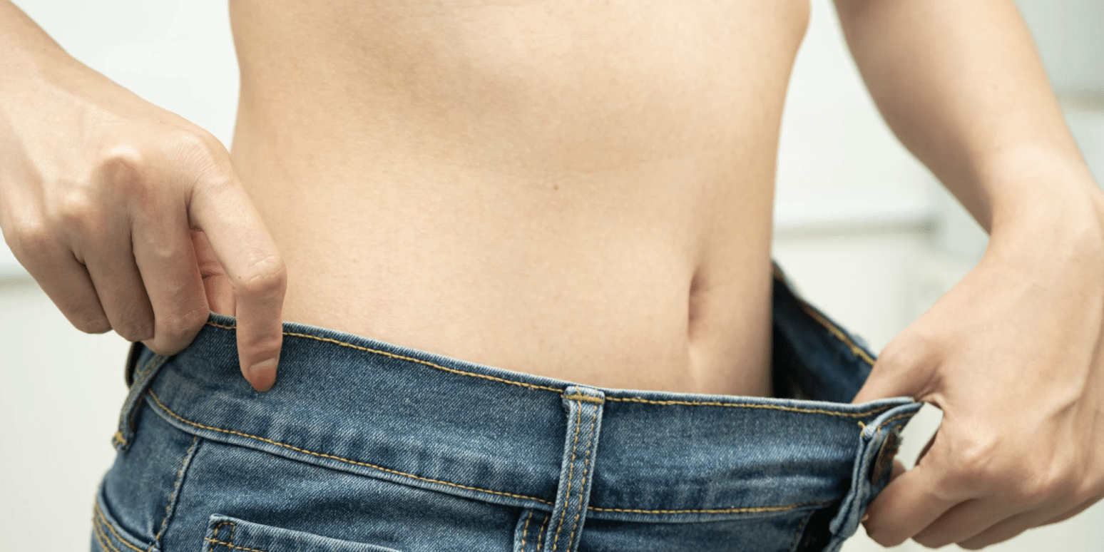
But here’s the situation a surprising number of hypermobile people find themselves in. They lose weight. Sometimes intentionally, sometimes not. Sometimes via medication, sometimes through sheer dietary discipline. And then their joints get worse. Subluxations they used to have twice a week start happening multiple times a day. Shoulders that were mostly manageable start sliding at the slightest provocation. Hips, ribs, fingers, things they’d almost forgotten about, begin acting up again. And when they mention this to their doctor or physiotherapist, they’re met with scepticism. Surely weight loss is supposed to help?
So they go online and find the same message repeated everywhere: lose weight, reduce joint stress. Nobody seems to be talking about what they’re experiencing. And they start wondering if they’re imagining it.
They’re not imagining it.
This article is going to make the case, as honestly as the research allows, for why weight loss can genuinely increase subluxation frequency in hypermobile people, what the biological mechanisms are, and what you can do about it. I should be upfront: there is no single study that neatly answers the question “does losing weight cause more subluxations in EDS?” That study hasn’t been done. But there is a body of adjacent evidence that is mechanistically coherent, clinically plausible, and points in one consistent direction. We’re going to work through it carefully, call out where the evidence is strong and where it’s thin, and try to give you something actually useful to take away.
We’ll also be looking at the wide-ranging ways hypermobility affects the body more broadly, because weight is never just about weight when connective tissue is involved. And if you want more context on the complicated relationship between connective tissue disorders and body weight before diving in, that’s worth reading alongside this.
Right. Let’s go.
The Weight Loss Paradox: Why Lighter Doesn’t Always Mean More Stable
The general population literature on weight loss and joint pain is pretty clear. In osteoarthritis (OA), where the problem is mechanical load grinding cartilage down over decades, losing weight reduces joint stress, reduces pain, and slows disease progression. There’s good evidence for this. It makes intuitive sense. Less weight pressing down on a damaged knee means less damage to that knee.
But osteoarthritis and hypermobility are fundamentally different conditions with fundamentally different problems. In OA, the joints are stiff and overloaded. In hypermobility, the joints are too loose and undercontrolled. The problem isn’t load. The problem is instability.
This distinction matters enormously, and it’s one that gets missed constantly when hypermobile patients receive generic joint pain advice. Weight loss helps OA patients because it reduces the thing that’s causing harm: excessive mechanical load on degenerated cartilage. For hypermobile patients, reducing load does not fix the underlying problem, and may actually remove things that were compensating for it.
It’s also worth noting that BMI and hypermobility aren’t meaningfully correlated. A 2014 study of 142 adolescents found no significant association between obesity and joint hypermobility (OR 0.79, p=0.31), and a 2024 cross-sectional study found no significant relationship between BMI and generalised joint hypermobility (chi-square p=0.112) [Wrotniak et al., 2014; Assessment of GJH and BMI, 2024]. You don’t become more hypermobile because you’re heavier. And you don’t become less hypermobile because you’re lighter. The laxity is in your connective tissue, not your BMI.
The most directly relevant study we have is a 2026 prospective cohort by Schubert-Hjalmarsson and colleagues, published in Obesity Science and Practice [Schubert-Hjalmarsson et al., 2026]. It followed 74 patients who underwent metabolic and bariatric surgery, comparing joint pain outcomes between those who had hypermobility (n=15) and those who did not (n=59). Both groups lost comparable amounts of weight, roughly 29-30% of their body weight over 12 months.
The non-hypermobile group did what you’d expect. They had significant reductions in pain at the neck, lumbar spine, hips, knees, ankles, and feet. Fewer painful body parts overall. The weight loss clearly helped them.
The hypermobile group showed none of this. No significant change in pain frequency at any joint site. No reduction in the number of painful body parts.
Now, there are real limitations here. The hypermobile group was small (n=15, which is severely underpowered for statistical analysis). Hypermobility was assessed by self-report questionnaire rather than clinical examination. And critically, the study measured pain frequency, not subluxation rate. So it doesn’t directly tell us that subluxations got worse. What it does tell us is that weight loss simply doesn’t deliver the joint benefit for hypermobile patients that it delivers for everyone else, and the authors’ interpretation is telling: they concluded that instability, not mechanical load, is what drives pain in hypermobile patients, and that losing weight does nothing to address instability.
This is the starting point. Weight is neutral at best for hypermobile patients when it comes to joint pain relief. But the more troubling question is whether it’s actively negative. And for that, we need to look at what actually gets lost during weight loss.
Three Reasons Weight Loss Can Increase Subluxations
There’s no single smoking gun here. Rather, there are three separate mechanisms that each independently push in the same direction, and when they compound, the effect can be significant. Understanding them is the key to understanding both why this happens and what you can do about it.
Mechanism 1: Muscle Loss, the Joint Stabiliser You Can’t Afford to Lose
Muscle is the primary dynamic stabiliser of hypermobile joints. When your ligaments are lax and your connective tissue isn’t doing the job that connective tissue is supposed to do, it falls to your muscles to pick up the slack. They act as active compression systems around joints, constantly making micro-adjustments to prevent the excessive range of motion that leads to subluxations. This isn’t a theory. It’s what every serious treatment programme for hypermobility and EDS is built around. Why building strength looks different when your joints are loose is a whole topic in itself, but the short version is: your muscles are working much harder than anyone without hypermobility can appreciate.
Here’s the problem. People with hEDS and HSD already have substantially weaker muscles than healthy controls, and this weakness runs deeper than just having less muscle mass. A 2022 study by Coussens and colleagues measured muscle strength in women with hEDS and HSD versus matched controls, using isokinetic dynamometry (a laboratory-grade strength testing machine). The results were striking. Knee extensor peak torque was around 87-91 Nm in the hEDS/HSD groups versus 134 Nm in controls. Knee flexor strength similarly reduced. Wall sit time: 31 seconds versus nearly 79 seconds for controls [Coussens et al., 2022].
Crucially, and this is the part that trips people up, the muscle mass between the groups was not significantly different. The hEDS and HSD patients had roughly the same amount of muscle as the healthy controls. But their muscles generated much less force. The weakness is neuromuscular in origin, not simply a case of having less muscle to work with. Something in the way the nervous system recruits and activates those muscles is compromised. A 2026 study on the medial gastrocnemius (calf muscle) found that HSD/hEDS participants were 36% weaker across the ankle range of motion, again without significant differences in muscle architecture between groups [Golden et al., 2026].
So before any weight loss happens, you are already working with a significant deficit. Roughly 35-40% less lower extremity strength than someone without hypermobility, despite having a comparable amount of muscle.
Now add diet-induced weight loss to this picture.
When people lose weight through caloric restriction, they don’t just lose fat. They lose muscle too. The proportion depends on how they lose the weight, but the numbers are sobering. Diet-only weight loss (no exercise) results in approximately 20-35% of the total weight lost coming from lean mass, including skeletal muscle [Bosy-Westphal et al., 2023; Stefanakis et al., 2024]. For bariatric surgery patients, about 21% of total weight lost is fat-free mass [Nóbrega et al., 2022]. So for someone losing 20kg through dietary restriction alone, we might be looking at 4-7kg of muscle gone.
There’s an additional wrinkle that’s particularly relevant to the hypermobile population. Leaner individuals lose proportionally more muscle during caloric restriction than obese individuals [Bosy-Westphal et al., 2023]. The fatter you are, the more your body preferentially burns fat during a deficit. Many people with hEDS have normal or low BMI, which means their bodies are more likely to cannibalise muscle when calories are restricted.
And then there’s GLP-1 medication. Semaglutide (Ozempic, Wegovy) and tirzepatide (Mounjaro) are increasingly prescribed for weight loss, and they can achieve 15-24% total body weight reduction. But a 2024 review by Stefanakis and colleagues found that GLP-1 agonists induce over 10% of total muscle mass loss during 68-72 week treatment periods, which they describe as approximately equivalent to 20 years of age-related muscle loss [Stefanakis et al., 2024]. Around 25% of the total weight lost with these medications comes from fat-free mass.
Let’s put those numbers together. You start with a 35-40% strength deficit versus a healthy control. You lose, say, 15kg through dietary restriction or GLP-1 medication. Roughly 3-5kg of that is muscle. Your neuromuscular system, which was already struggling to activate the muscle you have, now has less to work with and the same activation problems. The muscles keeping your hip from subluxating during a step, the rotator cuff muscles preventing your shoulder from sliding out of place, the deep spinal muscles stabilising your vertebrae: all of them are now weaker.
The compounding effect here is the thing to hold onto. You weren’t starting from a position of strength. Losing further muscle mass from an already-depleted baseline is not the same as a healthy person losing the same amount of muscle. For someone without hypermobility, losing 3-4kg of muscle during weight loss is inconvenient. For someone whose muscles are the primary thing standing between them and a subluxation, it can be the tipping point.
A 2017 study looking at muscle strength and activity limitations in EDS found that muscle strength was the dominant factor predicting functional limitation, though proprioception also played a significant role [Scheper et al., 2017]. A gait analysis study from 2024 found that hEDS participants had a 40% deficit in peak hip extensor strength versus controls, and that 91% of those participants reported weekly lower extremity subluxations [Ball et al., 2024]. These aren’t coincidences. The strength deficit and the instability are causally linked.
The mechanisms behind chronic pain in hypermobility are complicated enough without adding further muscle depletion to the picture. But this is the reality of what diet-only weight loss can do.
Mechanism 2: Fat as a Passive Joint Stabiliser
This one is less intuitive, and the evidence is weaker, but it’s worth taking seriously because the patient experience points to it consistently and the biology is plausible.
When most people think about fat and joints, they think of it as purely a burden: extra load pressing down on cartilage, increasing wear and tear. And in the general population with healthy connective tissue, that framing is largely correct. But fat isn’t inert. Particularly the fat that sits immediately around joints.
The knee is the most studied example. The infrapatellar fat pad (IFP) sits just below the kneecap, between the patellar tendon and the knee joint itself. A 2021 biomechanical study found that the IFP functions as a genuine mechanical shock absorber, distributing synovial fluid and absorbing impulsive forces during joint loading [Belluzzi et al., 2021]. Its mechanical behaviour is non-linear and viscoelastic, meaning it stiffens appropriately under increasing strain. A 2024 review of periarticular adipose tissue confirmed that these structures are “primary mechanical shock absorbers of the knee” that “reduce friction between intra-articular structures during joint movement” [Rouhezamin et al., 2024]. A more fundamental review noted that articular fat pad dimensions are “conserved across species,” suggesting these structures serve important functional roles rather than being evolutionary leftovers [Zugun-Eloae and Labuşca, 2018].
In other words, periarticular fat pads aren’t passengers. They’re doing something. When you lose body fat, you lose some of this periarticular cushioning too.
Now, does this matter differently in hypermobility? Here’s where it gets interesting. A 2025 retrospective cohort study by Boulu and colleagues examined compression garment use in 20 patients with hEDS and HSD [Boulu et al., 2025]. Compression garments work by providing external proprioceptive and mechanical feedback to joints. What they found was that treatment response was significantly associated with higher BMI. Patients who responded well to the garments had a mean BMI of 26.1, whilst non-responders had a mean BMI of 17.5 (p=0.005). This is a small study and we should be cautious about over-interpreting it. But the direction of the finding is consistent with the idea that having more body tissue around joints provides proprioceptive and mechanical feedback that compression garments are trying to mimic. Lower-BMI patients, who have less natural tissue providing that feedback, also got less benefit from the external version.
Think of it like using external feedback to remind your joints where they are. In hypermobility, proprioception (the body’s ability to sense joint position) is already compromised. Periarticular fat may provide some of the mechanical afferent signals that inform your nervous system where your joint is in space. Remove that fat, and you potentially remove some of those signals.
There’s also those rib subluxations that can catch you off guard, and patients in online communities have reported that fat loss around the trunk and torso seems to be particularly associated with worsening rib and spinal instability, as though the fat was providing a structural envelope around structures that needed it.
An angle that gets virtually no clinical attention is what fat loss does to internal organs in hypermobile patients. When fat is lost from the abdominal cavity, the supportive envelope that keeps organs in their anatomical positions is reduced. In people with already-lax connective tissue, there are reports of nephroptosis (kidney drooping) and increased visceral laxity following significant weight loss. This isn’t about joints in the traditional sense, but it illustrates that fat isn’t passive in hypermobile bodies. It’s doing structural work, and its removal has consequences that extend beyond what we typically see on a DEXA scan.
Mechanism 3: Collagen Under Siege During Caloric Restriction
The third mechanism is the one with the least direct human evidence, but it’s arguably the most mechanistically concerning for anyone with a connective tissue disorder.
Collagen is the primary structural protein in your tendons, ligaments, cartilage, and skin. If you have hypermobility, your collagen is already structurally abnormal in ways that make your connective tissue less stable and more prone to injury. What actually happens to collagen in hypermobile connective tissue is something we’ve looked at in detail elsewhere. The short version is that the collagen you have is doing a compromised job. Which makes the rate of collagen synthesis and repair more important, not less, than it would be in a healthy person.
So what happens to collagen synthesis during caloric restriction?
The most striking data here comes from animal models, and I want to be upfront about that limitation. A 1985 study by Spanheimer and Peterkofsky found that collagen production in guinea pigs dropped to 8-12% of normal values within 96 hours of fasting, even when the animals were given adequate vitamin C supplementation [Spanheimer and Peterkofsky, 1985]. The effect was specific to collagen: non-collagen protein production was much less severely affected. Procollagen mRNA was specifically decreased. And it took 96 hours of refeeding to restore collagen production to normal, even though non-collagen protein normalised within 24 hours. These are guinea pigs, not humans. Direct extrapolation isn’t appropriate. But the fact that the effect was observed across different tissues, different animal species, and different ages suggests a consistent underlying mechanism.
In humans, the evidence is indirect but consistent. A 2024 study found that massive weight loss significantly reduces elastic fibre content in skin connective tissue [Hany et al., 2024]. A separate proteomic analysis found decreased expression of collagen XIV (which regulates the physical properties of connective tissue) in post-bariatric patients [Bozola et al., 2019]. Weight cycling, which is common in patients who find sustained exercise difficult, appears to damage the extracellular matrix (ECM) repeatedly, with each cycle of loss and regain degrading and remodelling connective tissue and potentially leaving it in progressively worse structural condition [Caria et al., 2017].
Now add the nutritional picture. Collagen synthesis requires specific amino acids: glycine and proline are the most important. It also requires vitamin C, which is an essential cofactor for the enzyme that cross-links collagen strands (prolyl-4-hydroxylase). Without adequate vitamin C, collagen structure is weakened [DePhillipo et al., 2018]. A 2016 randomised trial found that vitamin C-enriched gelatin supplementation before exercise doubled collagen synthesis in engineered ligaments, demonstrating how strongly both the amino acid substrate and the cofactor matter [Shaw et al., 2016].
The problem is that caloric restriction reduces all of these inputs simultaneously: overall protein availability, specific amino acid intake, and micronutrient levels including vitamin C.
And this is compounded by a finding specific to the hypermobile population. A 2022 body composition study found that hEDS and HSD patients consumed significantly less protein than healthy controls (p=0.03), despite having matched BMI, and that both groups were in caloric deficit relative to their actual energy needs [DiFrancisco-Donoghue et al., 2022]. So the hypermobile population is already protein-deficient and calorie-insufficient before any deliberate weight loss begins. Adding a caloric deficit on top of this nutritional baseline further compromises the inputs needed for connective tissue maintenance and repair.
Standard protein supplementation doesn’t fully address this. A 2021 review by Holwerda and van Loon found that conventional protein sources like whey do not stimulate muscle connective tissue protein synthesis specifically [Holwerda and van Loon, 2021]. Connective tissue synthesis requires collagen-specific precursor amino acids, i.e., the glycine and proline that are abundant in collagen peptides but scarce in standard high-protein foods. So even if you’re hitting a reasonable protein target during caloric restriction, you may still be falling short of what your connective tissue specifically needs.
Fuelling your body properly when connective tissue is already compromised is genuinely difficult during weight loss, and it matters more in this population than most nutritional guidance acknowledges.
What About Medications? GLP-1 Drugs and Hypermobile Joints
This section deserves its own space, because GLP-1 receptor agonists like semaglutide (Ozempic, Wegovy) and tirzepatide (Mounjaro) are now being prescribed widely for weight management, and the hypermobile community is starting to encounter them in increasing numbers.
The weight loss these medications achieve is real and often substantial: 15-24% of body weight in clinical trials, which is comparable to some surgical interventions. But the muscle loss that accompanies them is a serious concern. As mentioned above, the evidence suggests that GLP-1 agonists cause approximately 10% or more of total muscle mass to be lost during the treatment period, amounting to what one 2024 review described as “approximately equivalent to 20 years of age-related muscle loss” [Stefanakis et al., 2024]. For a 70kg person, that could mean losing around 6kg of muscle.
For the general obese population, this trade-off might be acceptable. The metabolic benefits of significant weight loss in obesity are substantial, and 6kg of muscle loss in a person with healthy joints and normal neuromuscular function is a manageable cost. But for someone with hEDS who is already 35-40% below the strength threshold of healthy controls, losing another 6kg of muscle could represent a functional collapse of their joint stability system. It’s the difference between already low and completely insufficient.
There are currently no published studies examining GLP-1 effects specifically in hEDS or HSD populations. This is another genuine evidence gap. We do not know whether the muscle loss pattern is different in hypermobile people, whether the rate of subluxations increases during treatment, or whether the weight loss itself confers any compensating benefit. Patients in online communities report worsening symptoms on these medications, but these are anecdotal accounts, not controlled data.
If you are taking or considering GLP-1 medication and you have hypermobility, the available evidence suggests that resistance training should be considered non-negotiable alongside it, not an optional add-on. A 2024 clinical review by Locatelli and colleagues specifically recommended concurrent resistance exercise to minimise muscle loss during incretin-based pharmacotherapy [Locatelli et al., 2024]. Protein targets need to be at the higher end: 2.2-3.0g per kg of body weight per day. And the specific concern about connective tissue amino acids, described above, applies here with additional force.
Those autonomic symptoms that can derail progress, particularly POTS, have also been reported to worsen with significant weight loss in some hypermobile patients, and this is an additional layer of complexity worth being aware of. The ways medication can shift your body composition more broadly is another topic we’ve covered if you want further context on the medication side of things.
When Weight Loss Does Help, and When It Doesn’t
It would be wrong to leave the impression that weight loss is always harmful for hypermobile people. It isn’t, and the picture is more nuanced than that.
The clearest scenario where weight loss is likely to help is someone who is significantly overweight and losing weight with a concurrent exercise programme. In this case, the reduction in mechanical load on joints like the knees and hips can reduce pain, particularly if those joints are carrying disproportionate stress. And if the weight loss is achieved partly through exercise rather than dietary restriction alone, muscle mass can be largely preserved. A 2007 study found that exercise-induced weight loss preserved or improved lower extremity muscle size, strength, and aerobic capacity, whilst equivalent caloric restriction-based weight loss significantly reduced all of these [Weiss et al., 2007]. The number on the scale was similar; the impact on the joints was completely different.
The scenario where weight loss most commonly makes things worse is someone at a healthy or lean BMI who loses further weight, particularly through dietary restriction alone, and particularly rapidly. In this group, the mechanical load on joints was not the problem to begin with. The instability was. And the weight loss, by removing both muscle mass and periarticular support, removes what little compensation existed.
Rapid weight loss, regardless of starting BMI, appears to be consistently associated with worse outcomes in hypermobile patients. The method matters at least as much as the destination. Going from 100kg to 75kg over three years, with consistent resistance training throughout, is a fundamentally different biological process from losing 25kg in 8 months through caloric restriction or medication.
One useful way to frame this: for OA patients, the primary problem is load. Weight loss reduces the thing causing harm. For hEDS patients, the primary problem is instability. Weight loss doesn’t reduce the thing causing harm, and can actively remove things that were compensating for it. These are different problems that don’t respond to the same solution.
How these conditions tend to change over the years is relevant context here too, because the interplay between natural age-related muscle loss and deliberate weight loss deserves consideration.
You’re Not Imagining It: Why So Many Patients Get Dismissed
Let me be direct about something.
Almost every resource that exists on joint pain and weight management is drawn from the OA literature. That literature is large, well-funded, and clear in its conclusions: excess weight damages joints, and weight loss helps them. These conclusions are correct for osteoarthritis. They are not automatically correct for hypermobility. But because the hypermobility-specific literature on this topic is so sparse, most clinicians, including most specialists, are applying OA-based conclusions to a different condition with different biology.
Patients in online communities report this experience with striking consistency. They’re told that weight loss will improve their symptoms. They lose weight, their symptoms worsen dramatically. They go back and report this. They’re told it can’t be right, that they must be doing something else wrong, that weight loss always helps joint pain. The cognitive dissonance between the generic advice and their lived experience is real and documented. It’s a form of medical dismissal that sits alongside the lasting impact of not being believed by the people meant to help you more broadly.
The Schubert-Hjalmarsson 2026 study gives us something important: a prospective, peer-reviewed anchor for this experience. It’s small and imperfect, but it’s real. It shows, in a controlled setting, that hypermobile patients do not get the same benefit from weight loss that everyone else does. And the mechanistic case for why weight loss can actively make things worse, drawn from the muscle loss, fat padding, and collagen synthesis literature, is coherent and plausible even without a direct interventional study.
The absence of direct evidence is not the same as evidence of absence. The research gap exists because this question hasn’t been prioritised, not because researchers looked and found nothing to worry about. It hasn’t been studied in any adequate way.
What this means practically is that if your symptoms worsened after weight loss, you are not imagining it. The biology supports the possibility. And you deserve a clinical conversation that starts from that premise rather than dismissing it.
So What Can You Actually Do? A Practical Framework
This is where we need to be careful not to overcorrect. The point isn’t “never lose weight if you’re hypermobile.” The point is that the method, the rate, and the context matter enormously, and the standard weight loss advice was not written with your body in mind.
Here’s what the evidence actually supports:
Build muscle before creating a deficit. This is probably the most important principle. Before you attempt to lose weight, get your strength as high as possible through resistance training. The more muscle mass you have going into a weight loss phase, the more you can afford to lose proportionally without falling below the threshold where joints start to destabilise. The complicated relationship between exercise and hypermobility is real, and getting this right takes time, but it’s not optional if weight management is your goal. Building a foundation of trunk control is a sensible starting point.
If you lose weight, lose it slowly. The research on resistance-trained individuals suggests 0.5-1.0% of body weight per week as the maximum rate that allows meaningful muscle preservation alongside fat loss [Aragon et al., 2021]. In an already-compromised population like hEDS, erring toward the lower end of this is sensible. Rapid weight loss almost invariably means more muscle loss. There is no shortcut here that doesn’t carry a cost.
Resistance training is non-negotiable during weight loss. A 2018 systematic review and meta-analysis found that resistance training during caloric restriction prevented nearly 100% of the muscle loss that would otherwise occur, whilst achieving comparable fat and body mass reductions [Sardeli et al., 2018]. This is one of the clearest findings in the weight management literature. Diet alone removes muscle. Resistance training during diet preserves it. For someone whose muscles are the primary joint stabiliser, this isn’t a nice addition to a weight loss programme. It’s the foundation of one. Practical modifications that actually work for hypermobile bodies are different from standard gym guidance, and it’s worth approaching this with someone who understands the population. Building stability around hypermobile knees and hips takes a specific approach.
Protein targets need to be higher than standard recommendations. Aim for at least 1.6-2.2g of protein per kg of body weight per day during any weight loss phase. If you’re using GLP-1 medication, the upper end of that range (2.2-3.0g/kg/day) is more appropriate given the stronger muscle-loss signal. And because standard protein sources don’t drive connective tissue synthesis specifically, supplementing with collagen peptides (rich in glycine and proline) alongside your general protein intake is worth considering [Holwerda and van Loon, 2021].
Don’t neglect vitamin C. This is a simple but often-missed point. Collagen synthesis is vitamin C-dependent, and the evidence suggests that even supplemental gelatin doesn’t perform optimally without adequate vitamin C present [Shaw et al., 2016]. Given that caloric restriction tends to reduce overall micronutrient intake, being intentional about vitamin C during any weight loss phase makes mechanistic sense.
There may be additional value in [nutritional strategies that may support your training](https://www.thefibroguy.com/blog/creatine-benefits-for-hypermobility-and-eds/) beyond basic protein. Creatine, for instance, has evidence for preserving muscle mass and function during periods of reduced training or illness, which may be relevant during a weight loss phase when some training days are inevitably disrupted.
Monitor your symptoms actively. If subluxation frequency starts increasing as you lose weight, that is a meaningful signal. Slowing the rate of loss, increasing protein intake, or temporarily pausing the caloric deficit to consolidate your strength are all reasonable responses. The number on the scale is not the most important variable here. Joint stability is.
If you’ve already lost weight and things got worse: the path forward is not to regain the weight. It’s to build strength through resistance training. The goal is to restore the neuromuscular capacity that was compromised, so that your muscles can take over the joint stabilisation job that the lost tissue was partially doing. It takes time, but it works. Some general principles worth following apply, but working with a clinician who understands hypermobility specifically makes a significant difference.
Work with someone who understands hypermobility. This is not just a box-ticking suggestion. Generic PT and dietitian advice is designed for the general population. The biomechanics of hypermobile joints are genuinely different, and the nutritional considerations during weight management are more specific than standard guidance covers. Most practitioners haven’t encountered the research reviewed in this article. That’s not a criticism of individual clinicians; it’s a reflection of how thin this literature currently is.
One final note on this point. The framing of “muscle first, weight second” can feel counterintuitive when you’re being told by every healthcare professional to lose weight as a priority. But the evidence supports it. You cannot build muscle and lose weight at the same time with the efficiency you can achieve sequentially, and for hypermobile patients, the muscle is the non-negotiable variable.
What We Don’t Know (And Why That’s Important to Acknowledge)
I want to be honest about the gaps here, because they’re significant.
No study has prospectively measured subluxation or dislocation frequency before and after weight loss in people with hEDS or HSD. Not one. The Schubert-Hjalmarsson 2026 paper is the closest thing we have, and it measured pain frequency, not instability, in a small sample with self-reported hypermobility.
The fat padding evidence is mostly from osteoarthritis research, extrapolated to hypermobility. The collagen synthesis data is partly from animal models. The proprioceptive data is largely from general population studies, not hypermobility-specific ones.
The GLP-1 literature on muscle loss is strong, but there are no studies specifically in hEDS or HSD populations. Whether the muscle loss pattern is different in connective tissue disorder patients, and what the functional consequences are for joint stability specifically, remains unknown.
This matters because it means we are building a case from mechanistically adjacent evidence, not direct proof. The case is coherent and it points in a consistent direction, but it is not the same as a randomised controlled trial that definitively shows weight loss increases subluxation frequency in hEDS.
Patients deserve to know that. But they also deserve to know that mechanistic plausibility, combined with consistent patient experience and a prospective clinical study showing no benefit of weight loss for joint pain in hypermobile patients, is not nothing. It’s enough to warrant a fundamentally different clinical conversation than the one most of them are currently having.
In Closing
The experience of losing weight and having your joints deteriorate is real, documented in patient communities, mechanistically plausible, and supported by the one prospective study we have. The fact that your doctor may not know about any of this isn’t a failing of your doctor as a person. It’s a reflection of how recently this research has begun to emerge, and how far the average clinical conversation about weight and joint pain still has to travel.
What we can say with reasonable confidence: the method of weight loss matters more than the number on the scale. Muscle must be treated as non-negotiable. Gradual loss with resistance training and adequate protein is categorically different from rapid loss through dietary restriction alone or medication without exercise. And if you are lean or normal weight already, the calculus around further weight loss is genuinely different than it is for someone significantly overweight.
There is practical hope here. Those sleep disturbances that so many with hypermobility struggle with are one part of the picture; the physical foundation is another. Building strength, being deliberate about nutrition, and moving carefully through any weight loss phase are not glamorous recommendations. But they’re the ones the evidence actually supports.
If you’ve been told to lose weight and found that it made things worse, you weren’t wrong. Your joints were right.
Stay strong out there.
The Fibro Guy Team
References
1. Schubert-Hjalmarsson E, Albertsson L, Englund E, Fagevik Olsén M. Joint Pain Outcomes After Metabolic and Bariatric Surgery in Patients With and Without Hypermobility. Obesity Science and Practice. 2026. DOI: 10.1002/osp4.70115
2. Wrotniak BH, et al. Adolescent obesity, joint pain, and hypermobility. Pediatric Rheumatology. 2014. DOI: 10.1186/1546-0096-12-11
3. Assessment of Generalised Joint Hypermobility and Its Association with Osteoarthritis, BMI, and Age. Cureus. 2024. DOI: 10.7759/cureus.55990
4. Coussens M, et al. Muscle Strength, Muscle Mass and Physical Impairment in Women with hypermobile Ehlers-Danlos syndrome and Hypermobility Spectrum Disorder. PMC: PMC8919651. 2022.
5. Golden DW, et al. In-Vivo Force-Length Relationship of the Medial Gastrocnemius Muscle in Hypermobile Ehlers-Danlos Syndrome. Journal of Musculoskeletal and Neuronal Interactions. 2026. DOI: 10.22540/jmni-26-001
6. Bosy-Westphal A, et al. Proportion of caloric restriction-induced weight loss as skeletal muscle. Obesity. 2023. DOI: 10.1002/oby.23910
7. Stefanakis K, Kokkorakis M, Mantzoros CS. The impact of weight loss on fat-free mass, muscle, bone and hematopoiesis health. Metabolism. 2024. DOI: 10.1016/j.metabol.2024.156057
8. Nóbrega E, et al. Musculoskeletal effects of obesity and bariatric surgery. Archives of Endocrinology and Metabolism. 2022. DOI: 10.20945/2359-3997000000551
9. Scheper M, et al. Muscle strength and activity limitations in EDS-HT: the impact of proprioception. Disability and Rehabilitation. 2017. DOI: 10.1080/09638288.2016.1196396
10. Ball LN, et al. Assessment of Gait Mechanics and Muscle Strength in hEDS. Clinical Biomechanics. 2024. DOI:10.1016/j.clinbiomech.2024.106210
11. Belluzzi E, et al. Mechanical behavior of infrapatellar fat pad. Journal of Biomechanics. 2021. DOI: 10.1016/j.jbiomech.2021.110931
12. Rouhezamin MR, et al. Adipose tissue around the knee. Current Problems in Diagnostic Radiology. 2024. DOI: 10.1067/j.cpradiol.2024.11.001
13. Zugun-Eloae F and Lăbușcă L. The Unexplored Role of Intra-articular Adipose Tissue. Frontiers in Veterinary Science. 2018. DOI: 10.3389/fvets.2018.00035
14. Boulu X, et al. Compression garments in hEDS/HSD. BMC Musculoskeletal Disorders. 2025. DOI: 10.1186/s12891-025-09318-z
15. Spanheimer R and Peterkofsky B. A specific decrease in collagen synthesis in acutely fasted guinea pigs. Journal of Biological Chemistry. 1985. DOI: 10.1016/s0021-9258(18)89215-8
16. Hany M, et al. Histological Skin Changes After Massive Weight Loss. Obesity Surgery. 2024. DOI: 10.1007/s11695-024-07066-y
17. Bozola AR, et al. Skin Protein Profile after Major Weight Loss. Plastic and Reconstructive Surgery Open. 2019. DOI: 10.1097/GOX.0000000000002339
18. Caria CRP, et al. Extracellular matrix remodeling during weight cycling in mice. Experimental Cell Research. 2017. DOI: 10.1016/j.yexcr.2017.08.026
19. DePhillipo NN, et al. Efficacy of Vitamin C Supplementation on Collagen Synthesis. Orthopaedic Journal of Sports Medicine. 2018. DOI: 10.1177/2325967118804544
20. Shaw G, et al. Vitamin C-enriched gelatin supplementation augments collagen synthesis. American Journal of Clinical Nutrition. 2016. DOI: 10.3945/ajcn.116.138594
21. DiFrancisco-Donoghue J, et al. GI function and body composition in hEDS/HSD. Journal of Clinical Densitometry. 2022. DOI: 10.1016/j.jocd.2022.07.005
22. Holwerda AM and van Loon LVC. The impact of collagen protein ingestion on connective tissue remodeling. Nutrition Reviews. 2021. DOI: 10.1093/nutrit/nuab083
23. Locatelli JC, et al. Incretin-based pharmacotherapy and resistance exercise. Diabetes Care. 2024. DOI: 10.2337/dci23-0100
24. Weiss EP, et al. Lower extremity muscle size and strength decrease with caloric restriction but not exercise-induced weight loss. Journal of Applied Physiology. 2007. DOI: 10.1152/JAPPLPHYSIOL.00853.2006
25. Sardeli A, et al. Resistance Training Prevents Muscle Loss Induced by Caloric Restriction. Nutrients. 2018. DOI: 10.3390/nu10040423
26. Aragon AA, et al. Achieving an Optimal Fat Loss Phase in Resistance-Trained Athletes. Nutrients. 2021. DOI: 10.3390/nu13093255