You’ve been told, at some point, that your fascia is the problem. Maybe a practitioner said it. Maybe you read it online. Maybe someone in a hypermobility support group mentioned “fascial release” and it felt like the first explanation that actually made sense of your body. It connected dots. Finally, something that could explain why you hurt everywhere, why your joints feel both too loose and somehow restricted at the same time, why stretching doesn’t work the way everyone says it should, why your pain is so far out of proportion to what imaging shows.
And honestly, when it comes to the hypermobility community specifically, the fascia narrative hits harder than it does for almost any other group. Because those with hypermobility often spend years being told their symptoms are functional, exaggerated, or simply unexplained, and many carry the weight of that medical dismissal, the idea that there’s a real, physical, biological structure that can be pointed to as “the thing” is enormously powerful. It validates the experience. It gives it a name and a place in the body. That’s not nothing.
I get it, when you’ve been in pain for years, and when standard explanations keep falling short, any framework that seems to hold everything together is going to appeal. And the fascia narrative is very good at holding things together. It’s got anatomy, it’s got imaging, it’s got a set of treatments, and it’s got a theory of suffering that takes the body seriously. For a community that has historically been dismissed or disbelieved by healthcare, that’s enormously appealing.
The problem is that somewhere between the research laboratory and the Instagram carousel, a lot has been lost in translation. Those with hypermobility are, arguably, the group most aggressively targeted by the gap between what the science actually shows and what’s being sold. When it comes to claims this specific, that discrepancy matters. Not just philosophically, but practically. Because treatment built on the wrong model doesn’t just fail to help. It can actively send people in the wrong direction.
So this is the post I’ve wanted to write for a while. Not a takedown of anyone. Not a defence of a competing school. Just a proper, thorough look at what the fascia literature actually says, where the hEDS-specific research sits right now, what the treatment evidence shows when you read the methods sections rather than the abstracts, and what all of that means practically for those of us working with hypermobile bodies every day.
By the time you’ve finished, you’ll know more about the fascia research than most practitioners currently charging for fascial release sessions. You’ll know what the imaging studies actually found (including the 2025 Wang review that’s been doing the rounds), what those findings can and can’t tell us, why the “your fascia is stuck” model is largely backwards, and where the real mechanisms live for most of what drives pain and dysfunction in those with hypermobility.
A quick scope note before we go further. This post is specifically about fascia as it relates to systemic connective tissue conditions, hypermobility, hEDS (hypermobile Ehlers-Danlos Syndrome), and chronic pain. It does not cover plantar fasciitis or foot-specific fascial issues (those deserve their own detailed treatment), gynaecological or pelvic floor fascial anatomy, surgical fascia, or post-surgical adhesion management. Those are real topics with their own evidence bases. They’re just not what this post is for.
So, if you’re ready to actually read the papers, let’s get to work.
What Fascia Actually Is, and Why None of This Is News
Key takeaways
- Fascia is a real, sensory-innervated connective tissue, not just packaging.
- Deep fascia, superficial fascia, visceral fascia, and aponeuroses are all different structures with different properties.
- The Fascia Research Congresses and the FRC nomenclature committee have agreed working definitions, the science isn’t new and isn’t fringe.
- When a therapist says ‘your fascia is stuck’, they mean something much narrower than what the science describes.
Before anything else, we need a definition, because the word “fascia” is used in at least three different ways in any given conversation about it, and that ambiguity does a lot of heavy lifting for the wellness industry.
In the broadest sense, fascia refers to the soft tissue component of the connective tissue system that permeates the human body, the working definition adopted by the Fascia Nomenclature Committee of the international Fascia Research Congresses [52][53]. This includes deep fascia (the dense fibrous layers wrapping muscles and muscle groups), superficial fascia (the loose connective tissue between skin and muscle), visceral fascia (surrounding organs), aponeuroses (flat tendinous sheets), and the connective tissue wrapping individual muscle fibres. When researchers say “fascia,” they might mean any of these distinct structures. When a therapist says “your fascia is stuck,” they typically mean something much more specific: a localised restriction in deep fascia that’s causing your symptoms. Whether those two usages are compatible is a question we’ll come back to repeatedly.
So what actually is fascia? At its most basic level, fascia is collagen, elastin, and ground substance (the gel-like matrix these fibres sit in), organised in layers and planes that give different fascial tissues their different mechanical properties. Tendons and ligaments are dense, highly organised fascial structures built for high tensile load. The thoracolumbar fascia (TLF, the flat sheet of dense connective tissue across the lower back, probably the most-studied piece of fascia in the pain literature) sits somewhere in the middle: tough, multi-layered, mechanically significant, and quite heavily innervated. That innervation piece is going to matter a lot when we get to the pain question.
The biology here is genuinely interesting, and it’s worth spending some real time on it, because this is where the fascia field has made legitimate contributions that the evidence actually supports. A 2022 systematic review of fascial innervation across 23 studies (in rats, mice, horses, and humans) confirmed that fascial tissue is genuinely innervated, but the picture is more region-specific than the slogans imply [13]. Free nerve endings are present in deep fascia generally, including the thoracolumbar fascia (TLF). These are your primary nociceptors, the nerve terminals that respond to tissue damage or potential damage and feed into pain processing. Pacinian corpuscles (which respond to vibration and rapid pressure) and Ruffini endings (which respond to sustained tissue deformation and shear) have been identified in the upper limb fasciae such as the brachial fascia, flexor retinaculum, lacertus fibrosus, and palmar aponeurosis. The same review notes that nobody has yet identified Pacini, Ruffini, or Golgi-Mazzini corpuscles in the TLF samples, which is a useful precision. Free nerve endings are enough on their own to make the TLF a candidate pain source, but the wider corpuscular receptor story belongs to the upper limb fasciae rather than the TLF specifically. An earlier cadaveric study confirmed the rich innervation of upper limb deep fascia at the same anatomical sites [9]. The density and type of innervation varies by region. Fascia is a sensory organ, just not a uniform one.
There’s also the hyaluronan story, which gets far less attention than it deserves. In 2011, a histological and ultrasound study found that between the layers of deep fascia there sits a sublayer of loose connective tissue containing a particularly high concentration of hyaluronan (HA, the same molecule used in some joint injections) [6]. This HA layer is what allows fascial planes to glide on each other. When you flex your trunk, your thoracolumbar fascia doesn’t stretch in isolation. It slides relative to the layers above and below it. The HA makes that sliding possible. When the HA becomes densified (thickened, viscous, less fluid) gliding is impaired. The researchers called this “hyaluronan densification” and proposed it as one mechanism underlying myofascial pain. This model has histological support and it’s relevant to the hEDS picture in ways I’ll return to shortly.
There’s also the myofibroblast question, which sits at the edge of what’s established and what’s still speculative. A 2005 paper proposed (and the authors are careful to use “may” throughout) that fascia contains smooth-muscle-like cells called myofibroblasts that can generate active contractile tone in response to autonomic nervous system (ANS) signals [30]. If that’s true, fascia isn’t just passively deformable, it’s actively modulated by the nervous system. It’s a hypothesis paper, published in a journal that explicitly accepts speculative proposals, and subsequent work suggests the forces generated by myofibroblast contraction in fascia are several orders of magnitude lower than skeletal muscle. But the basic point (that fascia may be neurologically reactive rather than purely structural) is consistent with a lot of other evidence and is relevant to how we think about why hands-on treatments feel like they do something. More on that later.
So, when it comes to assessing the fascia field fairly, the starting point has to be acknowledging that the foundational biology is real and has been built by serious researchers doing careful work. None of this is new. Serious fascia science has been building for over two decades. Robert Schleip’s 2003 foundational paper argued that the “release” felt during manual therapy couldn’t be explained by tissue mechanics and laid out a neurobiological case instead [7]. Carla Stecco’s group at the University of Padua have been mapping fascial anatomy and innervation for fifteen years. The Fascia Research Congresses have been running since 2007. This is an established field with real findings, real methodological debates between researchers, and real scientists who are careful about what they claim.
The problem isn’t the science. The problem is the layer of marketing built on top of it, and the way those with hypermobility have become a particularly targeted audience for claims that go substantially beyond what the evidence supports. When it comes to connective tissue conditions specifically, getting the diagnosis right is quite important. And things like this matter because if we can’t be precise about what the evidence shows, we build treatment approaches on shaky foundations, and the people who pay for that are the ones sitting in front of us in genuine pain.
The hEDS Fascia Imaging Studies: What They Actually Show
Key takeaways
- Three primary imaging papers (2021, 2023, 2025) from one research group find thicker, less-glide-friendly fascia in hEDS compared to controls.
- Sample sizes are small and the design is cross-sectional, which can show difference but not direction of cause.
- The 2025 paper is a literature review, not new evidence.
- Thicker fascia is a finding, not a pain mechanism.
This is the section that’s been most actively circulated in hypermobility communities lately, so let’s go through it carefully. As of mid-2025, there are three primary imaging papers on fascia specifically in hEDS, and they all come from the same research group. They matter. They also have significant limitations that tend to disappear when people share them on social media.
The first is a 2021 cross-sectional observational study that used B-mode ultrasound and strain elastography to compare fascia in three groups: people with hEDS meeting the 2017 international criteria, people with chronic pain who didn’t have hEDS, and healthy controls [3]. The headline finding surprised a lot of people, because the naive expectation had been that “hypermobile” connective tissue would be thin and structurally loose. Instead, hEDS subjects had significantly higher mean deep fascia thickness at the sternocleidomastoid (SCM, the muscle running down the side of your neck) compared to both other groups: 1.8 mm on average versus 1.5 mm in the non-hEDS pain group and 1.3 mm in controls. There were no significant differences in iliac fascia or iliotibial tract (ITT) thickness between groups. The elastography found different stiffness patterns in hEDS compared to non-hEDS pain, with each group showing distinct characteristics rather than a single “hypermobility fascia phenotype.”
What does that actually mean? The researchers interpreted the thickening as possible reactive or fibrotic remodelling of the extracellular matrix (ECM, the structural scaffolding between cells). The fascia thickening, not because collagen is overabundant in a healthy way, but because it’s responding to mechanical stress or molecular disorganisation rather than simply being thinner due to lax collagen. This is an important point. Joint laxity in hEDS doesn’t mean everything is looser. The fascia may be responding to the mechanical environment created by that laxity in ways that produce paradoxical stiffening of the interfascial planes. This is the opposite of what most people are told to expect, and it has real implications for treatment approach.
The second paper, published in 2023, used ultrasound cross-correlation (a technique for measuring how two adjacent tissue layers slide on each other during movement) to look at the iliotibial tract in a small pilot study: nine subjects total across three groups [4]. The findings are striking, if preliminary. hEDS subjects showed shear strain of 46.2% compared to 89.5% in the non-hEDS pain group and 121.1% in controls. In plain terms, the fascial planes in hEDS were sliding about 38% as much as they do in healthy subjects during the same movement. That’s a substantial reduction in interfascial gliding, consistent with the HA densification model: less fluid, less slide. But nine people is nine people (or lack of statistical power, really). A single anatomical site can’t be extrapolated to the whole body. This is a pilot study in the most literal sense, not a definitive finding.
The third paper is the 2025 narrative review by Wang, Stecco, Hakim, and Schleip [5]. This is the most comprehensive synthesis currently available and the one most likely to be cited as if it settles the debate. It proposes a “fascia-centred framework” for hEDS pathogenesis and integrates the imaging findings with molecular data: transcriptomic studies of dermal fibroblasts in hEDS showing upregulation of ECM-degrading enzymes and pro-inflammatory pathways, evidence for fibroblast-to-myofibroblast transition in hEDS tissue, mast cell dysregulation, and TGF-beta signalling as a possible driver of fascial remodelling. It brings together a lot of threads. It’s also explicitly a narrative review without formal risk-of-bias assessment (not a systematic review), and the authors themselves acknowledge that the direct hEDS fascial imaging data remains sparse, cross-sectional, and small in sample size.
So, what do we actually have, when it comes to hEDS-specific fascia data as of 2025?
We have three imaging studies from one group, two of them with sample sizes so small that replication is essential before drawing clinical conclusions, and one narrative review synthesising those findings with molecular data. That’s interesting and worth taking seriously. It’s not the tsunami of evidence that some corners of the hypermobility space have treated it as. Let me be precise about what these papers collectively do and don’t show. They show that hEDS-affected fascial tissue looks measurably different on imaging compared to healthy and pain-only controls. They show a pattern of thickening and reduced gliding that is consistent with pathological ECM remodelling and HA densification. They do not show that the fascial changes are the primary cause of pain, instability, or dysfunction in hEDS. They cannot, because they’re cross-sectional studies. Cross-sectional means everyone was measured at one point in time, so you can see that two things go together, but you can’t tell which came first. Did the fascial changes precede the pain, or is the altered fascia a downstream consequence of the pain and altered movement patterns that come with years of chronic central sensitisation? That’s a critical question, and the current data can’t answer it.
There’s also the diagnostic uncertainty layer. hEDS is the only one of the 13 Ehlers-Danlos subtypes without a confirmed molecular genetic cause [2]. All other EDS subtypes have identified genetic mutations (or collagen-processing enzyme defects). hEDS is diagnosed entirely on clinical criteria (the 2017 international criteria require generalised joint hypermobility, plus systemic features and family history, plus exclusion of other connective tissue conditions [1]). That means all of the imaging research is being done in populations defined by clinical presentation, not by a molecular biomarker. The imaging findings reflect a clinically-defined group, with whatever diagnostic inconsistency that involves, not a genetically-homogeneous population. The researchers are themselves careful about this in their papers. When someone on social media says “the research proves hEDS is fundamentally a fascia disorder,” they’re making a causal and mechanistic claim that the research explicitly does not support, and that the researchers who conducted it have not made.
Why “Thicker Fascia” Doesn’t Mean “Fascia Is the Problem”
Key takeaways
- Cross-sectional data shows two groups are different, not which difference came first.
- Years of altered movement, central sensitisation, and reduced activity can all change connective tissue downstream.
- Shared upstream connective tissue biology in hEDS affects fascia and many other tissues, the fascia is one expression, not the cause.
- The ‘fascia is the problem’ interpretation is the least supported of the available explanations.
Here’s the thing about cross-sectional imaging data that the wellness industry consistently skips past: correlation is not causation, and it never will be, regardless of how many before and after ultrasound scans are presented at conferences. This isn’t a methodological technicality, It’s a fundamental limit of what this type of evidence can tell you about the real world.
Think about what we know from the low back pain fascia literature, which has been running longer and has more studies than the hEDS literature. In 2009, a study of 107 subjects (60 with chronic or recurrent low back pain and 47 without) found that the LBP group had approximately 25% greater perimuscular connective tissue thickness and echogenicity at the L2-L3 region compared to pain-free controls [36]. In 2011, a follow-up study of 121 subjects found that TLF shear strain was approximately 20% lower in the chronic LBP group than in pain-free controls [37]. Both findings are real. Both are consistent with the hEDS imaging data, and both carry the same fundamental limitation: we can’t tell which direction causality runs.
Have you noticed how the conversation about fascia almost always starts with the imaging finding and works backwards to the cause? The thickened TLF therefore causes the pain. The reduced gliding therefore causes the instability. But that’s not how you establish causation. That’s working from observation to conclusion without the mechanistic evidence to bridge the gap. Here are three entirely plausible explanations for thickened, less-gliding fascia in someone with chronic pain, none of which require the fascia to be the primary driver of that pain:
First, altered movement patterns driven by central sensitisation (CS) and fear-avoidance behaviour. When pain makes a person guard a joint, stiffen a movement, or avoid loading a body part, the tissues in that region experience a different mechanical environment. Sustained guarding and reduced sliding movement are exactly the inputs that would produce hyaluronan densification over time. The fascia stops gliding, the HA thickens, the fascia glides even less. The pain created the movement change, the movement change created the fascial change. Not the other way around.
Second, the neurogenic inflammation pathway. We know from rat studies (and from the human injection experiments we’ll come to shortly) that sustained nociceptive signalling from any source triggers sprouting of new CGRP-positive (calcitonin gene-related peptide) and substance P-positive fibres in fascia, and that local inflammation drives further remodelling [11][12]. In someone with years of chronic pain and CS, the fascial tissue is being bathed in pro-inflammatory neuropeptides on an ongoing basis. Of course it changes its structure over that timescale.
Third, the ECM molecular disorganisation present in hEDS may produce fascial changes independent of pain entirely, as part of the underlying connective tissue biology rather than as a response to mechanical stress or pain. This is the most “tissue-centric” explanation and probably contains some truth. But even if it’s correct, it doesn’t automatically follow that targeting the fascia clinically is the right approach. A structural difference doesn’t tell you it’s a tractable structural target. A difference you can see on a scan doesn’t tell you the intervention to address it is fascial release.
The 2025 review acknowledges all three of these possibilities [5]. The researchers are not claiming that fascia is the sole driver of hEDS symptoms. They’re proposing a framework that positions fascial pathology as one component of a complex picture. That’s a reasonable scientific position. It’s very different from the clinical message that’s been derived from this work in some prominent fascia educators in the hypermobility space, which is that your pain is essentially a fascia problem requiring fascia-specific treatment. When it comes to the distance between what a careful narrative review proposes and what practitioners are doing with it clinically, that gap can be significant. And for those with hypermobility who’ve already been through years of misdiagnosis and ineffective treatment, another misdirected framework is the last thing they need.
Pain is about protection, not damage. That’s the most important single sentence in modern pain neuroscience. It applies here too. Imaging differences tell you something is structurally different. They don’t tell you the difference is generating your pain. They don’t tell you fixing the difference will reduce your pain. And they certainly don’t tell you that any particular clinical intervention will change the imaging finding in a direction that matters to the person in front of you.
Semantics is quite important when we’re building treatment plans for people in significant pain. If we tell someone “your fascia is stuck and that’s why you hurt,” we’ve given them an explanatory model that implies structural damage, implies passive manual treatment is the fix, and implies their body is in a broken state that requires ongoing external intervention to manage. That model has consequences for how that person behaves, how much they fear movement, and how much their nervous system remains on high alert. Those consequences are not neutral.
The Innervation Question: Yes, Fascia Can Hurt. Here’s the Catch.
Key takeaways
- The thoracolumbar fascia contains free nerve endings (nociceptors) and can be a peripheral pain source.
- Pacinian and Ruffini corpuscles have been identified in upper limb fasciae but not in the TLF.
- Hypertonic saline injection studies show fascial pain has its own pattern (longer duration, wider radiation), and muscle injection produces local pressure hyperalgesia.
- In a sensitised nervous system, even modest fascial input gets amplified well beyond what the tissue state would predict.
One of the most important experiments in the fascia field was published in 2014 [10]. The Schilder group injected hypertonic saline (a reliably painful chemical stimulus used in pain research to activate nociceptors without creating lasting tissue damage) into three sites in 12 healthy subjects: the thoracolumbar fascia (posterior layer), the erector spinae muscle, and the overlying subcutis (the loose tissue just under the skin). The subjects were blinded to which site received which injection. All three produced pain, but the fascial injections produced pain that lasted significantly longer, radiated more widely, and carried qualitative descriptors (burning, throbbing, stinging) suggesting innervation by both A-fibre and C-fibre nociceptors. Pressure pain thresholds dropped only after the muscle injection, not the fascia or subcutis. So the fascia was the most pain-sensitive of the three deep tissues to a chemical stimulus, but it didn’t produce the classic local pressure hyperalgesia signature that the muscle injection did. Two distinct findings, both real, and both worth holding onto.
This paper gets cited constantly by people arguing that fascia is a key pain generator. In one sense, they’re right. It does demonstrate that deep fascia is genuinely nociceptive. It contains the neural architecture to send pain relevant signals to the brain. When stimulated chemically, it hurts, and in a qualitatively distinct way from muscle stimulation. The fascia is not irrelevant to pain. Full stop.
But, here’s what almost always gets dropped when this paper is cited. And this is the bit that matters most for those with hypermobility. Even in this landmark study, with healthy volunteers in a controlled laboratory setting, the mechanism for the spreading, disproportionate pain response was central sensitisation, not ongoing fascial tissue pathology. The fascia acted as one peripheral nociceptive input. The wider radiation of pain, the longer duration, the qualitative descriptors that resembled skin pain more than deep muscle pain, all of that was a central nervous system response to an initial peripheral input from the fascia. The fascia triggered the alarm. The alarm system is what generated the experience. In a person who already has chronically elevated CS (as those with hypermobility typically do, as we’ll see), even a modest fascial input gets amplified far beyond what the tissue state would predict, which is exactly what the research on hyperalgesia in chronic pain would lead you to expect.
A follow-up study from the same group in 2018 extended the finding and added an important clinical warning. The pain quality descriptors from fascial stimulation overlap substantially with descriptors used for neuropathic pain. Which means that in clinical practice, burning back pain that feels like it originates from fascial tissue might actually be coming from dorsal ramus nerve irritation (neuropathic in origin) and being incorrectly classified as fascial. The fascia is not always the source even when the pain has a burning, spreading quality.
A 2022 dose-response follow-up by the same research group looked at this more systematically [51]. Sixteen healthy subjects received three injection volumes (50, 200, and 800 microlitres) of hypertonic saline into the thoracolumbar fascia and the multifidus muscle in a single-blinded, cross-over design. Fascial stimulation produced 86% higher peak pain and 65% larger pain areas than the muscle, at the same dose. Peak pain and area roughly tripled going from 50 to 200 microlitres, but barely moved at 800. That saturation plateau is interesting: pumping more chemical irritant into the tissue stops linearly producing more pain at some point, which is consistent with central pain recruitment running into its own limits rather than the peripheral input being the bottleneck. Higher doses didn’t compensate for the lower sensitivity of muscle either. So the pain wasn’t simply about how much nociceptive input you generated, it was about which tissue you stimulated, and that tells you something useful about how the nervous system weights different deep tissues in the back.
The histological side of this is well established. Free nerve endings with nociceptor function have been confirmed in human and rat TLF [11]. A detailed review of TLF innervation confirmed that approximately 40% of fascial nerve fibres are postganglionic sympathetic (likely serving vasomotor function), and that nociceptor density increases markedly when the fascia is inflamed [12]. This provides a straightforward mechanism for why a fascial injury or chronic mechanical stress could sensitise the peripheral nociceptive apparatus and feed ongoing input into a central sensitisation cycle. The fascia is part of the nociceptive loop. It’s not the destination of the loop.
For those with hypermobility, the CS context is not theoretical. There is solid case-control evidence. A 2015 study measured pressure pain thresholds at multiple body sites (including locations with no symptoms at the time of testing) in 23 women with hEDS and 23 age-matched healthy female controls [16]. The hEDS group showed significantly reduced pressure pain thresholds not just locally but at that distant site too. Reduced pain thresholds at a body location with no obvious peripheral pathology is the signature of central sensitisation, not peripheral tissue pathology. The pain has become a whole-system phenomenon.
A 2022 case-control study went further [17]. Twenty women with hEDS and twenty age-matched healthy controls were tested using temporal summation of pain (TSP, a measure of how the nervous system amplifies repeated identical stimuli over time), conditioned pain modulation (CPM, a measure of the brain’s own pain-dampening capacity: you apply a painful stimulus to one body part and measure whether it reduces pain sensitivity elsewhere, which normally it does), and exercise-induced hypoalgesia (EIH, the analgesic effect that exercise normally produces in healthy systems). The hEDS group showed reduced pressure pain thresholds and significantly more temporal summation than controls. They also showed less exercise-induced hypoalgesia at the quadriceps test location after a submaximal cycling protocol. Interestingly, CPM did not differ significantly between groups in this particular cohort, which the authors call inconclusive and flag as needing more research. So the central sensitisation picture in hEDS is real but it isn’t a uniform “all the brakes are gone” story: the gain side (TSP) is clearly elevated, the inhibition side (CPM) is more nuanced and probably depends on the protocol and the cohort, and the exercise-induced inhibition is reduced in muscles that have actually done work. None of this is mechanistically about fascial tissue stiffness or HA gliding properties.
The Lancet Rheumatology’s 2021 precision medicine review of CS across chronic pain conditions explicitly lists EDS in its table of conditions with documented CS features [15]. Not as a theoretical possibility, but as a documented finding. The fascia may well be one peripheral input into a sensitised system. But CS, once established, runs on its own. This is Clifford Woolf’s definitive 2011 characterisation of CS: “increased responsiveness of nociceptive neurons in the CNS to their normal or subthreshold afferent input” [14]. The peripheral tissue state (including the fascial tissue state) is no longer the dominant determinant of pain once CS is established. The central nervous system has taken over the amplification job. When it comes to understanding why those with hypermobility hurt as much as they do, this is probably the single most important concept in the whole field. The evidence for CS in hEDS is not weak or preliminary. It’s case-control level evidence, with direct physiological measurements, showing a fundamentally altered pain processing system.
The Fascial Release Evidence: What Foam Rolling, MFR, IASTM, Cupping, and Dry Needling Actually Do
Key takeaways
- The Chaudhry 2008 model showed roughly 925 kg of force is needed for 1% deformation of dense fascia, which is well outside what manual contact achieves.
- Foam rolling has small short-term effects on flexibility and DOMS, but no advantage over a standard warm-up.
- MFR has mixed low-to-moderate quality evidence; Cochrane has declined to support it as primary treatment in chronic pain.
- IASTM showed an effect size of -0.05 versus active controls in the largest meta-analysis, essentially zero.
- Effects, when they exist, are neurological and contextual, not structural.
Let’s start with the mechanical question, because everything else flows from it. Can you actually change the physical state of fascia using your hands, a foam roller, a Graston tool, or a cupping set?
The answer is: not the deep, dense fascial structures, no. And we know this because a co-author of some of the most important fascia research in the world has already done the maths, and the answer is unambiguous.
In 2008, a research group including Robert Schleip published a three-dimensional mathematical model of fascial deformation under manual therapy [8]. They applied finite deformation theory (the same branch of solid mechanics used to model engineering materials under high stress) to the fascia lata, the plantar fascia, and the superficial nasal fascia. What forces are needed to produce even 1% compression and 1% shear in these structures? Can a human hand actually deliver enough force to permanently change deep fascial tissue? For the fascia lata: approximately nine thousand Newtons of normal load, roughly 925 kilograms of force. For the plantar fascia: around eight thousand Newtons. For permanent plastic deformation (the kind that would actually constitute structural remodelling), the stresses required are in the range of 1,275 to 1,949 Newtons per square centimetre. The authors’ own conclusion: “The palpable sensations of tissue release that are often reported by osteopathic physicians and other manual therapists cannot be due to deformations produced in the firm tissues of plantar fascia and fascia lata.”
That’s not a critic attacking the field from outside. That’s Schleip, in a peer-reviewed paper in a mainstream osteopathic journal, with a co-author list of maths and biomechanics specialists, demonstrating that the foundational mechanical claim of fascial release therapy is physically impossible at the forces any human hand, thumb, foam roller, or Graston tool can generate. Softer, more superficial fascial structures (like the superficial nasal fascia, which is what some lighter touch techniques interact with) can be mechanically deformed. Dense deep fascial structures cannot.
So, what actually happens during a “fascia release” session that leaves people feeling genuinely better? Schleip’s 2003 paper provides the most coherent answer [7]. Fascia is densely innervated by mechanoreceptors (Ruffini endings, Pacinian corpuscles, and interstitial types III and IV). Stimulation of these receptors triggers neurally mediated changes: Ruffini stimulation is associated with reduced sympathetic nervous system tone; Pacinian stimulation influences vibration and rapid pressure processing; interstitial receptor stimulation modulates local vasodilation and the viscosity of the ground substance through what’s called thixotropy (the property of becoming less viscous under mechanical stress). None of this requires the fascia to be physically deformed. It requires sensory input from the fascia, which travels up the nervous system and produces top down effects on pain processing, muscle tone, and movement quality. That’s a real and interesting mechanism. It just has nothing to do with “releasing” anything.
A comprehensive mechanistic review published in 2009 formalised this as a cascade [27]: mechanical input from manual therapy leads to peripheral neurophysiological effects, which lead to spinal cord effects (including pain gating at the dorsal horn), which feed into brainstem descending pain modulation, which reach cortical processing, producing clinical outcomes. The specific tissue technique is the very first step in that cascade, and it contributes probably the smallest portion of the overall effect. Subsequent work has confirmed that sham manual therapy (a convincing but mechanistically neutral touch) achieves somewhere between 60 and 80% of the clinical effect of “real” manual therapy in controlled trials [28]. A 2022 meta-analysis of 53 placebo-controlled manual therapy trials confirmed that sham manipulation itself produces significant pain reductions [29]. The effect is real. The proposed fascial mechanism is largely irrelevant. What’s doing the work is the neurological and contextual response to being touched in a meaningful therapeutic setting by someone who knows what they’re doing and communicates that they do.
This matters because it predicts something important, and this is where the evidence gets quite interesting for those thinking about their treatment choices. Pretty much every manual therapy that involves sustained, intentional touch will produce short-term pain relief and improved movement quality, regardless of the stated mechanism. When it comes to manual therapy research, this is the finding that shows up consistently across conditions, treatment types, and populations. Fascial release, deep tissue, myofascial trigger point therapy, sports massage, shiatsu, even therapeutic touch at the lighter end of the spectrum. The hands on contact, the meaningful clinical interaction, the descending pain modulation, the temporary increase in movement tolerance: that’s the engine. The explanatory story attached to the technique is largely independent of whether it works in the short term.
Now let’s look at specific techniques, because the evidence varies.
Foam rolling
A 2019 meta analysis of 21 studies is probably the most cited evidence base here [22]. It found small effects on sprint performance and flexibility before exercise (Hedges’ g = 0.28 and 0.34 respectively, where g below 0.5 is considered small) and similarly modest effects on delayed onset muscle soreness (DOMS) after exercise. The authors characterised these effects as “rather minor and partly negligible.” Not one of the 21 included studies used a placebo or sham condition, so there’s no way to separate technique-specific effects from general warm-up effects or expectation effects. A separate 2019 systematic review found small-to-moderate acute range of motion (ROM) gains from foam rolling compared to rest. But here’s the part that consistently gets left out: when a 2024 systematic review compared foam rolling directly to other warm-up activities (walking, cycling, light calisthenics), there was no meaningful advantage from the roller [24]. The ROM gains from foam rolling are real and consistent. They’re just not better than doing a decent warm-up, and they evaporate within minutes to an hour, which is the opposite of what structural fascial remodelling would look like.

Ready to work through this with structure and support?
The Hypermobility Live Workshop is four weeks of breaking down the frameworks we use in our studios, with live Q&A so you can ask about your specific situation. Two free courses are included.
You would honestly be better off trying to stretch the tyres on your car than trying to mechanically remodel your deep fascia with a foam roller. The case for stretching has the same mechanical limitation. What you can do is give your nervous system a sensory input that temporarily modulates tone and increases movement tolerance. That’s useful in the right context. Just call it what it is, and don’t plan your whole rehabilitation programme around it.
Myofascial release (MFR)
A systematic review of RCTs on practitioner-applied MFR found mixed evidence of low to moderate quality [45]. Most included studies lacked sham controls (blinding the therapist is impossible in manual therapy research), had small sample sizes, and showed benefits that were modest and short-lived. The Cochrane Collaboration has stated that current evidence on MFR is insufficient to support it as a primary treatment in chronic musculoskeletal pain. That doesn’t mean MFR does nothing. It means we can’t attribute the observable benefits to specific fascial mechanisms, and the neurological and contextual effects described above are adequate explanatory alternatives.
IASTM (Instrument-Assisted Soft Tissue Mobilisation, including Graston technique and similar tools). This is where the evidence becomes particularly difficult to defend. An updated 2022 systematic review with meta-analyses covering 46 RCTs found a pooled standardised mean difference (SMD) for pain of negative 0.05 compared to active controls [47]. For reference, an SMD of 0 means no effect at all. An SMD of negative 0.05 is, for all practical purposes, zero. These instruments don’t work through the proposed mechanism of breaking down fascial adhesions (because the mechanical forces involved still can’t meaningfully deform deep fascia), and when tested against other active interventions, there is no measurable advantage. The marketing for these tools is considerably further ahead of the evidence than almost anything else in this space.
Cupping
The 2016 Olympics made cupping visible in a way that far outpaced the evidence for it. Systematic reviews find small positive effects on pain versus passive controls (rest, no treatment), but effects comparable to acupuncture, which suggests non-specific mechanisms. The proposed tissue mechanism (suction creating fascial release in deep structures) is mechanically implausible: suction cups produce effects on skin and subcutaneous tissue only. They cannot reach the depth of the dense fascial structures most relevant to the pain and movement claims made for cupping. The effect, to the degree it exists and is real rather than contextual, is probably thermally and neurologically mediated through skin and superficial tissue stimulation.
Dry needling
A systematic review of RCTs found small short term pain reductions from dry needling for myofascial trigger points [46]. The complication here is the same as with trigger point therapy more generally: the target (the trigger point) may not exist reliably as a structural entity, and sham needles (superficial needling at locations that don’t correspond to the claimed trigger point sites) produce effects comparable to “real” trigger point needling. A 2020 systematic review on dry needling for neck pain found small short-term benefit with effect sizes similar to other manual therapies and no long term advantage over other approaches. The mechanism is almost certainly neurological, with needle stimulation of local tissues providing an afferent input that modulates CNS pain processing through the same cascade described above, not through disruption of any specific fascial structure.
Trigger Points: The Construct and Why It’s Contested
Key takeaways
- The trigger point construct has been seriously challenged in the peer-reviewed literature, most notably by Quintner, Bove, and Cohen 2015.
- Inter-rater reliability of trigger point palpation is consistently poor (kappa below 0.5 across studies).
- Dry needling and other trigger point therapies show small short-term effects similar to sham needling.
- Effective treatments may work, but the proposed mechanism (a discrete taut band of dysfunctional muscle) is not well supported.
Trigger points deserve their own section because they’re so central to the myofascial pain narrative, and because the evidence situation is probably worse than most practitioners who work with them realise. When it comes to how widely trigger point therapy is used versus how well the underlying construct holds up to scrutiny, the gap is quite large. So let’s look at it properly.
The myofascial trigger point (MTrP) concept was developed by Travell and Simons and proposes that localised areas of hypersensitive muscle tissue exist in “taut bands,” produce characteristic referred pain patterns when compressed, and are treatable by direct compression, dry needling, or manual release. This model has been enormously influential in physical therapy, osteopathy, and manual medicine for fifty years. It’s also, when examined critically, built on a weaker evidential foundation than most practitioners using it realise.
In 2015, a critical evaluation by two pain physicians and a neurologist systematically reviewed the scientific basis for the MTrP construct [25]. Does the trigger point concept hold up? Their central conclusions: the hypothetical MTrP construct lacks a credible scientific basis. The “taut band,” “local twitch response,” “referred pain pattern,” and “spontaneous electrical activity” that supposedly define MTrPs have not been reliably demonstrated or distinguished from normal muscle phenomena in controlled studies. The referred pain patterns described by Travell are also produced by needle stimulation of normal, non-pathological muscle and subcutaneous tissue. They are not specific to pathological trigger points.
The palpation reliability problem is the most practically significant issue here. A systematic review of all reliability studies on palpation-based MTrP identification found consistently poor inter-rater reliability across the whole literature [26]. Kappa values (the statistical measure of agreement above chance, where 1.0 would be perfect agreement) in most studies were below 0.5, with many below 0.3. What that means in practice: if you ask two experienced clinicians to independently locate the same trigger point on the same patient, they frequently can’t agree on where it is. This isn’t a minor methodological footnote. The entire trigger point treatment model depends on clinicians being able to reliably identify the thing they’re treating. If they can’t, any trial comparing “trigger point treatment” to a control is testing a poorly defined intervention applied to an unreliably identified target.
A published response to that critical evaluation argued that it failed to engage with neurochemical evidence: studies showing elevated substance P and CGRP in tissue regions identified as MTrPs. That’s a legitimate scientific counterpoint, and the debate has continued. But the response doesn’t resolve the palpation reliability problem, and the debate itself confirms something important: trigger point science is genuinely contested at the fundamental mechanistic level. It’s not settled. When it comes to treatment decisions for those with hypermobility, treating a contested construct via a technique with a disputed mechanism using a diagnostic approach that can’t be reliably replicated is a significant gap between what’s being offered and what’s actually known.
For those with hypermobility specifically, there’s a further complication. CS means that pressure pain thresholds are reduced across the whole body, not just at symptomatic sites [16]. A tender point under compression in an hEDS patient may well not be a structural tissue abnormality at that location. It may simply be a point where a sensitised nervous system is expressing its heightened reactivity. Treating that as a trigger point to be mechanically compressed or needled, rather than as a manifestation of CS requiring central-mechanism management, is directing the intervention at the wrong level of the system entirely.
The “Trauma Stored in Fascia” Claim: What the Evidence Shows (or Doesn’t)
Key takeaways
- The ‘trauma is stored in the fascia’ claim has essentially no direct primary evidence.
- Bessel van der Kolk’s actual writing is about the nervous system, autonomic memory, and bodily states, not about fascial tissue specifically.
- Somatic experiencing therapies have some evidence for trauma, but their mechanism is nervous system regulation, not fascial release.
- Conflating ‘the body keeps the score’ with ‘the fascia keeps the trauma’ is a marketing move, not a scientific finding.
When it comes to claims in the fascia space that are furthest from their evidence base, this one is probably the most egregious. There’s a claim circulating widely in hypermobility and chronic pain communities that traumatic experiences are literally stored in fascial tissue, and that fascial release therapy can access and release that stored trauma. It comes packaged with references to “The Body Keeps the Score” and to Schleip’s myofibroblast hypothesis. It’s presented as emerging science. It’s important to be precise here, because the science being cited doesn’t actually support the specific claim being made.
Bessel van der Kolk’s central argument in “The Body Keeps the Score” is that trauma affects the body through the brain and nervous system: through the limbic system, the amygdala, the hippocampus, the prefrontal cortex, and the autonomic nervous system. His concept of the body “keeping the score” is explicitly about neurological and autonomic dysregulation. Chronic muscular tension, altered posture, visceral hypervigilance, and blunted interoception are the body-level manifestations he describes, and he attributes them to top down and bottom up nervous system processing. Van der Kolk mentions Rolfing as one of many somatic approaches that some trauma survivors find helpful for body awareness and grounding. He does not claim that trauma is stored in fascial collagen specifically, or that fascially manipulating tissue directly processes or releases traumatic memory. A 2025 critical review of the neurobiological claims in that book found that while the cultural and clinical influence has been extraordinary, many of the specific neurobiological claims have rarely been subjected to systematic critical evaluation [44]. The trauma in fascia narrative has taken van der Kolk’s metaphorical language about the body and turned it into a tissue specific mechanistic claim he never made.
There’s also a very basic neuroscience problem with the “trauma stored in tissue” model. To store memories (including somatic threat memories) you need neurons. Neurons have the electrochemical and synaptic architecture that makes memory encoding possible. Connective tissue doesn’t have neurons in it. The free nerve endings in fascia are peripheral terminals of neurons whose cell bodies live in the dorsal root ganglia and whose connections extend through the spinal cord to the brain. The memory of a traumatic experience, including all its somatic components, lives in the nervous system. Not in fascial collagen. There is a lot of stuff on the internet that says trauma is stored in fascial tissue, but that’s not how it works. To store memories in tissue you need neurons, and we don’t have neurons in fascia. Do you want to know what those neurons are actually doing? They’re reporting sensory information upward. They’re not storing biographical experiences. The storage happens in the brain. The nervous system is the whole point.
What actually has preliminary evidence? Somatic Experiencing (SE, a body oriented trauma therapy developed by Peter Levine that works through the nervous system and interoceptive awareness) has a scoping review suggesting early evidence for benefits in PTSD symptoms [43]. The mechanism in SE is nervous system regulation, gradual completion of incomplete defensive movements, and expansion of the window of tolerance, not breaking up fascial adhesions. EMDR, yoga, and mindfulness-based interventions have stronger evidence bases for trauma outcomes than any fascia specific approach, and the mechanism in all of those cases is neural, not connective tissue.
As of the date of this post, there are zero peer-reviewed RCTs or systematic reviews testing whether fascial manipulation (Rolfing, MFR, IASTM) reduces objectively measured trauma symptoms (using validated PTSD scales, cortisol reactivity, or HPA axis function) compared to sham treatment or non fascial somatic approaches. This is not a contested area of science. It’s an untested one. The claim has simply never been put to an adequate empirical test. For those with hypermobility who also have trauma histories (and the hEDS community includes a significant number of people who have experienced medical trauma through years of diagnostic delays, and many more whose early life experiences have shaped their pain systems), the “trauma in fascia” narrative may feel validating because it takes the body seriously. The problem is that it mislabels the mechanism and points toward an intervention category that hasn’t been validated for this purpose. Those people deserve better than an untested framework.
Why Most “Fascia” Wins Are Actually Wins on Something Else
Key takeaways
- Hands-on work modulates central sensitisation through novel safe sensory input.
- Slow sustained pressure activates parasympathetic regulation through Ruffini endings and skin receptors.
- Manual contact provides proprioceptive input that temporarily improves cortical body maps.
- Therapeutic context, expectation, and the relationship account for 60 to 80% of the effect of ‘real’ manual therapy.
- Guided movement exposure reduces fear-avoidance, which is one of the most evidence-supported approaches in chronic pain.
Here’s the thing that always sits with me when I spend enough time with this literature. When it comes to the evidence for fascial treatment outcomes, the honest picture is that something is happening when people feel better after these sessions. The clinical benefits that people attribute to fascia work are real. The reduction in pain, the improvement in movement quality, the feeling of increased body awareness, the sense that something has shifted. I’m not going to tell someone their experience didn’t happen. It did. And some of those experiences are quite significant.
What I am going to say is that what we think is producing those benefits is probably not what’s actually producing them. And that distinction matters enormously when it comes to building a durable rehabilitation programme. If you misidentify the mechanism, you build a treatment model that requires ongoing external intervention indefinitely and never builds the person’s own capacity to regulate their system. You’re not building a map. You’re just making them dependent on someone else’s GPS.
So what are the real mechanisms behind those wins? Let me walk through the four most important ones, because each of them has direct practical implications for those with hypermobility.
Central sensitisation modulation
Have you ever noticed that you feel better after a session not just locally but throughout your whole body? That’s CS modulation in action. Any input that the brain categorises as safe rather than threatening has the potential to temporarily down-regulate CS. Think of CS like a dimmer switch that’s been turned too high: the right input doesn’t just affect the one switch, it recalibrates the whole system slightly. Manual therapy, foam rolling, and movement all generate sensory inputs. When those inputs are processed as safe (not as confirming that the tissue is damaged or dangerous), the descending pain inhibition system activates slightly more, the spinal cord dorsal horn processes subsequent inputs with slightly less amplification, and the person feels better. This is central sensitisation modulation. It’s a real and clinically important mechanism. It doesn’t require any fascial tissue to structurally change.
Autonomic regulation
For those with hypermobility, autonomic dysregulation is one of the best-documented co-occurring features. Case-control studies using cardiovascular autonomic testing have found baroreflex dysfunction and sympathetic dominance at rest in hEDS [31]. Comparison studies found that hEDS patients had the highest autonomic symptom burden of all groups tested, including fibromyalgia and two other EDS subtypes [50]. A comprehensive review of POTS in EDS found that the mechanism in most patients is functional (venous pooling from vascular laxity, volume depletion) and that treatment targets physical conditioning, compression, and volume management rather than fascial release [32]. When manual therapy produces a sense of calm, improved breathing, reduced fightnor flight activation, or a noticeable shift in heart rate: that’s the parasympathetic system being upregulated via skin and mechanoreceptor stimulation. Ruffini endings in the fascia contribute to this through the sympatholytic effect of slow sustained manual pressure. The fascia is part of the route. It’s not the destination.
Proprioceptive recalibration
Why do those with hypermobility so often describe feeling “more connected” to their bodies after hands-on work? This mechanism explains a lot of that. It’s particularly important for those with hypermobility and particularly underappreciated in the fascia conversation. Joint position sense (JPS, your body’s ability to know where its joints are without looking) is measurably impaired in hypermobility syndromes. A systematic review and meta-analysis confirmed significantly greater JPS error in hypermobility compared to controls, with worse errors at larger joint angles [19]. A study specifically in hEDS found this deficit at all target angles, along with elevated vibration perception thresholds, suggesting impairment in large-fibre afferent function (the Aβ fibres that carry both vibration and proprioceptive signals) [20]. A further study found that the deficit in hEDS wasn’t just a systematic offset in position sense, it was greater variability: noisier internal maps, less reliable information about where the joints actually are [21]. When you have noisy proprioception, your brain’s ability to predict and manage joint movement is reduced, and any sensory input that increases the quality of positional information (for example, manual pressure, compression, taping, or deliberate slow movement with attention) temporarily improves signal clarity. Better signal clarity means more accurate motor predictions, more coordinated movement, less compensatory guarding, and a more comfortable movement experience. This is proprioceptive recalibration. The fascia’s mechanoreceptors contribute to the afferent signal, but the win is a nervous system one, not a tissue one.
Placebo, expectation, and therapeutic context
These are words that tend to put practitioners on the defensive, so let me be precise about what I mean. Placebo is a real neurobiological mechanism, not a trick or a failure. When a person expects improvement from a treatment and the treatment is delivered in a meaningful therapeutic relationship, the brain generates real neurochemical changes: endogenous opioid release, oxytocin production, activation of descending pain inhibition pathways. These produce measurable, real pain reduction. Sham manual therapy achieves 60 to 80% of the effect of “real” manual therapy in controlled trials [28]. That’s not the “real” technique being weak. That’s the context and expectation being powerful. For someone who has been told “your fascia is stuck and we’re going to fix it” and who has waited years for an explanation that takes their symptoms seriously, the experience of a practitioner confidently applying a technique framed as targeting their specific problem is a very powerful context. That context is producing real neurochemical change. The fascial tissue being worked on is not the active ingredient. And the person deserves to know that, so they can build their own capacity rather than depending on the next session to feel okay.
Exposure to safe movement
A lot of fascia-branded work involves moving the body in ways the person has been avoiding. Slow, guided, supported movement within a therapeutic frame. For someone whose nervous system has become hypervigilant about a body part (who has been guarding a hip or a shoulder for years because movement produces pain), being guided through that movement in a safe, supported context is graded exposure: experience of a previously threatening movement category that the nervous system comes to categorise as manageable. This is the mechanism behind graded exposure to movement, and it’s one of the most evidence supported approaches we have. It’s not fascia-specific. But, it’s real, and when it works, it works at the level of the nervous system’s threat evaluation, not at the level of tissue mechanics.
The Stecco Method: The Cleanest Fascia Work and What Its Evidence Shows
Key takeaways
- Fascial Manipulation has the most coherent anatomical model of the fascia-specific approaches.
- It uses focused friction at specific anatomical points to address localised hyaluronan densification.
- Trial evidence shows promising effects in low back pain, but sample sizes are small and there are no validated sham controls.
- The mechanism is plausible but not proven; specific FM effects can’t yet be cleanly separated from placebo and therapeutic alliance.
Not all fascia-branded therapy is equivalent, and it would be intellectually dishonest to treat it as if it is. When it comes to the more rigorous end of the fascia clinical spectrum, the Fascial Manipulation (FM) approach developed by Luigi Stecco and researched extensively by his daughter Carla Stecco at the University of Padova deserves separate treatment, because it has a more developed research programme than almost any other fascia specific clinical approach. And when it comes to deciding which approach to use clinically with those who have hypermobility and are exploring manual therapy options, knowing the difference between FM and the rest of the field is genuinely useful information.
The Stecco method starts from a specific anatomical and histological model. Each muscular segment has particular “centres of coordination” (CCs, areas where myofascial unit vectors converge and where HA densification is most likely to produce movement dysfunction) and “centres of fusion” (CFs, areas of diagonal force convergence for biplanar movements). Treatment involves precise manual assessment and localised deep friction at specific CCs and CFs, with the explicit goal of restoring HA fluid dynamics between fascial layers rather than mechanically deforming the dense tissue itself. The model is internally consistent, the underlying anatomy is well documented, and the clinical reasoning is more sophisticated than “push here until it releases.”
A 2014 randomised clinical trial used ultrasound both for diagnosis and for treatment monitoring in patients with myofascial neck pain [49]. After FM treatment, fascial thickness at the SCM decreased significantly, and this thickness reduction correlated with pain reduction scores. That correlation is genuinely interesting: if you accept the HA densification model, a reduction in fascial thickness post-treatment is consistent with the proposed mechanism (improved fluid dynamics, less densified HA between the layers). A small single-blind RCT of FM for chronic low back pain found statistically and clinically significant improvements across pain and disability measures compared to standard physiotherapy, with benefits persisting at one-month and three-month follow-up [38].
The same limitations that apply to all manual therapy research apply here. You can’t blind the therapist to whether they’re delivering FM or standard physiotherapy, the sample size in the LBP RCT was 24 people (which is severely underpowered for subgroup analysis), and there’s no validated sham FM condition in any of the trials. A sham FM condition would need to involve the same contact time, the same therapeutic relationship, and the same ritual, but at anatomically meaningless sites. Without that, specific FM effects can’t be distinguished from non-specific therapeutic contact effects. A systematic review found low to moderate quality evidence for FM across 13 eligible studies, with all included RCTs showing risk of bias from inability to blind therapists.
For those with hypermobility specifically, the FM model is interesting for two reasons. First, it’s compatible with what the imaging studies actually show in hEDS (thicker, less gliding fascia, rather than the “everything is loose” model [3][4]), and it doesn’t rely on the mechanically untenable breaking-down-adhesions theory [8]. Second, Hakim (one of the leading hEDS clinical researchers) is a co-author on the 2025 Wang review [5], suggesting the FM/Stecco group is aware of and interested in the hEDS application. Whether FM is specifically the right tool for hEDS populations specifically needs properly powered trials in hEDS-diagnosed patients using adequate sham controls. Those trials don’t yet exist.
Fibromyalgia, Hypermobility, and the Shared Biology
Key takeaways
- Fibromyalgia and hypermobility co-occur far more often than chance would predict.
- Both conditions show widespread hyperalgesia, central sensitisation, and autonomic dysregulation.
- Mast cell biology and neurodivergence (autism, ADHD) cluster with this picture in a non-trivial proportion of cases.
- The explanation is shared central sensitisation architecture, not shared fascial pathology.
Before we get to practical takeaways, it’s worth stepping back and looking at the broader picture of why those with hypermobility are so vulnerable to the fascia marketing narrative in the first place, because it’s connected to a co-occurrence pattern that most clinicians are aware of but few fully explain. When it comes to understanding hypermobility as a systemic condition rather than a collection of loose joints, this data is some of the most important we have.
Generalised joint hypermobility (GJH, the broader category that includes hEDS and hypermobility spectrum disorder, or HSD) co-occurs with fibromyalgia at rates well above what you’d expect, and distinguishing between the two is not always straightforward. A study of 120 women with fibromyalgia found GJH in 40.8% of them compared to 10% in healthy controls [48]. A separate case-control study found GJH in 56% of fibromyalgia patients versus 17% in controls, with hypermobility correlating with fibromyalgia impact scores on fatigue, depression, and physical function. These are solid findings pointing to a shared underlying biology.
The most parsimonious explanation for this co-occurrence is not shared fascial pathology. It’s shared central sensitisation architecture. Both hEDS and fibromyalgia show widespread hyperalgesia, impaired CPM, enhanced temporal summation, and autonomic dysregulation [17][18]. A 2022 cohort study found that 51% of neurodivergent adults (autism spectrum, ADHD, Tourette syndrome) had GJH compared to approximately 20% in the general population, and that the number of hypermobile joints mediated the statistical association between neurodivergence and both dysautonomia and pain symptoms [39]. This mediation finding positions hypermobility as a biological bridge between neurodevelopmental differences (likely related to connective tissue and interoceptive biology at a deep level) and the autonomic pain complex. It’s a systemic biology problem that shows up in the fascia, among many other places. Not a fascia problem that happens to produce systemic biology changes.
Pain in hEDS was found in a large multi-subtype EDS study to be comparable in severity to pain in more structurally severe EDS subtypes, despite hEDS having considerably less tissue fragility [41]. This mismatch between structural severity and pain experience is the pattern you see in central sensitisation conditions: the nervous system’s amplification is doing the heavy lifting, not the degree of tissue damage or structural abnormality. A 2015 narrative review of pain in hEDS articulated the cascade model that best fits the evidence [42]: joint instability from ligamentous laxity leads to repetitive microtrauma, which drives chronic peripheral nociception, which over time establishes CS. Pain catastrophising and kinesiophobia (fear of movement, a real and measurable feature in this population) then amplify the CS once it’s established. The fascia is part of the peripheral nociceptive load in that cascade. It’s not the perpetuating mechanism.
And the MCAS piece. Mast cell activation syndrome (MCAS) co-occurs in hEDS at rates estimated between 10 and 66% across studies, depending on diagnostic criteria [33]. Mast cells release histamine, prostaglandins, tryptase, and cytokines, all of which can sensitise nociceptors and contribute to peripheral and central pain amplification. The mast cell microenvironment sits in the connective tissue (near the fascia), but the therapeutic implication is antihistamine management, low-histamine dietary approaches, and mast cell stabilisers. Not foam rolling. Not fascial manipulation. These are different problems that need different solutions, and they’re frequently getting attributed to “fascial dysfunction” in clinical settings where the connective tissue picture isn’t being considered in its full complexity.
What This Means for Those with Hypermobility: Practical Takeaways
Key takeaways
- Your fascia may be different, but it’s not your primary pain driver.
- Proprioceptive work is one of the most undervalued tools in hypermobility rehab.
- Graded loading is the primary tissue intervention you actually have, and it works through structural adaptation over weeks and months.
- Address the whole picture: central sensitisation, proprioceptive deficit, autonomic dysregulation, and where present, mast cell biology.
- Manual therapy can be part of a wider rehab approach but shouldn’t be framed as ‘fixing your fascia’.
After all of that, after going through the imaging studies, the innervation work, the treatment evidence, the mechanism literature, the CS picture, and the co-occurring biology: what does it actually mean for someone managing hypermobility day to day? This is the part that matters most, so let’s be direct. When it comes to translating research evidence into practical decisions, this is where everything else needs to land.
Your fascia is probably different, and that matters for how you move
The Wang and Stecco research [3][4][5] gives us the most direct imaging evidence available for hEDS-specific populations. If you have hEDS, your fascia may well be paradoxically thicker at certain sites and demonstrate reduced interfascial gliding compared to healthy connective tissue. That’s a real structural difference with real movement implications. It means approaches that support interfascial gliding are plausibly useful: varied, loaded movement through range (rather than sustained static postures), hydration (which directly affects HA viscosity and therefore fascial gliding properties), and approaches that work through the nervous system to reduce HA densification via neurally mediated tone changes. It means that sustained immobility (a sedentary lifestyle, prolonged bed rest) is probably worse for your fascial gliding than a graded movement programme, regardless of whether that movement programme is labelled as “fascia work.”
The fascia is not your primary pain driver, and that’s actually good news
The CS evidence in hEDS is too strong and too directly measured to accept a peripheral tissue explanation for the bulk of the pain experience. Generalised hyperalgesia at remote body sites, impaired CPM, enhanced temporal summation [16][17]: these are central nervous system findings. They respond to central mechanism approaches: pain neuroscience education, graded exposure to movement, sleep quality improvement, nervous system regulation, and where appropriate, CNS-modulating medications. Treatment aimed only at peripheral fascial tissue and ignoring the CS picture will produce short-term relief that doesn’t accumulate into lasting change. Understanding your pain as a nervous system running at elevated sensitivity (rather than as damaged tissue that needs releasing) isn’t a dismissal of your symptoms. It’s the opposite: it’s a model that actually connects to tractable interventions that build capacity over time. Pain neuroscience education approaches that specifically reframe pain experience have consistent evidence across multiple systematic reviews for reducing pain, disability, and catastrophising [34][40][35].
Proprioceptive work is probably the most undervalued tool you have
Impaired joint position sense [19][20][21] is constitutional to the hypermobility condition. It isn’t fixed by any amount of fascial work. But it can be trained. The brain’s internal body maps (think of them like your body’s GPS for where each joint is in space and time) are built through repeated, attended movement with good sensory feedback. Slow, deliberate, sensory-rich movement (paying careful attention to where a joint is, what its position feels like, how it moves through range) builds more accurate and precise cortical maps. Better maps mean less unexpected joint movement, less joint stress, less nociceptive load feeding the CS system, and over time, a quieter alarm system. This is the mechanism behind a significant portion of what works in hypermobility rehabilitation, regardless of what label the programme carries.
Graded loading is the primary tissue intervention you actually have
When it comes to changing connective tissue properties over time (including fascial tissue), the most evidence-supported tool is progressive mechanical loading. Tendons remodel under load. Fascial tissue responds to and remodels in response to mechanical input, as both the LBP research and the general connective tissue biology literature confirm. This doesn’t mean heavy lifting from day one. For someone with deconditioning, pain sensitivity, and CS, graded loading means starting at a level the nervous system finds manageable and building systematically from there. The load teaches the tissue to handle load, and it does this through real structural adaptation over weeks and months. You cannot shortcut this with passive treatment. And crucially, improving the mechanical competence of the musculoskeletal system (particularly the muscular support systems around hypermobile joints) reduces the nociceptive load feeding CS, not by “fixing the fascia” as a primary target, but by making the whole system more resilient to the demands placed on it.
Manual therapy can be part of a programme, but it shouldn’t be the whole programme. If foam rolling, MFR, or any other hands-on approach temporarily reduces your pain, improves movement quality, or allows you to do more in your exercise session, it’s a legitimate tool. Use it for what it is: a sensory input that modulates the nervous system state and temporarily improves movement tolerance. The mistake is making it the primary strategy, attending sessions weekly for years without any change in the underlying CS or proprioceptive picture, or believing that the tissue is being structurally corrected. As always, use it as one part of your toolkit, not the whole solution. The goal is to build enough proprioceptive clarity, physical resilience, and nervous system regulation that you need the hands-on work less over time, not more.
Address the whole picture, not just the connective tissue
When it comes to those with hypermobility, the evidence points to four intersecting mechanisms that all need attention: CS, proprioceptive deficit, autonomic dysregulation, and where present, mast cell or immune dysregulation. These don’t all have the same solution. CS responds to pain neuroscience education, graded exposure, movement, sleep, stress management, and sometimes CNS-modulating medications. Proprioceptive deficit responds to sensory-rich, attended progressive movement with increasing load. Autonomic dysregulation responds to graduated aerobic conditioning, compression, volume management, and lifestyle regulation. MCAS responds to its own specific clinical protocols. None of these is a fascia problem. But if you understand your body as an interconnected system rather than a collection of stuck tissues, you’re working with a map that can actually guide you somewhere useful.
The Honest Summary: What Fascia Work Can Reasonably Help With, and Where the Marketing Breaks
Key takeaways
- Fascia work can reasonably help with: sensory input for nervous system regulation, improving proprioceptive signal clarity, reducing fear-avoidance through graded movement exposure, and providing therapeutic context.
- Where the marketing breaks: claims of permanent structural change in fascia from manual contact, claims of trauma stored in fascia, claims that fascia work is the missing piece in hypermobility rehab.
- The most evidence-supported framework for hypermobility is multi-system, mapping the central sensitisation, proprioceptive, autonomic, and where present mast cell pieces, then addressing each through approaches with their own dedicated evidence base.
Let me draw this together clearly, because clarity matters more here than protecting any particular therapeutic approach.
What the evidence supports:
- Fascia is a genuine sensory and structural tissue. It’s innervated, it contributes to proprioception, it can generate nociceptive signals, and it remodels in response to mechanical and inflammatory inputs. The science is real.
- In hEDS, cross-sectional imaging studies suggest fascia may be paradoxically thicker at some sites and shows reduced interfascial gliding. This is consistent with ECM disorganisation and HA densification, not simple tissue laxity.
- Fascial stimulation can trigger central sensitisation even in healthy volunteers. The fascia is one nociceptive input into a system that, in hEDS, is running at elevated sensitivity.
- Manual therapy and hands-on fascia work produce genuine short-term benefits through neurological mechanisms: CS modulation, autonomic regulation, proprioceptive recalibration, and therapeutic context. Not structural tissue change.
- Of the fascia-specific manual therapy approaches, the Stecco Fascial Manipulation method has the most developed evidence base and the most mechanistically coherent model, but adequately powered sham-controlled trials in hEDS populations don’t yet exist.
- Foam rolling has small short-term benefits comparable to any other warm-up method. It’s a reasonable warm-up tool.
Where the marketing goes well beyond the evidence:
- The claim that manual therapy permanently remodels deep fascia. Physically impossible at achievable forces [8]. The evidence from Schleip’s own group settles this.
- The claim that IASTM breaks down fascial adhesions. The updated systematic review shows SMD of negative 0.05 versus active controls [47]. Essentially no effect.
- The claim that cupping releases deep fascial restrictions. Suction cups reach skin and subcutaneous tissue only.
- The claim that trigger points are reliable, diagnosable, treatable structures. Palpation reliability is poor across the entire literature [26], and the fundamental construct is contested [25].
- The claim that trauma is stored in fascial tissue. Zero controlled trials. The underlying neuroscience explicitly attributes trauma physiology to the nervous system [44]. The claim hasn’t even been formulated as a testable hypothesis yet.
- The claim that hEDS is fundamentally a fascia problem. The most mechanistically supported explanation for the pain experience in hEDS is CS plus proprioceptive deficit plus autonomic dysregulation, all of which are tractable through rehabilitation approaches that don’t require structural fascial change as their primary mechanism.
The wellness industry loves a simple story: your fascia is stuck, we can release it, you’ll feel better. That story has enough partial truth in it (fascia is real, sensory input does modulate pain, bodies do respond to touch and movement) to be convincing. What it lacks is mechanistic precision, and in the absence of precision, it sells treatments that can work for reasons other than the stated ones, and leaves people without the understanding they need to build something durable for themselves. Understanding how the brain shapes the body’s experience of pain is where the durable work begins.
It’s mechanics and nervous systems, not magic. The fascia is part of the picture, not the whole picture. And once you know which part, you can start working on the whole thing properly.
Your brain can change. Your pain can improve. Not because someone releases your fascia in a session, but because you gradually build a nervous system that feels safer, a proprioceptive system that is clearer, and a body that is physically more capable of handling what you ask it to do. That change is yours. Not the practitioner’s. Yours.
Frequently Asked Questions
Both apply, to different degrees. The imaging research from Wang, Stecco, Hakim, and Schleip does show real measurable differences in fascial thickness and gliding in hEDS compared to healthy controls [3][4][5]. These are cross-sectional findings (you can see differences but can’t establish causation) from small studies at specific anatomical sites. The finding that hEDS fascia is paradoxically thicker and slides less, rather than simply being looser, is genuinely important and fits a model of ECM disorganisation driving HA densification. What’s marketing is the leap from “your fascia looks different on imaging” to “your fascia is causing all your symptoms and releasing it will fix them.” The fascia may well be a contributor to the broader picture. It is almost certainly not the primary driver of chronic pain in hEDS, which the CS evidence addresses much more directly and consistently [16][17].
It can be a reasonable warm-up tool, but its effects are modest and short-lived, and there’s nothing fascia-specific about what it actually does. A 2019 meta-analysis found small effects on flexibility and DOMS with no placebo controls in any included trial [22]. A 2024 systematic review found foam rolling produced no greater ROM benefit than any other warm-up activity like walking or cycling [24]. For those with hypermobility specifically, the sensory input from foam rolling may temporarily improve proprioceptive clarity, which is a legitimate benefit worth having in your warm-up. The mechanism is neural, not structural. Use it as part of your warm-up if you find it helpful. Don’t use it as a primary treatment strategy, and don’t expect it to change your connective tissue architecture in any lasting way.
Yes, with one important caveat. If manual therapy reliably reduces your pain and improves movement quality enough that you can do more in your rehabilitation sessions, it’s a legitimate part of a programme regardless of the actual mechanism. The question to ask is whether it’s helping you build something durable, or providing temporary relief that requires you to keep returning. The most evidence-supported use of manual therapy is as an adjunct to active rehabilitation: something that temporarily reduces pain and improves movement tolerance enough for you to do the exercise and movement work that produces lasting change [34][40]. If manual therapy is the whole programme and you’re not building physical capacity and nervous system regulation alongside it, the evidence suggests you’ll keep needing it indefinitely without meaningful long-term improvement. The goal is to need the hands-on work less over time, not more.
There is no peer-reviewed, controlled evidence that trauma is stored in fascial tissue or that fascial manipulation specifically processes or releases traumatic experience. The neuroscience underlying trauma physiology (the work of van der Kolk and others) explicitly attributes trauma to limbic system, autonomic nervous system, and CNS dysregulation, not to fascial collagen [44]. To store memories, you need neurons. Fascia doesn’t have neurons in it. What does have preliminary evidence is body-oriented trauma therapy that works through nervous system regulation and interoceptive awareness (for example, Somatic Experiencing [43]). These work through the nervous system, not the fascia specifically. For those with hypermobility who also have trauma histories, the most evidence-consistent path involves working with the nervous system directly, not attributing trauma to tissue and trying to mechanically access it.
This is a genuinely important question, and the imaging research addresses it directly. The assumption that hypermobility means everything is looser turns out to be too simple. The Wang and Stecco studies found that hEDS fascia can be paradoxically thicker at some sites and shows reduced interfascial gliding compared to healthy controls [3][4]. The proposed explanation is reactive ECM remodelling: the fascia thickens and the hyaluronan-rich layer between fascial planes becomes more viscous and less fluid in response to the abnormal mechanical environment created by joint laxity. So joints are loose (or lack appropriate active stability) while fascial planes may be stiff and poorly gliding. These coexist. When a clinician tells you your fascia is “tight,” the most mechanically accurate interpretation is probably impaired interfascial gliding. The appropriate response is movement that promotes gliding, not aggressive compression that still can’t mechanically deform the dense tissue anyway [8].
Of the fascia-specific manual therapy approaches, Fascial Manipulation has the most developed research programme and the most mechanistically coherent theoretical model. It has RCT data showing benefits over standard physiotherapy for chronic low back pain [38], a specific anatomical framework built around HA dynamics that is at least partially consistent with the hEDS imaging findings [3][4][6], and Hakim (a leading hEDS clinical researcher) is a co-author on the 2025 Wang review [5]. Whether FM is specifically beneficial for hEDS populations has not been studied in adequately powered trials. The same non-specific effect limitation applies here as to all manual therapy: without a validated sham FM condition, specific versus non-specific contributions can’t be separated. If you have access to a properly trained FM practitioner and want to include it as part of a broader programme, the evidence doesn’t argue against it. It should be one tool in a wider programme, not a standalone treatment, and the goal should be building your own capacity over time.
Several intersecting reasons. First, stretch-based programmes designed for neurotypical connective tissue often push hypermobile joints into ranges that are already excessive, increasing joint stress and nociceptive load rather than reducing it. Second, for those with CS, the nocebo effect of being told “your fascia is tight and restricted and you need to work harder to release it” increases threat perception, which directly amplifies CS. Third, passive end-range stretching doesn’t address the proprioceptive deficit constitutional to the condition. You can hold a hypermobile joint at end range indefinitely without improving joint position sense. What improves JPS is controlled, attended movement with progressive load [19][20][21]. Fourth, where MCAS is a factor, prolonged mechanical stimulation of connective tissue can trigger mast cell degranulation and worsen symptoms through the histamine and prostaglandin pathway [33]. Understanding why standard approaches don’t work isn’t a reason for despair. It’s a reason to use a framework that’s actually built for the physiology you’re dealing with.
Where to Go From Here
If you’ve read this far, you now have a more detailed, more nuanced, and more evidence grounded picture of fascia and hypermobility than is available in most of what’s circulating on this topic. That’s worth something. Not because the goal is to be sceptical of everything (it isn’t), but because when you know what the evidence actually supports and what it doesn’t, you can make better decisions about where you invest your time and effort.
The honest picture, after going through all of this, is the following. Your fascia is a real and biologically significant tissue that is probably behaving differently in hEDS than in healthy connective tissue [5]. That difference matters for how you move and how you load your body. The approach you use should support interfascial gliding through varied, loaded, sensory-rich movement rather than sustained static postures. It should not attempt to mechanically crush or deform deep fascial structures, because the forces required are far outside what any human hand or tool can generate [8]. And it should sit within a programme that also addresses CS, proprioceptive training, and where applicable, autonomic and immune management.
The pain you experience is real. It’s not made up, not disproportionate for its underlying biology, and not a personal failing. The mechanisms driving it are mostly central and neurological rather than peripheral and structural. That’s not a dismissal. That’s the opposite: it means your pain has tractable biological mechanisms that respond to the right interventions. The anxiety and nervous system hypervigilance that so often accompanies hypermobility are part of that same picture, and they respond to the same approach. It means change is possible, not because someone releases your fascia in a session, but because you gradually build a nervous system that processes the world with less threat, a proprioceptive system that gives you clearer positional information, and a musculoskeletal system that can handle what life asks of it. The cognitive effects, including the brain fog and cognitive fatigue, are part of the same picture and respond to the same approach.
That work starts with an honest map. Not a marketing map. A map that reflects what the papers actually say, what the mechanisms actually are, and what the evidence actually supports. You’ve got that map now. Use it well.
If you want to work with us on building a programme around the actual evidence base, you can find out more about our studios and online programmes at The Fibro Guy. We work with those with hypermobility, hEDS, fibromyalgia, POTS, and chronic pain using a framework built from this kind of evidence, consistently, and without the insane marketing.
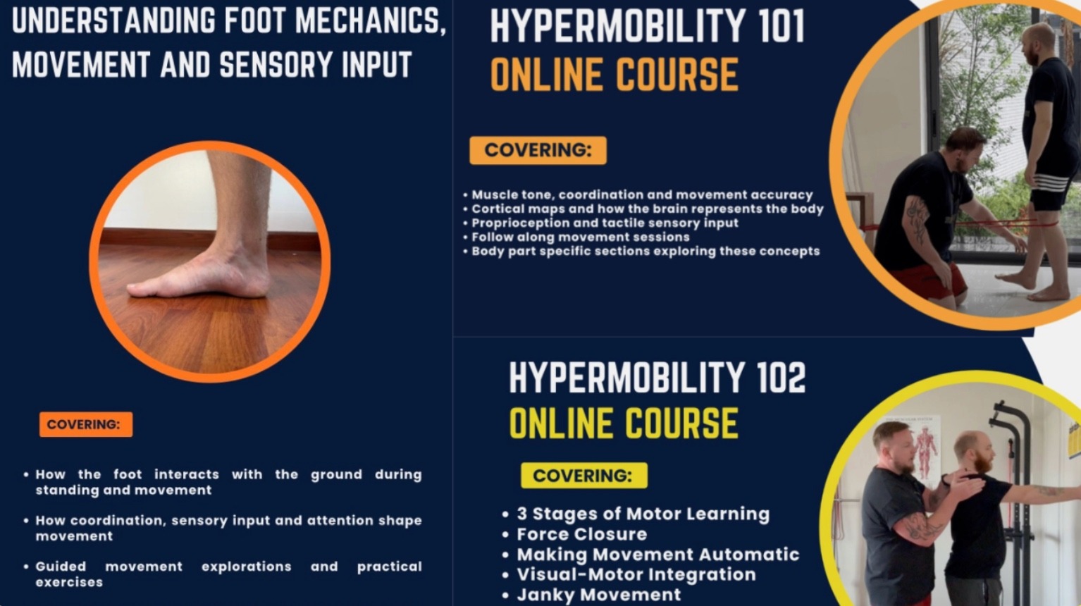
The Hypermobility Fundamentals Bundle
The Hypermobility Fundamentals Bundle is a structured education and movement pathway that starts at the bottom and builds upwards, grounded in motor learning and sensory motor science. Designed for people with hypermobility and persistent pain, it focuses on how strength and stability are organised and learned by the nervous system, beginning with the feet and progressing through sensory input, coordination, and motor learning. The courses are intended to be completed in sequence, with each one establishing the concepts and skills needed for the next.
— The Fibro Guy Team —
References
[1] Tinkle B, Castori M, Berglund B, Cohen H, Grahame R, Kazkaz H, Levy H (2017) ‘Hypermobile Ehlers-Danlos syndrome (a.k.a. Ehlers-Danlos syndrome Type III and Ehlers-Danlos syndrome hypermobility type): Clinical description and natural history’, American Journal of Medical Genetics Part C: Seminars in Medical Genetics, 175(1), pp. 48-69. doi: 10.1002/ajmg.c.31538 [2] Malfait F, Francomano C, Byers P, Belmont J, Berglund B, Black J et al. (2017) ‘The 2017 International Classification of the Ehlers-Danlos Syndromes’, American Journal of Medical Genetics Part C: Seminars in Medical Genetics, 175(1), pp. 8-26. doi: 10.1002/ajmg.c.31552 [3] Wang TJ, Stecco A (2021) ‘Fascial thickness and stiffness in hypermobile Ehlers-Danlos syndrome’, American Journal of Medical Genetics Part C: Seminars in Medical Genetics, 187(4), pp. 446-452. doi: 10.1002/ajmg.c.31948 [4] Wang TJ, Stecco A, Schleip R, Stecco C, Pirri C (2023) ‘Change in gliding properties of the iliotibial tract in hypermobile Ehlers-Danlos syndrome’, Journal of Ultrasound, 26(4), pp. 809-813. doi: 10.1007/s40477-023-00775-7 [5] Wang TJ, Stecco A, Hakim AJ, Schleip R (2025) ‘Fascial Pathophysiology in Hypermobility Spectrum Disorders and Hypermobile Ehlers-Danlos Syndrome: A Review of Emerging Evidence’, International Journal of Molecular Sciences, 26(12), p. 5587. doi: 10.3390/ijms26125587 [6] Stecco C, Stern R, Porzionato A, Macchi V, Masiero S, Stecco A, De Caro R (2011) ‘Hyaluronan within fascia in the etiology of myofascial pain’, Surgical and Radiologic Anatomy, 33(10), pp. 891-896. doi: 10.1007/s00276-011-0876-9 [7] Schleip R (2003) ‘Fascial plasticity – a new neurobiological explanation: Part 1’, Journal of Bodywork and Movement Therapies, 7(1), pp. 11-19. doi: 10.1016/s1360-8592(02)00067-0 [8] Chaudhry H, Schleip R, Ji Z, Bukiet B, Maney M, Findley T (2008) ‘Three-Dimensional Mathematical Model for Deformation of Human Fasciae in Manual Therapy’, Journal of Osteopathic Medicine, 108(8), pp. 379-390. doi: 10.7556/jaoa.2008.108.8.379 [9] Stecco C, Gagey O, Belloni A, Pozzuoli A, Porzionato A, Macchi V, Aldegheri R, De Caro R, Delmas V (2007) ‘Anatomy of the deep fascia of the upper limb. Second part: study of innervation’, Morphologie, 91(292), pp. 38-43. doi: 10.1016/j.morpho.2007.05.002 [10] Schilder A, Hoheisel U, Magerl W, Benrath J, Klein T, Treede R-D (2014) ‘Sensory findings after stimulation of the thoracolumbar fascia with hypertonic saline suggest its contribution to low back pain’, Pain, 155(2), pp. 222-231. doi: 10.1016/j.pain.2013.09.025 [11] Tesarz J, Hoheisel U, Wiedenhöfer B, Mense S (2011) ‘Sensory innervation of the thoracolumbar fascia in rats and humans’, Neuroscience, 194, pp. 302-308. doi: 10.1016/j.neuroscience.2011.07.066 [12] Mense S (2019) ‘Innervation of the thoracolumbar fascia’, European Journal of Translational Myology, 29(3), p. 8297. doi: 10.4081/ejtm.2019.8297 [13] Suarez-Rodriguez V, Fede C, Pirri C, Petrelli L, Loro-Ferrer JF, Rodriguez-Ruiz D, De Caro R, Stecco C (2022) ‘Fascial Innervation: A Systematic Review of the Literature’, International Journal of Molecular Sciences, 23(10), p. 5674. doi: 10.3390/ijms23105674 [14] Woolf CJ (2011) ‘Central sensitization: Implications for the diagnosis and treatment of pain’, Pain, 152(3 Suppl), pp. S2-S15. doi: 10.1016/j.pain.2010.09.030 [15] Nijs J, George SZ, Clauw DJ, Fernández-de-las-Peñas C, Kosek E, Ickmans K et al. (2021) ‘Central sensitisation in chronic pain conditions: latest discoveries and their potential for precision medicine’, The Lancet Rheumatology, 3(5), pp. e383-e392. doi: 10.1016/S2665-9913(21)00032-1 [16] Rombaut L, Scheper M, De Wandele I, De Vries J, Meeus M, Malfait F, Engelbert R, Calders P (2015) ‘Chronic pain in patients with the hypermobility type of Ehlers-Danlos syndrome: evidence for generalized hyperalgesia’, Clinical Rheumatology, 34(6), pp. 1121-1129. doi: 10.1007/s10067-014-2499-0 [17] De Wandele, I., Colman M, Hermans L, Van Oosterwijck J, Meeus M, Rombaut L, Brusselmans G, Syx D, Calders P, Malfait F (2022) ‘Exploring pain mechanisms in hypermobile Ehlers-Danlos syndrome: A case-control study’, European Journal of Pain, 26(6), pp. 1355-1367. doi: 10.1002/ejp.1956 [18] Rombaut L, Malfait F, De Paepe A, Rimbaut S, Verbruggen G, De Wandele I, Calders P (2011) ‘Impairment and impact of pain in female patients with Ehlers-Danlos syndrome: A comparative study with fibromyalgia and rheumatoid arthritis’, Arthritis and Rheumatism, 63(7), pp. 1979-1987. doi: 10.1002/art.30337 [19] Smith TO, Jerman E, Easton V, Bacon H, Armon K, Poland F, Macgregor AJ (2013) ‘Do people with benign joint hypermobility syndrome (BJHS) have reduced joint proprioception? A systematic review and meta-analysis’, Rheumatology International, 33(11), pp. 2739-2746. doi: 10.1007/s00296-013-2790-4 [20] Rombaut L, De Paepe A, Malfait F, Cools A, Calders P (2010) ‘Joint position sense and vibratory perception sense in patients with Ehlers-Danlos syndrome type III (hypermobility type)’, Clinical Rheumatology, 29(3), pp. 289-295. doi: 10.1007/s10067-009-1320-y [21] Clayton HA, Jones SAH, Henriques DYP (2015) ‘Proprioceptive precision is impaired in Ehlers-Danlos syndrome’, SpringerPlus, 4, p. 323. doi: 10.1186/s40064-015-1089-1 [22] Wiewelhove T, Döweling A, Schneider C, Hottenrott L, Meyer T, Kellmann M, Pfeiffer M, Ferrauti A (2019) ‘A Meta-Analysis of the Effects of Foam Rolling on Performance and Recovery’, Frontiers in Physiology, 10, p. 376. doi: 10.3389/fphys.2019.00376 [23] Wilke J, Müller AL, Giesche F, Power G, Ahmedi H, Behm DG (2019) ‘Acute Effects of Foam Rolling on Range of Motion in Healthy Adults: A Systematic Review with Multilevel Meta-analysis’, Sports Medicine. doi: 10.1007/s40279-019-01205-7 [24] Warneke K, Plöschberger G, Lohmann L, Lichtenstein E, Jochum D, Siegel SD, Zech A, Behm D (2024) ‘Foam Rolling and Stretching Do Not Provide Superior Acute Flexibility and Stiffness Improvements Compared to Any Other Warm-Up Intervention’, Journal of Sport Health Science. doi: 10.1016/j.jshs.2024.01.006 [25] Quintner JL, Bove GM, Cohen ML (2015) ‘A Critical Evaluation of the Trigger Point Phenomenon’, Rheumatology, 54(3), pp. 392-399. doi: 10.1093/rheumatology/keu471 [26] Myburgh C, Larsen AH, Hartvigsen J (2008) ‘A Systematic, Critical Review of Manual Palpation for Identifying Myofascial Trigger Points: Evidence and Clinical Significance’, Archives of Physical Medicine and Rehabilitation, 89(6), pp. 1169-1176. doi: 10.1016/j.apmr.2007.12.033 [27] Bialosky JE, Bishop MD, Price DD, Robinson ME, George SZ (2009) ‘The Mechanisms of Manual Therapy in the Treatment of Musculoskeletal Pain: A Comprehensive Model’, Manual Therapy, 14(5), pp. 531-538. doi: 10.1016/j.math.2008.09.001 [28] Bialosky JE, Bishop MD, Penza CW (2017) ‘Placebo Mechanisms of Manual Therapy: A Sheep in Wolf’s Clothing?’, Journal of Orthopaedic and Sports Physical Therapy, 47(5), pp. 301-304. doi: 10.2519/jospt.2017.0604 [29] Molina-Álvarez M, Arribas-Romano A, Rodríguez-Rivera C, García MM, Fernández-Carnero J, Armijo-Olivo S, Goicoechea Garcia C (2022) ‘Manual Therapy Effect in Placebo-Controlled Trials: A Systematic Review and Meta-Analysis’, International Journal of Environmental Research and Public Health, 19(21), p. 14021. doi: 10.3390/ijerph192114021 [30] Schleip R, Klingler W, Lehmann-Horn F (2005) ‘Active fascial contractility: Fascia may be able to contract in a smooth muscle-like manner and thereby influence musculoskeletal dynamics’, Medical Hypotheses, 65(2), pp. 273-277. doi: 10.1016/j.mehy.2005.03.005 [31] De Wandele, I., Rombaut L, Leybaert L, Van de Borne P, De Backer T, Malfait F, De Paepe A, Calders P (2014) ‘Dysautonomia and its underlying mechanisms in the hypermobility type of Ehlers-Danlos syndrome’, Seminars in Arthritis and Rheumatism, 44(1), pp. 93-100. doi: 10.1016/j.semarthrit.2013.12.006 [32] Mathias CJ, Owens A, Iodice V, Hakim A (2021) ‘Dysautonomia in the Ehlers-Danlos syndromes and hypermobility spectrum disorders – With a focus on the postural tachycardia syndrome’, American Journal of Medical Genetics Part C, 187(4), pp. 551-570. doi: 10.1002/ajmg.c.31951 [33] Seneviratne SL, Maitland A, Afrin L (2017) ‘Mast cell disorders in Ehlers-Danlos syndrome’, American Journal of Medical Genetics Part C, 175(1), pp. 226-236. doi: 10.1002/ajmg.c.31555 [34] Louw A, Diener I, Butler DS, Puentedura EJ (2011) ‘The Effect of Neuroscience Education on Pain, Disability, Anxiety, and Stress in Chronic Musculoskeletal Pain’, Archives of Physical Medicine and Rehabilitation, 92(12), pp. 2041-2056. doi: 10.1016/j.apmr.2011.07.198 [35] Wood L, Hendrick PA (2019) ‘A systematic review and meta-analysis of pain neuroscience education for chronic low back pain: Short- and long-term outcomes of pain and disability’, European Journal of Pain, 23(2), pp. 234-249. doi: 10.1002/ejp.1314 [36] Langevin HM, Stevens-Tuttle D, Fox JR, Badger GJ, Bouffard NA, Krag MH, Wu J, Henry SM (2009) ‘Ultrasound evidence of altered lumbar connective tissue structure in human subjects with chronic low back pain’, BMC Musculoskeletal Disorders, 10, p. 151. doi: 10.1186/1471-2474-10-151 [37] Langevin HM, Fox JR, Koptiuch C, Badger GJ, Greenan-Naumann AC, Bouffard NA, Konofagou EE, Lee W-N, Triano JJ, Henry SM (2011) ‘Reduced thoracolumbar fascia shear strain in human chronic low back pain’, BMC Musculoskeletal Disorders, 12, p. 203. doi: 10.1186/1471-2474-12-203 [38] Branchini M, Lopopolo F, Andreoli E, Loreti I, Marchand AM, Stecco A (2015) ‘Fascial Manipulation for Chronic Aspecific Low Back Pain: A Single Blinded Randomized Controlled Trial’, F1000Research. doi: 10.12688/f1000research.6890.2 [39] Csecs JLL, Iodice V, Rae CL, Brooke A, Simmons R, Quadt L, Savage G, Dowell N, Prowse F, Themelis K, Mathias C, Critchley H, Eccles J (2022) ‘Joint Hypermobility Links Neurodivergence to Dysautonomia and Pain’, Frontiers in Psychiatry, 12, p. 786916. doi: 10.3389/fpsyt.2021.786916 [40] Louw A, Zimney K, Puentedura EJ, Diener I (2016) ‘The efficacy of pain neuroscience education on musculoskeletal pain: A systematic review of the literature’, Physiotherapy Theory and Practice, 32(5), pp. 332-355. doi: 10.1080/09593985.2016.1194646 [41] Voermans NC, Knoop H, Bleijenberg G, van Engelen BGM (2010) ‘Pain in Ehlers-Danlos Syndrome is Common, Severe, and Associated with Functional Impairment’, Journal of Pain and Symptom Management, 40(3), pp. 370-378. doi: 10.1016/j.jpainsymman.2009.12.026 [42] Scheper MC, de Vries JE, Verbunt J, Engelbert RHH (2015) ‘Chronic pain in hypermobility syndrome and Ehlers-Danlos syndrome (hypermobility type): it is a challenge’, Journal of Pain Research, 8, pp. 591-601. doi: 10.2147/JPR.S64251 [43] Kuhfuß M, Maldei T, Hetmanek A, Baumann N (2021) ‘Somatic Experiencing – Effectiveness and Key Factors of a Body-Oriented Trauma Therapy: A Scoping Literature Review’, European Journal of Psychotraumatology, 12(1), p. 1929023. doi: 10.1080/20008198.2021.1929023 [44] Scheeringa MS (2025) ‘Evaluating Evidence Behind Popular Trauma Narratives: Neurobiological and Treatment Claims in The Body Keeps the Score’, BJPsych Bulletin. doi: 10.1192/bjb.2025.10174 [45] Ajimsha MS, Al-Mudahka NR, Al-Madzhar JA (2015) ‘Effectiveness of Myofascial Release: Systematic Review of Randomized Controlled Trials’, Journal of Bodywork and Movement Therapies, 19(1), pp. 102-112. doi: 10.1016/j.jbmt.2014.06.001 [46] Espejo-Antúnez L, Fernández-Huertas Tejeda J, Albornoz-Cabello M, Rodríguez-Mansilla J, de la Cruz-Torres B, Ribeiro F, Silva AG (2017) ‘Dry Needling in the Management of Myofascial Trigger Points: A Systematic Review of Randomized Controlled Trials’, Complementary Therapies in Medicine, 33, pp. 46-57. doi: 10.1016/j.ctim.2017.06.003 [47] Nazari G, Bobos P, Lu SZ, Reischl S, Sharma S, Le CY, Vader K, Held N, MacDermid JC (2022) ‘Effectiveness of Instrument-Assisted Soft Tissue Mobilization for the Management of Upper Body, Lower Body, and Spinal Conditions – An Updated Systematic Review with Meta-Analyses’, Disability and Rehabilitation. doi: 10.1080/09638288.2022.2070288 [48] Sendur OF, Gurer G, Bozbas GT (2007) ‘The frequency of hypermobility and its relationship with clinical findings of fibromyalgia’, Clinical Rheumatology, 26(4), pp. 485-487. doi: 10.1007/s10067-006-0304-4 [49] Stecco A, Meneghini A, Stern R, Stecco C, Imamura M (2014) ‘Ultrasonography in myofascial neck pain: randomized clinical trial for diagnosis and follow-up’, Surgical and Radiologic Anatomy, 36(3), pp. 243-253. doi: 10.1007/s00276-013-1185-2 [50] De Wandele, I., Calders P, Peersman W, Rimbaut S, De Backer T, Malfait F, De Paepe A, Rombaut L (2014) ‘Autonomic symptom burden in the hypermobility type of Ehlers-Danlos syndrome: A comparative study with two other EDS types, fibromyalgia, and healthy controls’, Seminars in Arthritis and Rheumatism, 44(3), pp. 353-361. doi: 10.1016/j.semarthrit.2014.05.013The fascia industry isn’t the only wellness scene where the studies turn out to be largely written by people selling the product. Our deep-dive into grounding and earthing covers a near-identical pattern in the earthing literature.