
Last updated September 2026
There’s no shortage of exercise advice for hypermobility, and that’s most of the problem. Some of it is decent, a lot of it is recycled from populations that have nothing in common with you, and a fair chunk of it is somebody’s opinion on Instagram. So, you get told to strengthen, then told to stretch, then told to do Pilates, then told to rest, and every one of those came from a person who sounded confident.
What you almost never get told is why your body behaves the way it does. Why the muscles feel tight and weak at once. Why an ankle you’ve strengthened for eight months still rolls on a kerb. Why the same session you got through last week is almost undoable today. That’s the part that actually decides whether a programme works or not, and it’s the part this guide leads with.
This is your starting point. Every section opens with the short version, and expands if you want more. The blue boxes link to the full articles. Start at the top if you’re new to this, or jump to whatever’s hurting.
Contents
- Why Exercise Is Different When You’re Hypermobile
- Core Stability, and Why Engaging Your Core Is the Wrong Instruction
- Pacing: The Skill That Decides Whether Any of This Sticks
- Foot Arches: The Bit Everyone Skips
- Nutrition and Hypermobility: What Food and Supplements Actually Do
- KT Tape: What It’s Actually Doing for a Hypermobile Joint
- Why Sleep Is So Hard to Fix When You’re Hypermobile
- Stretching: Why It Feels Good and Never Holds
- Pilates and Yoga: What the Evidence Actually Says
- Braces, Compression and the Body Braid: Support, Not Dependency
- The Pelvic Floor, and Why the Standard Advice Gets Hypermobility Backwards
- Hypermobile Children and Exercise: What Actually Predicts Trouble
- Exercising With POTS: The First Step Everyone Skips
- Why Fear of Movement in Hypermobility Makes Sense
- Fascia and Hypermobility: What Your Hands Can Actually Reach
- Running With Hypermobility: It Isn’t the Flat No You Were Given
- Scoliosis and Hypermobility: What the Curve Is Actually Telling You
- References
Why Exercise Is Different When You’re Hypermobile
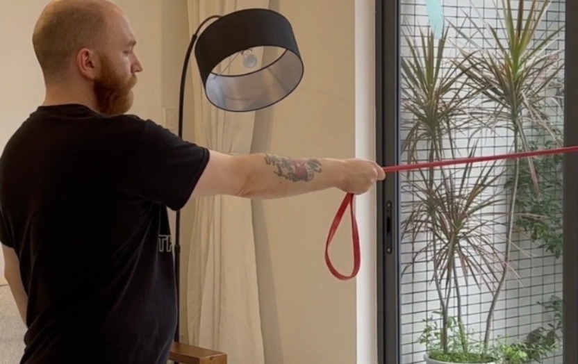
Most of those with hypermobility have already done the exercises. The bands, the clams, the sheet with the little stick figures on it, three times a week for months on end. And the annoying part is that it often does make you stronger. But the shoulder still slips out when you reach upwards, and the hip still slides when you roll over in bed. So either you’re doing it wrong, or nobody has quite explained what the problem is, and generally it’s the second one. A joint that travels further than average isn’t only a mechanical problem, it’s an information problem, as the sensors telling your brain where you are, live in that same tissue. Strength is still worth having, we’re not one of those places that’s against it. It’s just that force you can’t aim doesn’t get you very far on its own.
Read MoreThe first thing worth knowing is that being hypermobile and being symptomatic are two different states, and the research keeps dividing along exactly that same line. Those with hypermobility who feel absolutely fine tend to measure the same as everybody else for muscle and tendon properties. In ordinary community samples of hypermobile children there’s often no difference in joint position sense at all. The differences turn up in the symptomatic group, which makes how far your joints travel a much less interesting number than it looks.
What does turn up in that group sits in three layers.
The first is the signal from the tissues itself. Laxer tissue has to travel a little further before a sensor reaches the point where it fires, so the information about where your joint is, arrives a little vague. Measured properly, the fault is scatter, not aim. Think of an old GPS: it gets you where you want to be, just within a thirty metre radius. People land in roughly the right place on average and simply spread more widely around it. You can’t calibrate noise away either, you can only give the system more to work with.
The second is tone, which is where the contradiction most people carry around actually lives. Being told you have low tone while feeling permanently clenched isn’t an oxymoron, as the word is quietly doing two jobs. The resistance built into the tissue often gives way earlier than average, so the activity your nervous system adds on top has to do more. Something has to hold the joint in place. That’s compensation in specific tasks though, and it varies a fair bit from one person to the next. It isn’t every hypermobile body braced everywhere at once, and we’ve overstated that one ourselves in the past, and we’ve since said so.
The third is how movement gets learned in the first place, which is the most underused idea in hypermobility rehab by a distance. Patterns get built through variety, not through doing the identical rep on the identical mat. And there’s a finding on training while you hurt that changed how we run sessions, as pain during practice can leave a session looking perfectly good on the day, then take most of it back again by the morning. So we work signal first, then strategy, then load. That order is our read though, not a proven sequence, built on the mechanisms and on watching a great many people go through it in the studios. If the head to head study turns up and says we had it backwards, we’ll say so and teach the other thing.
One last thing worth setting expectations on. The first six to eight weeks generally look like nothing much from the outside, as what’s changing is the quality of the information, not the size of anything. That’s the stage where nearly everybody quits.
Go deeperHypermobility and Exercise: Why Traditional Strength Training Falls ShortStrength work has more behind it than anything else in this field, and it still gets badly oversold. What supervised heavy loading did and didn’t do for painful, unstable shoulders, and what actually tracks how limited somebody’s day is, which turns out to have very little to do with how far the elbows go back.Proprioception, Brain Maps and Why Your Body Feels LostWalking into door frames, fine in a bright kitchen and genuinely unsafe on the landing at two in the morning, then wiped out for the afternoon by an hour in a supermarket. What your position sense is really doing, and what happened to the popular brain based explanation once somebody pointed a better scanner at it.Why Your Hypermobile Muscles Feel Tight and Weak at the Same TimeSomebody in a clinic said low tone, and meanwhile your jaw is clenched before breakfast and your back aches after a day of doing very little. The two jobs that word is quietly doing, an honest limit on the gripping story including on our own older writing, and the strength finding that explains why building the muscle bigger was never all that likely to be the missing piece.Motor Learning for Hypermobility: How Your Nervous System Builds Reliable MovementYou were consistent, you did the work, and a year on very little of it stuck, which has rather less to do with discipline than it does with how movement gets learned. Why the identical rep on the identical mat mostly teaches your system that mat, the two things that can quietly switch a muscle off before a session even starts, and what training in pain does to the part you were hoping to keep.
Core Stability, and Why Engaging Your Core Is the Wrong Instruction

Ask anybody who’s been given exercises for a painful back, a hip that clicks on the stairs, or an SI joint that only plays up on certain days, and you’ll hear the same four words. Engage your core. Navel to spine, hold it through the supermarket and the car door and the reach for the kettle, and repeat until that’s simply how you stand now. What almost nobody explains is where the instruction came from. It traces back to one deep stomach muscle being found to come on a few milliseconds late in people with back pain, back in the mid nineties. Not a weak muscle, a slightly slow one. An entire industry got built on that gap, and in a body that already runs loose, the constant squeezing is generally the problem, not the fix.
Read MoreYour core isn’t the muscle you can see in the mirror, it’s closer to a pressurised cylinder: the diaphragm as the roof, the pelvic floor as the floor, the deep abdominal wall and obliques as the sides, and the deep spinal muscles as the back wall. What that structure manages is pressure, and the coordination between its parts matters a good deal more than how strong any one of them is on its own. In a spine that’s behaving, the deep abdominal wall fires before the shoulder muscle does when you go to lift your arm, so the system is anticipating the load instead of catching up with it. That timing, and not force, is closer to what stability actually means.
The original finding hasn’t been overturned, though it has been quietly reinterpreted since. Give somebody temporary back pain who didn’t have any five minutes earlier and the same muscle starts firing late, which turns the arrow round, as pain now looks like it’s causing the delay and not the other way about. Head to head, dedicated core training doesn’t beat general exercise or manual therapy for back pain, it mostly beats doing not very much at all. And rather awkwardly, plenty of the people who do improve show no matching change in the muscle itself on a scan.
In hypermobility the cylinder tends to run over active, not weak, which is the bit that changes the advice. Looser joints report back a fuzzier sense of where they are, so the nervous system compensates by turning tone up more or less permanently, instead of only when something needs holding. Adding a deliberate brace on top of a system that’s already gripping doesn’t add stability. In our experience it mostly adds a pelvic floor that won’t switch off, and a neck doing a job it was never built for.
Which is why the useful instruction isn’t a hold at all, it’s the breath. The diaphragm and the pelvic floor work as a pair, lengthening on the in breath and lifting on the out. A cylinder that can change pressure when the task asks for it will do more for you than one clamped shut all day. That’s also a far better fit for hypermobility, where the thing in short supply is almost never effort.
Core stability for hypermobility
Go deeperHypermobility Core Exercises: What Actually Works, and What Doesn’tWhy the breath does the job most people try to force with a brace, what goes wrong with a hypermobile pelvic floor when kegels get piled on top, and how the SI joint comes to depend on muscles picking up work the ligaments used to manage on their own.
Pacing: The Skill That Decides Whether Any of This Sticks

You already know the cycle without anybody needing to name it. A good day turns up, so you get through the washing, cook a proper meal and take the dog further than you have in weeks. By the second morning you’re barely upright and cross about it, and the pile of things still waiting hasn’t moved an inch. Somewhere in that story somebody will have told you to pace yourself, usually followed by listen to your body. Which sounds sensible, right up until you notice your body tends to report the damage a day or two after it happened, well past the point where you could still have done something about it. Pacing is meant to break that loop, and it’s a skill, not a polite word for doing less.
Read MoreThe first thing worth knowing is that pacing isn’t a gentler way of saying rest more. Pull apart what the word actually covers and you get five things: action, time, balance, learning and self management, and not one of them is passive. It means deciding in advance how long you’ll do something, and setting that limit before you start rather than after. It means building the rest in ahead of the point where symptoms force it on you, then watching what happens and adjusting. That’s something you build over months, not an instruction to sit down more often.
The obvious alternative, stopping when symptoms tell you to, turns out to be the less reliable version. In these conditions the signal tends to arrive well after the point where it would have been any use, so stopping once things have picked up usually means you went past your limit a while ago. The better supported approach sets the stopping point in advance, off a baseline measured on an ordinary or a bad day rather than a good one. You work off a timer, not a feeling.
Worth being straight about the evidence though, as pacing gets handed out as if it were settled and it isn’t. The most careful tally of it comes from the condition where it’s been studied hardest. Most trials leaned towards a benefit, a handful showed nothing at all and a couple made things worse, and the people doing the reviewing concluded there wasn’t enough there to guide practice either way. A more recent pooling of the stronger studies found much the same shape: real movement in the right direction, with ranges wide enough that no effect whatsoever stayed a genuine possibility.
There’s also a detail almost every pacing guide skips. Physical activity is only one of several things drawing on the same reserve. Cognitive effort, emotional load and a noisy, crowded environment all come out of the same account, so a supermarket trip or a difficult phone call can undo a day of carefully paced walking without you taking a single extra step. If your physical pacing is sound and you’re still crashing, that’s generally where to look before you decide pacing doesn’t work for you.
Go deeperPacing for Fibromyalgia, EDS and Chronic Pain: The Complete Evidence Based GuideWhat the trial evidence actually shows once you get past the leaflet version, how to find a baseline that holds up on a bad day rather than a good one, and why post exertional malaise changes the whole approach and not only the speed of it.
Foot Arches: The Bit Everyone Skips
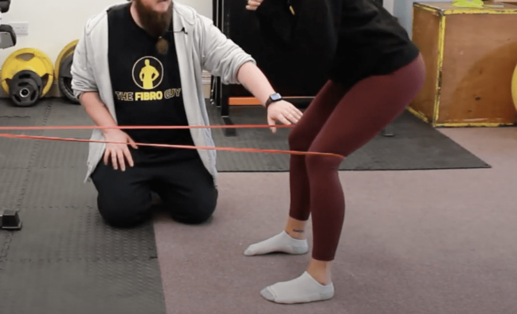
Somebody has probably stood you on a plinth, looked at your arch while you weren’t moving, and told you it had collapsed, usually handing over an insole in the same breath. That’s not a daft thing to say, it’s just aimed at the wrong thing, as your arch was never a shape holding still under the weight of you. It’s a movement. It drops to store a bit of energy in the tissue underneath, then rises again to give you something stiff to push off from, a few thousand times between the bed and the kettle before you’ve had breakfast. In a hypermobile foot it’s the dropping and the rising that goes wrong, and not the shape somebody measured while you stood there, obediently, not moving.
Read MoreThe sole of your foot is thick with sensors, feeding your balance both consciously and through reflex routes that never reach conscious thought at all. Model what happens in actual walking instead of standing still, and the toes, the heel and the outer border seem to carry a particularly large share of that reporting. None of that machinery is broken in a hypermobile foot though. What’s different is the tissue those sensors sit in, which is more compliant, deforms further for the same load and takes longer to pull tight enough to set them off. The signal still arrives, it just arrives a little late and a little vague about how much. Which is why this reads as a foot that gives way at a moment you weren’t expecting, and not a foot that’s simply weak.
The shape of your foot at rest matters less than you’d think. Hypermobile feet have only been watched properly while actually walking once, in adults with hEDS and HSD, and that work didn’t sort anybody into arch categories at all. What it found was the smaller bones of the foot moving more than they should, mostly through the inside of the forefoot. It’s small and exploratory though, so we’d hold it loosely rather than lean on it. Whether hypermobile feet run flatter as a rule is genuinely unsettled too, with some groups measuring more pronated and others finding no difference.
Then the orthotics question, which gets treated as either a cure or a trap and is honestly neither. Custom devices do measurably lift the arch while you’ve got them on, off the shelf ones rather less, and in several conditions they do bring pain down. What nothing does, orthotics or exercise, is rebuild the arch itself, as nothing in the pooled research reliably shifts how much it drops. We used to say orthotics weaken the foot over time, and that was too strong, so we’ve dropped it. The actual picture is one study finding the muscles shrank, another finding they grew and a third finding no change at all. Which says the device, your age and whether you’re training alongside it matter a good deal more than the insole on its own.
Arch control and loading pattern
Go deeperHypermobility and the Foot Arch: Why Yours Keeps Collapsing, and What to Do About ItThe three point test that shows which part of your foot has quietly stopped doing its job, an honest read on whether your insoles are helping, hurting or doing very little, and the two exercises we use with clients more than any others.
Nutrition and Hypermobility: What Food and Supplements Actually Do
Somebody in this community will have told you that your joints are made of collagen, so eating more of it, or drinking it in a tub, must be the answer. That logic sounds tidy enough to have survived being wrong for years. There’s no EDS diet, and the biology says a swallowed collagen supplement can’t override the genetics that decide how your own gets rebuilt. What food and a handful of supplements actually seem to change is the system underneath the joint: how much energy you’ve got left for rehab, whether your gut is settled enough to eat properly, and whether you’re getting the fairly boring building blocks, protein chief among them, that repair actually runs on. That’s a smaller claim than the one on the label, and it happens to be the one that holds up.
Read MoreCurrent gastroenterology guidance says changing what you eat belongs to an identified problem: IBS type symptoms, gastroparesis, reflux, suspected coeliac disease or mast cell triggers. It doesn’t belong to a hypermobility diagnosis sitting alone on a letter. That matters because gut symptoms are genuinely common here, and so is quietly cutting foods out to cope with them, which tends to grow year on year without anybody deciding it should. A diet shrunk that way for long enough tends to end in deficiencies that make everything else harder.
Protein is the one nutrient actually worth paying attention to, and not for the reason it gets sold to you. There’s no evidence hypermobility raises your requirement above the ordinary 0.8 to 1.2 grams per kilogram a day, though plenty of people aren’t meeting even that. Protein is the material the body uses to repair tissue during rehab, so what gets corrected is a shortfall, and not protein doing anything special for connective tissue. A duller sentence than the one on a shake tub, and the more useful one.
Supplements are meant to be exactly what the word says, added to eating properly and not a substitute for it, and almost none of the popular ones have actually been tested in hypermobility or EDS. Collagen is the big one, and the case against it is structural rather than a trial result. What you swallow gets broken down and rebuilt according to your own genetics, which a tablet can’t rewrite, so for most people it’s money going nowhere. That’s our reasoning though, not a cited study in this population.
Creatine is the exception with a real evidence base, just not the one usually claimed for it. In fibromyalgia, over sixteen weeks against a placebo, it reliably raised muscle energy stores and improved strength. Pain didn’t move at all, nor did sleep or quality of life. For hypermobility or EDS specifically, direct evidence is more or less absent, so anything written up about it borrows from fibromyalgia, ME/CFS or post viral fatigue, and it’s worth knowing which one you’re being handed. What it seems to help with is capacity, not pain: what you can physically do, rather than how much it hurts doing it. Which matters, because feeling capable of more while your tissue tolerance hasn’t changed is a fairly reliable way to turn a decent week into a rotten fortnight.
Go deeperHypermobility and EDS: What is the best diet?When a specific diet is actually justified rather than assumed from the diagnosis, what the low FODMAP diet can and can’t do for gut symptoms, and the point where cutting foods out turns into something worth naming out loud.Creatine for Hypermobility, Fibromyalgia and Chronic Pain: What the Research Actually ShowsWhat actually happens to the kidney worry when it’s checked properly, why the fatigue research is more interesting than the pain research, and how to dose it sensibly if you decide it’s worth trying.
KT Tape: What It’s Actually Doing for a Hypermobile Joint
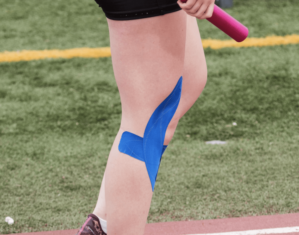
Half the people at any hypermobility clinic have a strip of brightly coloured tape somewhere: a shoulder, a wrist, a kneecap poking out from under a pair of jeans. Most were sold on it because the box says it lifts the skin, drains the lymph or holds the joint in place. None of that survives contact with the evidence, and a couple of the big brands have paid out in the US courts over claims along exactly those lines. What’s odd is that the tape still isn’t nothing. Those who tape a subluxing shoulder do tend to hurt less and do more afterwards, and that’s a real, measured change. It’s just that nobody can currently tell you whether the tape is doing it or something else is.
Read MoreAway from any one clinic, the general evidence for kinesiology tape is thin. Across a dozen randomised trials spanning shoulder, knee, neck, back and plantar fasciitis, no consistent benefit turned up against any comparison at all: sham tape, no treatment or ordinary physiotherapy. When the strength work was pooled separately, the only effect showed up in muscle that was already fatigued or already compromised by illness. So the case for tape was never really about healthy performance, whatever the packaging with the footballers on it implies.
The explanation usually offered, that it improves proprioception, your sense of where the joint actually is, has taken a knock lately. Pooled across taping studies in general there is a solid effect on joint position sense, and not one of the studies in that pool included anybody hypermobile. It’s only been properly tested once in hEDS, in a small group with hypermobile shoulders, and joint repositioning didn’t improve with either of the two taping techniques used. So the mechanism most people reach for is, in this exact population, the one that’s been looked at and come back empty.
What did move in that same work was pain and function, by a wide margin, and the direction the tape was applied in changed how much rotation people got back. Both taped groups improved though, with nobody untaped in the mix, so time, attention or the assessment itself could be doing some of that. Our read is that it’s closer to sensory competition. The tape on the skin gives the nervous system something else to process alongside the ache coming off a joint that already feels unreliable, not unlike the instinct to rub a bumped elbow. That’s a much smaller claim than structural support or drained lymph, and it still leaves tape with a genuine, if modest, part to play.
Worth knowing if your skin already reacts to things. Most tape adhesive contains colophonium, a pine resin derivative that has been shown to sensitise people who’d never reacted to anything like it before, purely through continuous wear. Where mast cell problems sit alongside the hypermobility, reports from those living with it describe reactions going well beyond the skin. There’s no published research on that combination yet though. None of which is a reason to avoid tape, it just isn’t the inert, harmless product the branding implies.
KT Taping for Hypermobility Part One
KT Taping for Hypermobility Part Two
Go deeperKT Tape for Hypermobility and Ehlers Danlos SyndromeThe full trial history for kinesiology tape in hEDS, what happened to range of motion once the tape crossed the joint, and a proper skin safe guide to barrier techniques, brands and removal for anybody whose skin already gives them grief.
Why Sleep Is So Hard to Fix When You’re Hypermobile
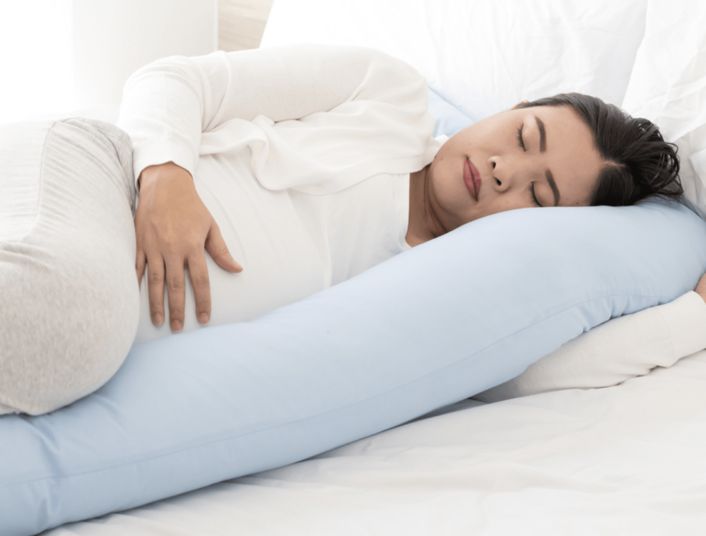
Most of the sleep advice aimed at hypermobility reads like it was written for somebody who is merely tired: dim the lights, leave the phone alone, don’t eat too late. None of that touches what’s actually waking you at three in the morning, which might be a shoulder that won’t settle, a stomach that objects to lying flat, or a heart that won’t stop announcing itself. What the advice sheets miss is that pain and sleep don’t take turns, they feed each other, and not gently. A single bad night is enough to make ordinary pressure and cold feel sharper the next day. And it isn’t only that the pain kept you up, as there’s a real route behind it, through lost deep sleep and a rise in inflammatory signalling. So the two problems are really one problem wearing different clothes depending on the hour.
Read MorePain interrupting sleep is the half of this most people already know. Subluxing joints, gut symptoms and anxiety all get in the way of dropping off and of staying under. In one survey of adults with EDS, most took over half an hour to nod off, and a good number were sleeping under six hours a night. The half that gets missed more often is sleep loss making the pain worse the next day, through lost deep sleep and inflammatory signalling, not tiredness alone. Pain sensitivity does settle once you’re allowed a proper run of nights, though the inflammatory and attention costs of a bad stretch seem to hang around rather longer.
Underneath a fair bit of this sits the autonomic nervous system, which in hypermobility often runs hot exactly when it’s meant to be winding down, with blood pressure failing to dip overnight the way it should. That’s the physiological version of tired but wired, and a decent explanation for why lying still in the dark doesn’t reliably turn into sleep. Obstructive sleep apnoea is also a good deal more common here than used to be assumed, with roughly a third of adults with EDS meeting the threshold against a small handful of matched controls. And the awkward finding is that even once it’s properly treated with CPAP, people often stay sleepier than the machine says they should.
There’s a repair argument too, worth stating plainly: deep sleep is when growth hormone gets released and tissue gets rebuilt, which matters rather more than usual if the tissue in question is already on the flimsy side and has spent the week subluxing.
On what actually helps, the generic hygiene sheet oversells itself in one direction and undersells in another. Even the tips that sound like common sense have a thinner record than you’d expect, as a later meal changes overnight stomach acidity but not the reflux itself, whatever we may have said here before. Cognitive behavioural therapy for insomnia is the one thing here with genuinely solid evidence behind it in chronic pain, reliably improving sleep in a way melatonin and magnesium do not. It is not remotely the same offer as being told to relax before bed. One last thing, because it might be the kindest fact here: accelerometer studies find no measurable difference in sleep between those with hypermobility and everybody else, despite the very real exhaustion. That gap is a measurement problem, not you exaggerating.
The Comprehensive Guide to Sleep with Hypermobility
Go deeperEverything About Sleep, Hypermobility, and Ehlers Danlos SyndromeWhat actually explains a tired but wired night, why a sleep study can come back perfectly normal while you still feel wrecked, and which of the mattresses, pillows, supplements and bedtime rituals doing the rounds online have anything behind them at all.
Stretching: Why It Feels Good and Never Holds
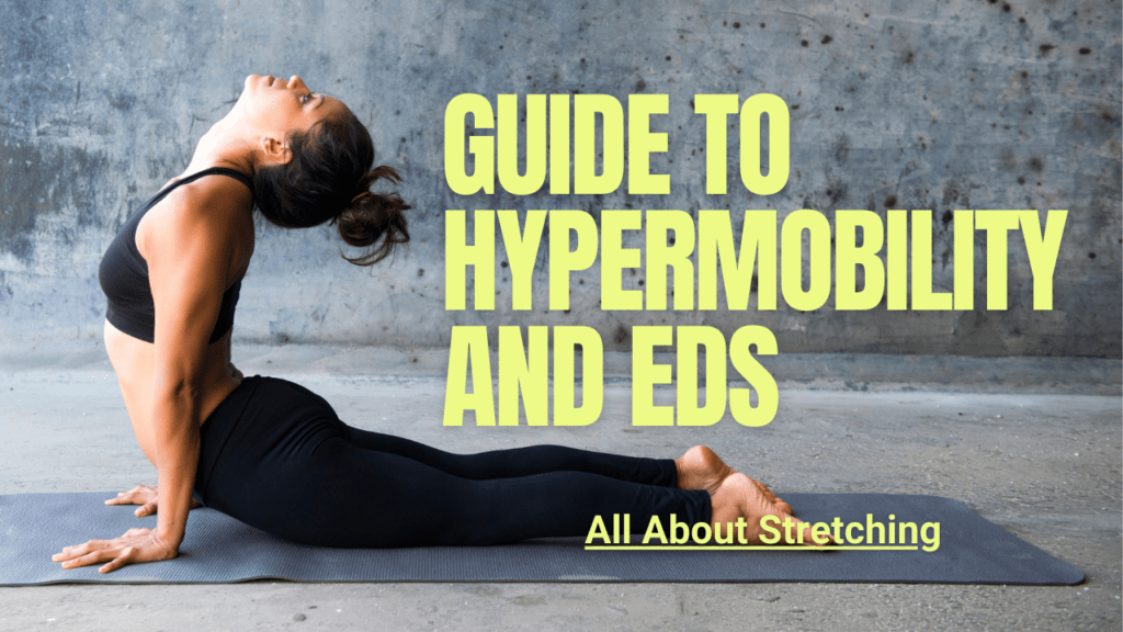
You stretch, it feels lovely, and about twenty minutes later you’re tight again, so you stretch again, and that loop can run for years without anyone quite asking what moved. It isn’t your muscle, mostly. Tightness is a sensation your brain produces, and it no more requires a short muscle than hunger requires an empty stomach, which is an odd thing to hear after a decade of being told the opposite. For those with hypermobility it gets stranger still, as the technique mostly works by persuading your nervous system to allow more travel, aimed at a joint that already travels further than it should. The relief is real though, that part isn’t in question. It’s just short for a reason nobody normally bothers explaining, and the reason is more interesting than “you didn’t hold it long enough.”
Read MoreStart with what a stretch reliably changes, which is how much your nervous system will let you travel before it says stop, plus a modest drop in stiffness. Run a stretching programme for six weeks, then go and measure the muscle itself. You can find a real increase in range with no measurable change in fibre length, muscle stiffness or tendon stiffness at all. The structural side, an actual lengthening of the tissue, is small at best and genuinely unsettled in the wider evidence. So most of what feels like progress is a negotiation you’ve won with your own nervous system, not a rope that’s got longer.
A muscle that’s guarding a joint your brain isn’t confident about can feel exactly like a short one from the inside: tight, resistant, the same pulling sensation. The two are not the same problem at all. One is a length. The other is your active muscle covering for a passive structure that gives a bit more than it should, and it’s a genuinely different thing to fix. That reading is ours, by the way, not a measured fact, though it is why we think the sensation turns up so consistently across hypermobile bodies. Stretch a joint that’s being guarded like that and you get relief for as long as the sensation stays novel, then the guard goes back on, because nothing has actually changed about the reason it was there.
Being hypermobile doesn’t make you immune to having one properly restricted spot, and the advice not to add range to already lax joints was never meant to cover it. If you can name the joint, the direction and the reason, stretching that one thing is perfectly reasonable. What doesn’t hold up is stretching everywhere because you feel tight everywhere. And the desk job version of that story, that sitting shortens your hip flexors the way a cast shortens a muscle, is borrowed from immobilisation research that has very little to do with an office chair.
Worth knowing too that you don’t need stretching to get the range anyway. Strength training done through a full range produces comparable gains as a side effect. And it does something a stretch can’t, which is build the control to own the range you’ve already got, instead of just handing you more of it.
Go deeperStretching and Hypermobility: What the Evidence Actually SaysThe four different sensations that all get called tightness and come from entirely different places, and which one is the only one where stretching a single named muscle is actually the right call.Fibromyalgia and StretchingA way of stretching that lets you explore new range without setting off the brace in the first place, and why chasing the feeling of a stretch isn’t automatically a mistake.
Pilates and Yoga: What the Evidence Actually Says
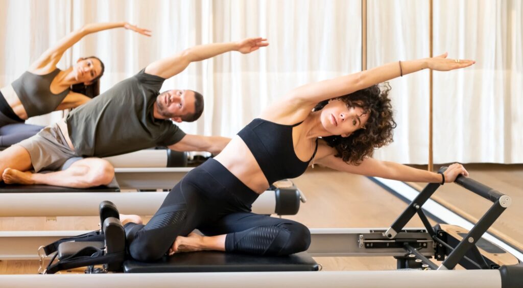
These two come up more than everything else combined, usually because a physio suggested one of them and then the appointments ran out. So people end up six months in, no better, wondering whether they’re doing it wrong or whether they were sent there because nobody had a better idea. The honest picture is more interesting than either the marketing or the backlash. Both can help, neither is doing what the label says, and for a hypermobile body the version that makes sense is close to the opposite of what most classes celebrate.
Go deeperPilates for hypermobility and fibromyalgiaYoga for chronic pain and hypermobility
Read MorePilates: better than nothing is a low bar
The claim on the tin is that Pilates activates the deep core stabilisers in a way other exercise doesn’t. A systematic review went looking for exactly that, measuring with ultrasound and surface electrode recordings, and every single outcome came back graded very low certainty. Worse, the largest effects came from the weakest trials, which is the relationship you never want to see, as it means the biggest numbers are the least trustworthy ones.
For back pain the Cochrane review is clear enough. Pilates beats minimal intervention, meaning a waiting list, a booklet or roughly nothing, for short term pain and disability. Against other exercise there’s no meaningful advantage. None of the individual trials reached the threshold for a clinically meaningful change in pain, and only three of ten did for disability. No trial in that review looked past six months.
For hypermobility specifically the position until very recently was stark. Five separate reviews across twenty three years of physiotherapy research in this population contained not one study testing Pilates as a named intervention. Not limited evidence. None. A 2026 study has finally put a number on it, showing a moderate to large improvement in hypermobility impact and body awareness holding at six months, which is genuinely encouraging. Take it with the caveats though. It wasn’t randomised, it was allocated by sign up order, the comparator was a waiting list, attrition was heavy, and the programme developer is a co-author who owns the intellectual property.
Yoga: strongest for backs, thinner everywhere else
Yoga’s best hand is chronic low back pain, where the Cochrane review found small to moderate gains in function and pain against not exercising, and little or no difference against other forms of exercise. Beyond the back it thins out quickly. The field also has a control group problem, as a large share of trials compare yoga against a waiting list, and there’s no such thing as a placebo downward dog. When trials do use a proper active comparator, the effects shrink.
For fibromyalgia the whole yoga evidence base rests heavily on one small pilot in fifty three women at a single centre against a waiting list. Within mind and body approaches, tai chi is currently better evidenced than yoga, and one recent review looking for decent yoga trials in fibromyalgia found a grand total of two that met inclusion. And anyone telling you, hand on heart, that yoga resets your nervous system is running a fair way past what the data can back.
The specific risk, and it’s the same one in both
A deep posture asks you to reach the full expression of a shape. Most bodies stop when a muscle reaches its limit. A hypermobile joint carries on travelling past where muscle can govern it. At the end of the range the load stops being carried by muscle and starts being carried by ligament and joint capsule, which don’t contract and can’t generate force. Add in a proprioceptive signal that’s late and imprecise, so the warning doesn’t arrive, and you’re achieving the shape by sacrificing stability rather than building it.
We’ve had people come to us after two years of reformer classes wondering why they feel worse, and when you look at what they were actually doing it’s usually obvious: light springs, fast tempo, and a lot of “go further” cueing. All of the things that matter for rebuilding a signal, done backwards. Heated rooms are the one specific thing we’d argue against, as heated yoga produced a greater acute increase in hip range than the same yoga at room temperature. And more range is not the thing you’re short of.
How to actually do either of them
If you love your class, none of this is a reason to stop. It’s a reason to do it with your eyes open. Heavier springs that limit how far the carriage travels, slow tempo, nothing cued at end range, small group or one to one with somebody who understands what a hypermobile joint does, and six to eight weeks of consistency before you judge it. Skip the party trick postures, because for you that’s not a strength, it’s the risk. Twice a week is the sweet spot in the dose response data for back pain, with once a week not reaching clinical significance and three times offering nothing extra.
The thing both practices tend to miss is the part in section one. They live in the early, thinking phase of motor learning, where you’re consciously placing your body in a shape somebody described. That’s a perfectly good place to start and it isn’t where control comes from, as control comes from varied, loaded, unpredictable practice that eventually stops needing your attention.
Braces, Compression and the Body Braid: Support, Not Dependency
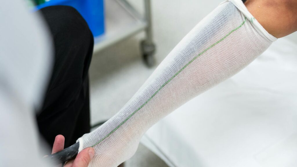
Somebody, usually a physio, will have warned you that leaning on compression or a brace risks making you dependent on it. That warning generally arrives before anybody has checked whether you had the right garment in the first place. Compression is being asked to do two completely different jobs depending on why you reached for it, and mixing them up is the commonest reason it disappoints. For POTS it’s a plumbing job, squeezing enough of you that your heart doesn’t have to work so hard when you stand. For a joint it’s a sensory one, giving the tissue a bit more to report on so your brain isn’t left guessing where the limb has got to. If you’ve got both, you probably need two garments, as a shoulder wrap does nothing for a resting heart rate.
Read MoreOn the POTS side, the clearest work so far put an abdominal garment on and off across stand tests on the same day. With it on, the rise in heart rate on standing came down by a meaningful amount. That’s a same day result with no control group and no blinding though, so it shows the physiology behaving as predicted rather than telling you what a year of wearing one does. The often quoted finding that hardly anybody rates compression as effective is mostly a location problem. Most of that group were wearing leg compression, not anything reaching the abdomen, which is where most of the pooling actually happens.
On the joint side, the largest group followed so far were people with hEDS tracked over two years. Pain came down meaningfully and dislocations fell at several joints. There was no comparison group though, and the garments were funded by the company selling them, which you should weigh accordingly. Adding compression to physiotherapy improved dynamic balance more than physiotherapy alone, while pain improved about the same either way, so that’s a balance finding, not a pain one. How well somebody responded also didn’t track with how hypermobile they were, so this isn’t only for the loosest joints in the room.
The dependency warning is worth picking apart. The effect is there while the garment is on and fades once it comes off, and where that’s been followed up at the shoulder, nothing was left over after the wearing period ended. That isn’t the garment weakening you, it behaves rather like a pair of reading glasses, useful while on and gone once removed. We used to suggest you could test whether compression suited you by watching for exactly that switch. That was a clinical hunch, not a validated test, and the trend underneath it wasn’t significant, so we’ve dropped it.
The Body Braid is a different matter, and the evidence is thinner again. Behind the marketing sits a single unpublished conference poster, sixteen people, one session with it and one without, using garments the manufacturer had donated. Some balance measures and a pain score moved, though it never measured proprioception, which is the exact claim it’s sold on. Running that many tests without correction also leaves a real chance some of it is noise. The fascial network it’s meant to load is anatomically real in cadavers, and whether it transmits any meaningful force in a living body wearing the thing is, by the reviewers’ own account, still an open question. If you own one and it helps, that’s genuinely fine. Just know what you’re buying.
Go deeperCompression Garments for Hypermobility, EDS and POTS: What the Evidence Actually SaysWhat pressure level matters and where, why the biggest survey saying compression fails is measuring the wrong garment, and the very ordinary reason most people give up on theirs before it’s had a fair trial.The Body Braid for Hypermobility: What the Evidence Actually Says, and What It Doesn’tWhat the one piece of research behind the marketing did and didn’t measure, and three things worth paying attention to while you wear it that have nothing to do with what’s printed on the box.
The Pelvic Floor, and Why the Standard Advice Gets Hypermobility Backwards
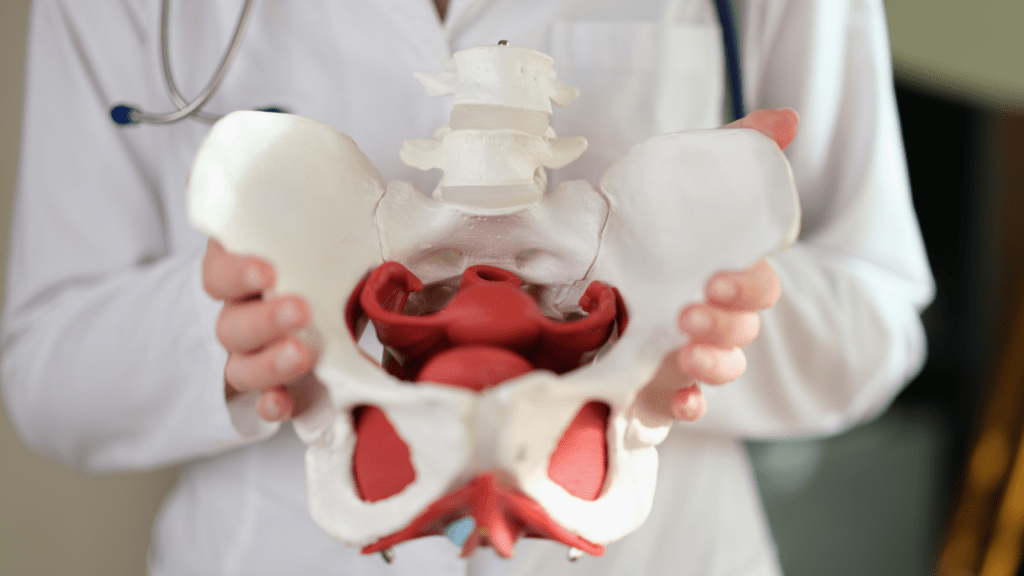
If you’ve been handed a sheet of pelvic floor squeezes, done them three times a day for months, and the urgency hasn’t shifted, the ache after sex is still there and you still don’t feel properly empty afterwards, you probably weren’t doing them wrong. It’s fairly likely you were squeezing the wrong problem. Being hypermobile tends to get read as everything being loose, floor included, so strengthening becomes the default. Often though, when the ligaments and fascia around the pelvis give more than they should, muscle picks up the slack, and a muscle asked to hold on all day eventually stops letting go properly. That isn’t weak, it’s a floor that’s short, tender and half switched on most of the time, and telling it to squeeze harder is a bit like telling a calf that’s already cramping to do more calf raises.
Read MorePelvic floor dysfunction is a label sitting over several different problems, and they don’t all respond to the same treatment. Tissue that isn’t holding as well as it should, muscle that’s overworking, poor sensory feedback from the pelvis and a nervous system running hot can all produce the same list: leaking, urgency, a dragging ache, pain with sex. Because that list overlaps almost completely, you can’t work out from your symptoms alone which one is yours. An internal exam by somebody trained in pelvic health is what actually separates a floor that isn’t being supported enough from one that’s overworking and tender, and those two need opposite programmes.
The overworking pattern is the one we’d flag first, as it’s common and, in our experience, the one that gets missed most often. Where it’s been measured properly, with a scored tenderness exam rather than a questionnaire, the women who came out high tone reported more constant pain, more pain brought on by activity and a harder time with sex. The symptom checklist on its own didn’t sort one group from the other at all. Direct comparisons of pelvic floor tone in hEDS and HSD against people without it are still thin though, so we’d call that a strong working pattern and not a settled finding. Something worth checking for, rather than assuming.
Pain with sex belongs in the same conversation and it deserves to be taken seriously, as it’s common and barely discussed anywhere. Roughly half of women with hEDS or HSD screen positive for probable vulvodynia, against something closer to one in ten women generally. A fair bit of that traces back to the same overworking floor, and not to anything wrong with the tissue. The best supported treatment, for vulvodynia and for pelvic floor pain more widely, is proper pelvic health physiotherapy aimed at releasing and coordinating the muscle, not strengthening it. Head to head, it beat topical numbing cream comfortably. Almost none of that evidence was gathered in hEDS or HSD specifically though, so treat it as a sound borrow rather than a proven result for this exact group.
The same logic runs well beyond sex. Bladder urgency, constipation that no amount of fibre shifts, and prolapse turning up in women in their twenties who’ve never been pregnant and get told they’re far too young for it, all sit on a system that’s either overworking or under supported. None of it gets sorted by guesswork, which is the main argument for getting it looked at properly rather than working off a leaflet.
Go deeperHypermobility, Ehlers Danlos Syndrome and Your Pelvic Floor: A Comprehensive GuideWhich of the several different problems is likely to be yours, what a proper pelvic floor exam actually checks, and why the bladder and bowel tests so often come back reassuringly normal while nothing feels right.Pelvic Floor Pain and Sex: A Practical Guide for Hypermobility, Fibromyalgia and Chronic PainWhat separates entry pain from deep pain, why dilators on their own are a different treatment from the one that works, and what’s worth asking a GP for when the answer so far has been to relax and persevere.
Hypermobile Children and Exercise: What Actually Predicts Trouble
You’ve probably watched her fold herself into a shape you recognise: the same knees that go back too far, the same finger that bends past where it should stop. And felt something between relief that it isn’t just you, and a low grade dread about what you might have passed on. The instinct is to get a Beighton score done and treat the number as a verdict, and it isn’t one. Two children can score identically and live completely differently, one scrambling up everything in sight without a thought and the other tripping over flat pavement. What predicts trouble is whether her nervous system has learned to control the range it’s been handed, not how far a joint travels, and that’s a different question for every child.
Read MoreStart with the distinction doing most of the work here: being hypermobile and being symptomatic are two different things, and it’s mostly the symptomatic group where the research finds anything worth reporting. Ordinary hypermobile schoolchildren, tested in their own classrooms instead of referred anywhere, generally look much like their classmates on movement and attention. That’s one look though, not a settled answer, and not a large one. Anxiety is more stubborn, that link keeps turning up even outside clinic samples. The score itself gets misapplied a fair bit too. Children need a higher cut off than adults do, six instead of four or five, so plenty of ordinary joints get flagged by a test built for grown ups.
The one combination that does seem to matter is a child who was both hypermobile and visibly behind as a baby. That pairing turned up again years later in the few children followed that long, while the ones without the early delay had simply caught up. Treat it as a lead, not a verdict, as the work behind it is small. It’s still a better place for your attention than a Beighton score, which tells you almost nothing about how a child actually moves. Fatigue is worth watching too, more than the aches are, since it has tracked how much of ordinary life a child can manage better than pain has.
There’s no single right exercise. Programmes mixing strength with balance, coordination and ordinary tasks hold up better than any one ingredient alone. Under ten, a structured programme rarely survives contact with an actual child, so play beats protocol. Older children can take an instruction and tell you what they feel, which is where precision earns its keep.
What we’d ask you to be careful of is teaching her that her body is dangerous while trying to keep it safe. A knee allowed to move through its full range in one trial hurt no more than one kept carefully in the middle, which sits oddly against the advice still handed out about never letting a joint go “too far”.
Go deeperHypermobility in children, growing pains and the Beighton scoreGrowing pains turns out to have real diagnostic criteria rather than a shrug, so this walks through the checklist worth taking to an appointment, and why the score your GP quotes may well be using the wrong age group.Hypermobility exercises for childrenNo single exercise fixes a hypermobile child, and the approach changes completely between six and sixteen, so this covers why play beats programmes early on, and what changes once a teenager can actually follow an instruction.Hypermobility and early developmentA hypermobile baby who’s behind on milestones isn’t the same story as one who isn’t, so this goes through what that combination actually predicts, and what it doesn’t.
Exercising With POTS: The First Step Everyone Skips

If you’ve got POTS, the standard advice is almost always more exercise, gently at first and then building towards proper cardio, because the underlying problem is supposedly deconditioning. So you try it, you push through the walking plan or the stationary bike, and instead of building tolerance you end up flat for two days feeling like you’ve been hit by a bus. That’s not you doing it wrong, and it isn’t a motivation problem either. It’s a fair way of getting the whole thing backwards. You weren’t deconditioned first with POTS arriving on top, as the deconditioning came after. Your body was never given the chance to sort out its own oxygen delivery before being asked to move more of it around.
Read MoreThe step that gets missed is carbon dioxide tolerance, which sounds an odd thing to train before a bicep curl, but bear with it. A lot of those with POTS have picked up a pattern of over breathing without ever noticing it, especially once they’re upright or under any kind of stress, and that quietly drags carbon dioxide down further than it should go. Low carbon dioxide does three things. It makes it harder for blood to release oxygen into the tissue that needs it, it narrows blood flow to the brain, and, independent of everything else going on, it pushes heart rate up on its own, by something like seventeen beats a minute where that’s been measured directly. So before the exercise part has even started, the system doing the exercising may already be running short.
Which is presumably a fair bit of why standing exercise programmes for POTS have such a poor completion record. Only around four in ten people finish a standard community exercise programme, and that isn’t a motivation figure, whatever it looks like from the outside. Starting upright hands the hardest problem in the whole condition to a system that hasn’t been given anything to solve it with yet, and when it fails, that tends to get read as the person’s fault rather than the plan’s.
So the order we’d use instead starts somewhere that doesn’t look much like exercise at all. Often a stretch of around two weeks if you’re doing this unsupervised, spent purely on building tolerance to carbon dioxide, mostly through controlled breath holds and nothing cardio. Only once that’s settled does movement come in, and even then it starts low body, tactile and slow, done lying down or reclined instead of standing, with breathing kept through the nose so carbon dioxide doesn’t drop further during the effort.
Cardio itself comes back in a strange looking way: a breath hold slotted into the odd repetition, not a bike or a jog. It adds a small, controlled dose of the very stress that’s normally avoided, without asking the system to cope with all of it standing up. Upright work, and the upper body, only get layered in once the lower body and the breathing hold steady. None of this is offered as something that resolves POTS on its own, only as the piece that decides whether the exercise everyone’s already being told to do stands a chance of being tolerated.
Go deeperPOTS and Exercise: The First Step Everyone MissesThe breathing based step used before anyone with POTS is asked to do a single upright rep of cardio, and why the usual push through the deconditioning advice gets the direction of cause and effect backwards.
Why Fear of Movement in Hypermobility Makes Sense

By the time a shoulder’s come out of its socket reaching into a cupboard for the third time, deciding to be careful about reaching into cupboards isn’t drama, it’s data. Fear of movement in hypermobility gets talked about as though it’s a mindset problem, something to be argued out of with reassurance or pushed through with willpower, and that’s exactly where most advice on it falls down. The textbook version of this fear assumes it’s disproportionate, that the danger passed and the nervous system simply failed to get the memo. Hypermobile joints don’t always give you that tidy ending. Sometimes the shoulder really does go again, and a body that’s learned to brace against something that keeps actually happening isn’t malfunctioning. It’s paying attention.
Read MoreThe clinical term for this is kinesiophobia, an excessive, irrational fear of movement built on a feeling of vulnerability to injury. That word “irrational” is doing a lot of work, though, and it doesn’t hold up cleanly here. Elsewhere in chronic pain research the fear counts as disproportionate by definition, the tissue’s healed, the movement’s safe, so what’s left is fear with nothing underneath it. A joint with a genuine history of coming part way out doesn’t offer that same reassurance. Telling somebody their fear is irrational when the shoulder actually has dislocated, more than once, isn’t correcting a distortion. It’s arguing with their own experience.
Underneath that sits a proprioception problem, and it’s a big part of why the caution feels justified rather than merely nervous. Proprioception is the sense that tells your brain where a joint is without looking, fed by receptors in the muscle and joint capsule. In looser tissue that signal has further to travel before it fires. The evidence points the same way each time it’s been checked, poorer position sense at the knee and elbow, worse the more mobile the joints measure. So the data on where a limb actually is arrives late and vague, and a brain working from that has every reason to be careful. That’s not catastrophising.
This costs something too. One tentative finding, from a single small study we’d hold loosely, is that kinesiophobia here tracks fatigue more closely than pain itself, which fits with how exhausting constant vigilance actually is. Fear of falling looks like its own separate problem, and where that’s been looked at directly it came out as one of the strongest predictors of disability, on a par with pain severity itself. Neither makes fear a side issue to tidy up once the physical work’s done.
None of this sorts neatly into a physical box or a psychological one. It’s biopsychosocial, biology, psychology and circumstance all loading the same system, and none of them makes the fear less earned or the pain less real. It also doesn’t mean every bit of caution needs fixing, as a lot of what looks like avoidance from outside a hypermobile body is actually competent self management, somebody weighing what today’s walk will cost tomorrow. Where the fear itself is the issue, the pattern was the right call when it was learned, usually after years of being told the pain wasn’t real. It just doesn’t update once the situation’s moved on, and that takes proper support rather than willpower.
Go deeperFear of Movement in Hypermobility and EDS: What It Is, Why It Happens, and What Actually HelpsWhy home exercise programmes can fix a shoulder’s function while leaving the fear of it exactly where it started, and how to tell a fear worth treating apart from a decision your body’s already got right.
Fascia and Hypermobility: What Your Hands Can Actually Reach
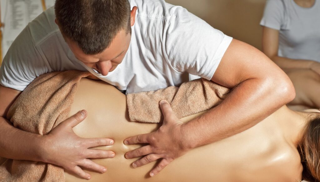
You’ve probably had the good session, a thumb working into your shoulder, or twenty minutes on a foam roller in front of the telly, and for a day or two you actually move better and hurt less. That part isn’t imagined, and it isn’t nothing. What’s harder to square is the explanation you’re usually handed for why it worked, that a stuck or tight layer of fascia has been physically freed. Fascia sits in dense, tough sheets under the skin and muscle, and it doesn’t move nearly as easily as a thumb pressing on it would suggest. The tissue itself is real though, laced with its own nerve endings, and it can hurt in its own right, so nobody is telling you the pain is invented. The trickier question is what a pair of hands is actually reaching, and what they aren’t.
Read MoreStart with hEDS specifically, since that’s the group asking hardest. The imaging that exists finds fascia that’s paradoxically thicker at some sites, with less sliding between its layers, than in people without hypermobility. Nearly the opposite of what you’d expect if hypermobile automatically meant loose everywhere. That’s a genuine and useful correction. It also comes from a small run of studies, cross sectional and mostly out of one research group, so they can show a difference without saying which came first. The altered fascia, or the years of guarding and altered movement that so often come before it.
The releasing part runs straight into physics. Somebody sat down and worked out how much force it would actually take to deform dense fascia, and the awkward part is that the number came out of the fascia world itself rather than from its critics. It’s in the region of 925 kilograms at one of the sites tested. A thumb, an elbow, a foam roller or a metal tool doesn’t get near that. So the tissue almost certainly isn’t being reshaped in the room, whatever it feels like. That doesn’t mean nothing is happening, it means the explanation on offer is wrong about the mechanism, not necessarily about the result.
What’s more plausible is a nervous system story rather than a tissue one. Manual contact can calm a nervous system that’s been running hot, nudge the autonomic balance towards rest, and sharpen the sense of where a joint actually is in space, all worth having in their own right. A fair share of the benefit is context and expectation too, since a convincing sham treatment gets most of the way to matching what the real technique produces in controlled trials. None of that makes the relief fake, it just means a nervous system responding to being touched with intent is doing the work, rather than fascia being physically altered.
A few of the claims riding alongside fascial release hold up even less well. Ask two clinicians to find the same trigger point on the same body and they’ll often disagree about where it even is. Tools sold as breaking down adhesions came out with next to no advantage over other active treatment in the largest analysis available. And the idea that trauma gets physically stored in fascia, waiting to be released by hand, has no trials behind it at all. It also runs into the basic problem that fascia doesn’t contain the neurons you’d need to store anything in.
Go deeperFascia and Hypermobility: What the Research Actually Says, and What’s Being SoldWhat the hEDS imaging actually measured and where its limits sit, the full breakdown of what foam rolling, cupping, dry needling and instrument tools can and can’t do, and why so many of the claims riding alongside fascial release don’t survive contact with the evidence.
Running With Hypermobility: It Isn’t the Flat No You Were Given
Somebody has probably told you not to run, and the chances are they never asked how you land, or how far you’d built up to before saying it. That advice tends to arrive with far more confidence than the evidence supports. Nobody has ever put a hypermobile runner on a force plate and measured what the leg does while it’s moving, not once. Which means the ban you were given, and any confident running plan pitched at hypermobility, are both borrowing from somebody else’s joints. What has been measured properly is what an ordinary landing needs to do in the half second a foot spends on the ground, a far more useful place to start than an argument about whether you’re allowed to run at all.
Read MoreHypermobile covers very different people, which is most of why blanket advice lands so badly. It takes in people who are simply lax and never have a day’s trouble. People with hEDS, which still has no confirmed molecular test, whatever you were told about faulty collagen. People whose laxity followed an injury. And a small number with skeletal conditions where caution is genuinely earned, mostly for reasons at the neck and not the legs. Treating that whole range as one group is how a rule meant for the rarest case reaches everybody. The evidence has actually moved away from avoid sport altogether, towards which activity, how much, and what’s built underneath it first.
The mechanics worth knowing are about what a landing is for, not what it survives. A leg in stance behaves like a spring, taking energy on through the first half of contact and handing a share back to push you along. The ankle and Achilles do most of that work, not the knee. Tendon tends to run more compliant than average in hEDS and HSD, which makes it a shock absorber of sorts as well as a spring. Cadence, a small rise in step rate at the same speed, is the change with the best return, whereas swapping foot strike mostly just moves the load elsewhere.
Impact gets more blame than it earns, as what a watch estimates maps poorly onto the load actually reaching the bone, most of which comes from your own muscles and not the ground. Fatigue is where it does go wrong, dropping leg stiffness and shifting work onto the knee and hip. Set that against a joint position sense that’s often a touch noisier in the symptomatic group already, and our guess, not a proven chain, is that it’s a fair part of why the last stretch catches people out.
None of this has actually been tested in hypermobile runners, worth saying plainly. A pattern learned on a treadmill doesn’t fully transfer to a towpath, so practising somewhere near what you’ll actually run on matters, and softer ground eases things a touch near your limit. There’s a gentler case too for simply being outside while you build it, since time outdoors on its own tracks with better mood and less reported pain across chronic pain groups.
Go deeperRunning with hypermobilityWhat a normal running leg is actually doing to absorb and return force, why the which part of your foot lands first debate is mostly asking the wrong question, and what our studios do differently before load gets added.Outdoor Exercise For FibromyalgiaThe wider case for taking movement outdoors rather than indoors when you’re dealing with chronic pain, and why the social and mental side of it might be doing as much work as the exercise itself.
Scoliosis and Hypermobility: What the Curve Is Actually Telling You
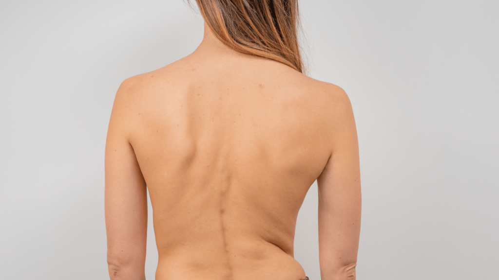
A fair few of those with hypermobility have had the same appointment. A scan turns up a curve in the spine, somebody says the word scoliosis, and from that day on it quietly becomes the explanation for every ache down one side, whether it earned that role or not. The truth is messier than that appointment let on. Scoliosis and hypermobility do turn up together, though how often depends entirely on which sort of hypermobility you’re talking about, and nobody has actually shown that being hypermobile causes the curve. A curve can be real, mild, and still not be the thing doing the damage, while the muscles either side of it are quietly telling a different story, and that’s the part almost nobody explains.
Read MoreThe distinction that actually matters isn’t hypermobile against not hypermobile, it’s between being generally flexible and having a symptomatic connective tissue condition, and then which one. In adults with hypermobile Ehlers-Danlos syndrome, roughly three in ten have a scoliosis, and most of those curves are mild enough that surgery was never on the table. In a rare subtype called musculocontractural EDS it’s closer to two thirds, and the spinal problems there are often severe. So “EDS causes scoliosis” is far too blunt a sentence to mean much on its own. At population level, hypermobile joints by themselves haven’t been convincingly tied to the ordinary teenage sort of scoliosis that turns up with no known cause. A fresh look at that question recently came back unresolved, not settled either way.
The muscles either side of a curve don’t behave the same. The outer, convex side tends to carry more bulk and more of the slow, enduring fibres, while the inner, concave side tends to carry more fat mixed through the muscle and more wasting, worst around the middle of the curve. Take that as a tendency though, not a rule, as the work behind it is small and mostly rated low quality. And which comes first is genuinely not known: a curve holding a muscle in a bad position, or a muscle that’s already stopped doing its job letting the curve happen. Worth sitting with, that same fat and wasting pattern turns up in ordinary persistent back pain with no curve anywhere in sight, so a scan showing wasted muscle next to a curve isn’t automatically telling you the curve is the culprit.
There’s a nervous system side to it too, which gets left out of most conversations. Teenagers with the idiopathic sort of curve measurably lean more on vision to work out where their own trunk is, and their balance is worse. Whether that’s a cause of the curve, or something that develops from living with one for years, is still unresolved. Mice bred with faulty joint position sensing grow curved spines, which is a genuine clue, though mice aren’t small people, so it’s a reason to suspect the mechanism rather than believe it. Hypermobility already comes with a noisier position sense of its own. So a spine that needs good positional feedback, sitting on a system that supplies a patchier version of it, is a plausible way the two problems feed each other. That’s our read though, not a demonstrated finding.
Which leaves two research literatures that have barely spoken to each other. The scoliosis side is full of muscle scans and balance testing, almost entirely in teenagers with no hypermobility diagnosis. The hypermobility side is full of pain, fatigue and walking that costs more energy than it should, and hardly ever puts anybody through a scanner. Very little sits in the middle, which is where you actually are.
Go deeperScoliosis and HypermobilityWhat scoliosis specific exercise has actually been shown to do to a curve, why the position you do an exercise in decides which muscle bothers to help, and why walking itself already costs you more than most programmes account for.
Want this as a programme rather than a reading list?
This guide is the reasoning. The Hypermobile Beginner Bundle is the same thinking built into something you can actually follow, pairing Hypermobility 101 with Arch and Ankle Control, so you start with the signal your brain is working from and the foot it’s coming through, before anything gets loaded. Self paced, no timetable to keep up with, and yours to work through on the days your body allows it.
Explore the Hypermobile Beginner BundleReferences
Every section above is a summary of a longer article, and these are the sources behind the claims in each one, grouped by section. The full reference list for any topic sits on its own article, linked in the blue box in that section.
Why Exercise Is Different When You’re Hypermobile
Scheper, M.C., Juul-Kristensen, B., Rombaut, L., Rameckers, E.A., Verbunt, J. and Engelbert, R.H. (2016) ‘Disability in Adolescents and Adults Diagnosed With Hypermobility-Related Disorders: A Meta-Analysis’, Archives of Physical Medicine and Rehabilitation. https://doi.org/10.1016/j.apmr.2016.02.015
van Meulenbroek, T., Huijnen, I.P.J., Simons, L.E., Conijn, A.E.A., Engelbert, R.H.H. and Verbunt, J.A. (2020) ‘Exploring the underlying mechanism of pain-related disability in hypermobile adolescents with chronic musculoskeletal pain’, Scandinavian Journal of Pain. https://doi.org/10.1515/sjpain-2020-0023
Liaghat, B., Skou, S.T., Søndergaard, J., Boyle, E., Søgaard, K. and Juul-Kristensen, B. (2022) ‘Short-term effectiveness of high-load compared with low-load strengthening exercise on self-reported function in patients with hypermobile shoulders: a randomised controlled trial’, British Journal of Sports Medicine. https://doi.org/10.1136/bjsports-2021-105223
Liaghat, B., Skou, S.T., Jørgensen, U., Sondergaard, J., Søgaard, K. and Juul-Kristensen, B. (2020) ‘Heavy shoulder strengthening exercise in people with hypermobility spectrum disorder (HSD) and long-lasting shoulder symptoms: a feasibility study’, Pilot and Feasibility Studies. https://doi.org/10.1186/s40814-020-00632-y
Liaghat, B., Juul-Kristensen, B., Christensen, F.H., Nissen, S.E., Skou, S.T., Søgaard, K. et al. (2025) ‘Pain trajectories and exercise-induced pain during 16 weeks of high-load or low-load shoulder exercise in patients with hypermobile shoulders: A secondary analysis of a randomized controlled trial’, Scandinavian Journal of Pain. https://doi.org/10.1515/sjpain-2024-0072
Liaghat, B., Juul-Kristensen, B., Faber, D.A., Christensen, E.O., Søgaard, K., Skou, S.T. et al. (2024) ‘One-year effectiveness of high-load compared with low-load strengthening exercise on self-reported function in patients with hypermobile shoulders: a secondary analysis from a randomised controlled trial’, British Journal of Sports Medicine. https://doi.org/10.1136/bjsports-2023-107563
Scheper, M., Rombaut, L., de Vries, J., De Wandele, I., van der Esch, M., Visser, B. et al. (2016) ‘The association between muscle strength and activity limitations in patients with the hypermobility type of Ehlers-Danlos syndrome: the impact of proprioception’, Disability and Rehabilitation. https://doi.org/10.1080/09638288.2016.1196396
Clayton, H.A., Jones, S.A.H. and Henriques, D.Y.P. (2015) ‘Proprioceptive precision is impaired in Ehlers-Danlos syndrome’, SpringerPlus. https://doi.org/10.1186/s40064-015-1089-1
Clayton, H.A., Cressman, E.K. and Henriques, D.Y.P. (2013) ‘Proprioceptive sensitivity in Ehlers-Danlos syndrome patients’, Experimental Brain Research. https://doi.org/10.1007/s00221-013-3656-4
Pacey, V., Adams, R.D., Tofts, L., Munns, C.F. and Nicholson, L.L. (2014) ‘Proprioceptive acuity into knee hypermobile range in children with Joint Hypermobility Syndrome’, Pediatric Rheumatology. https://doi.org/10.1186/1546-0096-12-40
Smith, T.O., Jerman, E., Easton, V., Bacon, H., Armon, K., Poland, F. et al. (2013) ‘Do people with benign joint hypermobility syndrome (BJHS) have reduced joint proprioception? A systematic review and meta-analysis’, Rheumatology International. https://doi.org/10.1007/s00296-013-2790-4
Clayton, H.A., Jones, S.A.H. and Henriques, D.Y.P. (2015) ‘Proprioceptive precision is impaired in Ehlers-Danlos syndrome’, SpringerPlus. https://doi.org/10.1186/s40064-015-1089-1
Mancini, F., Wang, A.P., Schira, M.M., Isherwood, Z.J., McAuley, J.H., Iannetti, G.D. et al. (2019) ‘Fine-Grained Mapping of Cortical Somatotopies in Chronic Complex Regional Pain Syndrome’, The Journal of Neuroscience. https://doi.org/10.1523/jneurosci.2005-18.2019
Dörig, M., Cole, D.M., Guekos, A., Stämpfli, P., Schütz, P., Schibli, L. et al. (2026) ‘Stable Mechanoreceptive Somatotopy in Chronic Low Back Pain: Is Cortical Map Reorganization a Myth?’, The Journal of Neuroscience. https://doi.org/10.1523/jneurosci.2069-25.2026
Delhaye, B.P., Long, K.H. and Bensmaia, S.J. (2018) ‘Neural Basis of Touch and Proprioception in Primate Cortex’, Comprehensive Physiology. https://doi.org/10.1002/j.2040-4603.2018.tb00045.x
Nagel, M. and Chesler, A.T. (2022) ‘PIEZO2 ion channels in proprioception’, Current Opinion in Neurobiology. https://doi.org/10.1016/j.conb.2022.102572
Smith, L., Norcliffe‐Kaufmann, L., Palma, J., Kaufmann, H. and Macefield, V.G. (2020) ‘Elbow proprioception is normal in patients with a congenital absence of functional muscle spindles’, The Journal of Physiology. https://doi.org/10.1113/jp279931
Rombaut, L., De Paepe, A., Malfait, F., Cools, A. and Calders, P. (2009) ‘Joint position sense and vibratory perception sense in patients with Ehlers-Danlos syndrome type III (hypermobility type)’, Clinical Rheumatology. https://doi.org/10.1007/s10067-009-1320-y
Long, M., Kiru, L., Kassam, J., Strutton, P.H. and Alexander, C.M. (2022) ‘An investigation of the control of quadriceps in people who are hypermobile; a case control design. Do the results impact our choice of exercise for people with symptomatic hypermobility?’, BMC Musculoskeletal Disorders. https://doi.org/10.1186/s12891-022-05540-1
Huft, K., Gaydos, E.F., White, J.N., Pascual-Diaz, S., Schubert-Hjalmarsson, E., Harrison, N. et al. (2026) ‘Hypermobility and chronic pain in adolescents: diverging functional and neural profiles without sensory differences’, Pain. https://doi.org/10.1097/j.pain.0000000000003999
Ganguly, J., Kulshreshtha, D., Almotiri, M. and Jog, M. (2021) ‘Muscle Tone Physiology and Abnormalities’, Toxins. https://doi.org/10.3390/toxins13040282
Masi, A.T. and Hannon, J.C. (2008) ‘Human resting muscle tone (HRMT): narrative introduction and modern concepts’, Journal of Bodywork and Movement Therapies. https://doi.org/10.1016/j.jbmt.2008.05.007
Cacciatore, T.W., Anderson, D.I. and Cohen, R.G. (2024) ‘Central mechanisms of muscle tone regulation: implications for pain and performance’, Frontiers in Neuroscience. https://doi.org/10.3389/fnins.2024.1511783
Rombaut, L., Malfait, F., De Wandele, I., Mahieu, N., Thijs, Y., Segers, P. et al. (2012) ‘Muscle-tendon tissue properties in the hypermobility type of Ehlers-Danlos syndrome’, Arthritis Care and Research. https://doi.org/10.1002/acr.21592
Tas, S., Dikici, T.F., Aktas, A. and Araci, A. (2021) ‘Mechanical Properties of Muscles and Tendons in Asymptomatic Individuals with Generalized Joint Hypermobility’, Muscle Ligaments and Tendons Journal. https://doi.org/10.32098/mltj.04.2021.05
Long, M., Kiru, L., Kassam, J., Strutton, P. and Alexander, C. (2022) ‘An investigation of the control of quadriceps in people who are hypermobile; a case control design’, BMC Musculoskeletal Disorders. https://doi.org/10.1186/s12891-022-05540-1
Rombaut, L., Malfait, F., De Wandele, I., Taes, Y., Thijs, Y., De Paepe, A. et al. (2012) ‘Muscle mass, muscle strength, functional performance, and physical impairment in women with the hypermobility type of Ehlers-Danlos syndrome’, Arthritis Care and Research. https://doi.org/10.1002/acr.21726
Coussens, M., Calders, P., Lapauw, B., Celie, B., Banica, T., De Wandele, I. et al. (2020) ‘Does Muscle Strength Change Over Time in Patients With Hypermobile Ehlers-Danlos Syndrome/Hypermobility Spectrum Disorder? An Eight-Year Follow-Up Study’, Arthritis Care and Research. https://doi.org/10.1002/acr.24220
Coussens, M., Lapauw, B., Banica, T., De Wandele, I., Pacey, V., Rombaut, L. et al. (2022) ‘Muscle Strength, Muscle Mass and Physical Impairment in Women with hypermobile Ehlers-Danlos syndrome and Hypermobility Spectrum Disorder’, Journal of Musculoskeletal and Neuronal Interactions.
Luder, G., Aeberli, D., Mebes, C., Haupt-Bertschy, B., Baeyens, J. and Verra, M. (2021) ‘Effect of resistance training on muscle properties and function in women with generalized joint hypermobility: a single-blind pragmatic randomized controlled trial’, BMC Sports Science, Medicine and Rehabilitation. https://doi.org/10.1186/s13102-021-00238-8
Therrien, A.S. and Wong, A.L. (2022) ‘Mechanisms of Human Motor Learning Do Not Function Independently’, Frontiers in Human Neuroscience. https://doi.org/10.3389/fnhum.2021.785992
Spampinato, D. and Celnik, P. (2020) ‘Multiple Motor Learning Processes in Humans: Defining Their Neurophysiological Bases’, The Neuroscientist. https://doi.org/10.1177/1073858420939552
Taylor, J.A., Krakauer, J.W. and Ivry, R.B. (2014) ‘Explicit and Implicit Contributions to Learning in a Sensorimotor Adaptation Task’, The Journal of Neuroscience. https://doi.org/10.1523/jneurosci.3619-13.2014
Schmidt, R.A. (1975) ‘A schema theory of discrete motor skill learning’, Psychological Review. https://doi.org/10.1037/h0076770
Van Rossum, J.H.A. (1990) ‘Schmidt’s schema theory: the empirical base of the variability of practice hypothesis’, Human Movement Science. https://doi.org/10.1016/0167-9457(90)90010-b
Schmidt, M., Kemena, M. and Jaitner, T. (2021) ‘Null Effects of Different Amounts of Task Variation in Both Contextual Interference and Differential Learning Paradigms’, Perceptual and Motor Skills. https://doi.org/10.1177/00315125211022302
Tsay, J.S., Kim, H.E., McDougle, S.D., Taylor, J.A., Haith, A., Avraham, G. et al. (2024) ‘Fundamental processes in sensorimotor learning: Reasoning, refinement, and retrieval’, eLife. https://doi.org/10.7554/elife.91839
Hodges, P.W. (2011) ‘Pain and motor control: From the laboratory to rehabilitation’, Journal of Electromyography and Kinesiology. https://doi.org/10.1016/j.jelekin.2011.01.002
van Dieen, J.H., Flor, H. and Hodges, P.W. (2017) ‘Low-Back Pain Patients Learn to Adapt Motor Behavior With Adverse Secondary Consequences’, Exercise and Sport Sciences Reviews. https://doi.org/10.1249/jes.0000000000000121
Core Stability
Lederman E (2010) ‘The myth of core stability’, Journal of Bodywork and Movement Therapies, 14(1), pp. 84-98. doi: 10.1016/j.jbmt.2009.08.001
Hodges PW, Richardson CA (1996) ‘Inefficient muscular stabilization of the lumbar spine associated with low back pain: a motor control evaluation of transversus abdominis’, Spine, 21(22), pp. 2640-2650. doi: 10.1097/00007632-199611150-00014
Hodges PW, Moseley GL, Gabrielsson A, Gandevia SC (2003) ‘Experimental muscle pain changes feedforward postural responses of the trunk muscles’, Experimental Brain Research, 151(2), pp. 262-271. doi: 10.1007/s00221-003-1457-x
Saragiotto BT, Maher CG, Yamato TP, Costa LOP, Menezes Costa LC, Ostelo RWJG, Macedo LG (2016) ‘Motor control exercise for chronic non-specific low-back pain’, Cochrane Database of Systematic Reviews, (1), CD012004. doi: 10.1002/14651858.CD012004
Mannion AF, Caporaso F, Pulkovski N, Sprott H (2012) ‘Spine stabilisation exercises in the treatment of chronic low back pain: a good clinical outcome is not associated with improved abdominal muscle function’, European Spine Journal, 21(7), pp. 1301-1310. doi: 10.1007/s00586-012-2155-9
Smith BE, Littlewood C, May S (2014) ‘An update of stabilisation exercises for low back pain: a systematic review with meta-analysis’, BMC Musculoskeletal Disorders, 15(1), p. 416. doi: 10.1186/1471-2474-15-416
Wong AY, Parent EC, Funabashi M, Kawchuk GN (2014) ‘Do changes in transversus abdominis and lumbar multifidus during conservative treatment explain changes in clinical outcomes related to nonspecific low back pain? A systematic review’, The Journal of Pain, 15(4), pp. 377.e1-377.e35. doi: 10.1016/j.jpain.2013.10.008
Reeves NP, Narendra KS, Cholewicki J (2007) ‘Spine stability: the six blind men and the elephant’, Clinical Biomechanics, 22(3), pp. 266-274. doi: 10.1016/j.clinbiomech.2006.11.011
Mulholland RC (2008) ‘The myth of lumbar instability: the importance of abnormal loading as a cause of low back pain’, European Spine Journal, 17(5), pp. 619-625. doi: 10.1007/s00586-008-0612-2
Long M, Kiru L, Kassam J, Strutton PH, Alexander CM (2022) ‘An investigation of the control of quadriceps in people who are hypermobile; a case control design’, BMC Musculoskeletal Disorders, 23(1), p. 607. doi: 10.1186/s12891-022-05540-1
Scheper MC, Pacey V, Rombaut L, Adams RD, Tofts L, Calders P (2017) ‘Generalized hyperalgesia in children and adults diagnosed with hypermobility syndrome and Ehlers-Danlos syndrome hypermobility type: a discriminative analysis’, Arthritis Care & Research, 69(3), pp. 421-429. doi: 10.1002/acr.22998
Hakimi A, Bergoin C, De Jesus A, Hermand E, Fabre C, Mucci P (2024) ‘Impairment of lung volume perception and breathing control in hypermobile Ehlers-Danlos syndrome’, Scientific Reports, 14(1). doi: 10.1038/s41598-024-58890-2
Faubion SS, Shuster LT, Bharucha AE (2012) ‘Recognition and management of nonrelaxing pelvic floor dysfunction’, Mayo Clinic Proceedings, 87(2), pp. 187-193. doi: 10.1016/j.mayocp.2011.09.004
Talasz H, Kremser C, Talasz HJ, Kofler M, Rudisch A (2022) ‘Breathing, (S)Training and the Pelvic Floor: a basic concept’, Healthcare, 10(6), p. 1035. doi: 10.3390/healthcare10061035
Hwang U, Lee M, Jung S, Ahn S, Kwon O (2021) ‘Effect of pelvic floor electrical stimulation on diaphragm excursion and rib cage movement during tidal and forceful breathing and coughing in women with stress urinary incontinence’, Medicine, 100(1), p. e24158. doi: 10.1097/MD.0000000000024158
Bastiaenen CHG, de Bie RA, Vlaeyen JWS, Goossens MEJB, Leffers P, Wolters PMJC, Bastiaanssen JM, van den Brandt PA, Essed GGM (2008) ‘Long-term effectiveness and costs of a brief self-management intervention in women with pregnancy-related low back pain after delivery’, BMC Pregnancy and Childbirth, 8(1), p. 19. doi: 10.1186/1471-2393-8-19
Chohan K, Mittal N, McGillis L, Lopez-Hernandez L, Camacho E, Rachinsky M, Santa Mina D, Reid WD, Ryan CM, Champagne KA, Orchanian-Cheff A, Clarke H, Rozenberg D (2021) ‘A review of respiratory manifestations and their management in Ehlers-Danlos syndromes and hypermobility spectrum disorders’, Chronic Respiratory Disease, 18. doi: 10.1177/14799731211025313
Pacing
Jamieson-Lega, K., Berry, R. and Brown, C.A. (2013) ‘Pacing: A concept analysis of a chronic pain intervention’, Pain Research and Management, 18(4), pp. 207-213. doi: 10.1155/2013/686179
Antcliff, D., Keeley, P., Campbell, M., Woby, S., Keenan, A.-M. and McGowan, L. (2018) ‘Activity pacing: moving beyond taking breaks and slowing down’, Quality of Life Research, 27(7), pp. 1933-1935. doi: 10.1007/s11136-018-1794-7
Antcliff, D., Keenan, A., Keeley, P., Woby, S. and McGowan, L. (2021) ‘Testing a newly developed activity pacing framework for chronic pain/fatigue: a feasibility study’, BMJ Open, 11(12):e045398. doi: 10.1136/bmjopen-2020-045398
Murphy, S.L. and Kratz, A.L. (2014) ‘Activity pacing in daily life: A within-day analysis’, Pain, 155(12), pp. 2630-2637. doi: 10.1016/j.pain.2014.09.028
Murphy, S.L. (2015) ‘Overactivity in daily life: a crucial step in understanding how to tailor treatments’, Pain, 156(10), pp. 1831-1832. doi: 10.1097/j.pain.0000000000000291
Barakou, I., Hackett, K.L., Finch, T. and Hettinga, F.J. (2023) ‘Self-regulation of effort for a better health-related quality of life: a multidimensional activity pacing model for chronic pain and fatigue management’, Annals of Medicine, 55(2):2270688. doi: 10.1080/07853890.2023.2270688
Woolf, C.J. (2011) ‘Central sensitization: Implications for the diagnosis and treatment of pain’, Pain, 152(3 Suppl), pp. S2-S15. doi: 10.1016/j.pain.2010.09.030
Williams, D.A. and Clauw, D.J. (2009) ‘Understanding fibromyalgia: lessons from the broader pain research community’, The Journal of Pain, 10(8), pp. 777-791. doi: 10.1016/j.jpain.2009.06.001
Galvez-Sánchez, C.M., Muñoz Ladrón de Guevara, C., Montoro, C.I., Fernández-Serrano, M.J., Duschek, S. and Reyes del Paso, G.A. (2018) ‘Cognitive deficits in fibromyalgia syndrome are associated with pain responses to low intensity pressure stimulation’, PLoS One, 13(8), e0201488. doi: 10.1371/journal.pone.0201488
Geraghty, A.W.A. et al. (2021) ‘Self-management for chronic widespread pain including fibromyalgia: A systematic review and meta-analysis’, PLoS One, 16(7), e0254642. doi: 10.1371/journal.pone.0254642
Scheper, M., Rombaut, L., de Vries, J., De Wandele, I., van der Esch, M., Visser, B., Malfait, F., Calders, P. and Engelbert, R. (2017) ‘The association between muscle strength and activity limitations in patients with the hypermobility type of Ehlers-Danlos syndrome: the impact of proprioception’, Disability and Rehabilitation, 39(14), pp. 1391-1397. doi: 10.1080/09638288.2016.1196396
Ruiz Maya, T., Fettig, V., Mehta, L., Gelb, B.D. and Kontorovich, A.R. (2021) ‘Dysautonomia in hypermobile Ehlers-Danlos syndrome and hypermobility spectrum disorders is associated with exercise intolerance and cardiac atrophy’, American Journal of Medical Genetics Part A, 185(12), pp. 3754-3761. doi: 10.1002/ajmg.a.62446
Fu, Q. and Levine, B.D. (2018) ‘Exercise and non-pharmacological treatment of POTS’, Autonomic Neuroscience, 215, pp. 20-27. doi: 10.1016/j.autneu.2018.07.001
Vøllestad, N.K. and Mengshoel, A.M. (2023) ‘Post-exertional malaise in daily life and experimental exercise models in patients with myalgic encephalomyelitis/chronic fatigue syndrome’, Frontiers in Physiology, 14, 1257557. doi: 10.3389/fphys.2023.1257557
Sanal-Hayes, N.E.M. et al. (2023) ‘A scoping review of “Pacing” for management of Myalgic Encephalomyelitis/Chronic Fatigue Syndrome (ME/CFS): lessons learned for the long COVID pandemic’, Journal of Translational Medicine, 21:720. doi: 10.1186/s12967-023-04587-5
Jason, L.A. et al. (2013) ‘Energy conservation/envelope theory interventions’, Fatigue: Biomedicine, Health & Behavior, 1(1-2), pp. 27-42. doi: 10.1080/21641846.2012.733602
Wormgoor, M. and Rodenburg, S.C. (2021) ‘The evidence base for physiotherapy in myalgic encephalomyelitis/chronic fatigue syndrome when considering post-exertional malaise: a systematic review and narrative synthesis’, Journal of Translational Medicine, 19(1), 1. doi: 10.1186/s12967-020-02683-4
National Institute for Health and Care Excellence (2021) ‘Myalgic encephalomyelitis (or encephalopathy)/chronic fatigue syndrome: diagnosis and management’, NICE guideline NG206, published 29 October 2021. Recommendation 1.11.14 and Box 4. Available at: https://www.nice.org.uk/guidance/ng206
Sanal-Hayes, N.E.M. et al. (2025) ‘“Pacing” for management of myalgic encephalomyelitis/chronic fatigue syndrome (ME/CFS): a systematic review and meta-analysis’, Fatigue: Biomedicine, Health & Behavior, 13(1), pp. 36-53. doi: 10.1080/21641846.2024.2433390
Foot Arches
Strzalkowski, N.D.J., Peters, R.M., Inglis, J.T. and Bent, L.R. (2018) ‘Cutaneous afferent innervation of the human foot sole: what can we learn from single-unit recordings?’, Journal of Neurophysiology. https://doi.org/10.1152/jn.00848.2017
Katic, N., Siqueira, R.K., Cleland, L., Strzalkowski, N., Bent, L., Raspopovic, S. et al. (2023) ‘Modeling foot sole cutaneous afferents: FootSim’, iScience. https://doi.org/10.1016/j.isci.2022.105874
Vermeulen, S., De Mits, S., De Ridder, R., Calders, P., De Schepper, J., Malfait, F. et al. (2022) ‘Altered Multisegment Ankle and Foot Kinematics During Gait in Patients With Hypermobile Ehlers‐Danlos Syndrome/Hypermobility Spectrum Disorder: A Case-Control Study’, Arthritis Care & Research. https://doi.org/10.1002/acr.24526
Alahmari, K.A., Kakaraparthi, V.N., Reddy, R.S., Samuel, P.S., Tedla, J.S., Rengaramanujam, K. et al. (2021) ‘Foot Posture Index Reference Values among Young Adults in Saudi Arabia and Their Association with Anthropometric Determinants, Balance, Functional Mobility, and Hypermobility’, BioMed Research International. https://doi.org/10.1155/2021/8844356
Martinez-Sebastian, C., De La Fuente, M.C.C., Gijon-Nogueron, G., Gomez-Carrion, A., Ramos-Petersen, L. and Evans, A.M.F. (2026) ‘Ankle muscle strength, joint hypermobility, and foot posture in children aged 5-10 years: A cross-sectional study’, European Journal of Pediatrics. https://doi.org/10.1007/s00431-026-07313-7
Fischer, K.M., Willwacher, S., Arndt, A. and Brüggemann, G. (2018) ‘Calcaneal adduction and eversion are coupled to talus and tibial rotation’, Journal of Anatomy. https://doi.org/10.1111/joa.12813
Jaffri, A.H., Koldenhoven, R., Saliba, S. and Hertel, J. (2023) ‘Evidence for Intrinsic Foot Muscle Training in Improving Foot Function: A Systematic Review and Meta-Analysis’, Journal of Athletic Training. https://doi.org/10.4085/1062-6050-0162.22
Balsdon, M., Dombroski, C., Bushey, K. and Jenkyn, T.R. (2019) ‘Hard, soft and off-the-shelf foot orthoses and their effect on the angle of the medial longitudinal arch’, Prosthetics & Orthotics International. https://doi.org/10.1177/0309364619825607
Hoang, N.T.T., Chen, S. and Chou, L.W. (2021) ‘The Impact of Foot Orthoses and Exercises on Pain and Navicular Drop for Adult Flatfoot: A Network Meta-Analysis’, International Journal of Environmental Research and Public Health. https://doi.org/10.3390/ijerph18158063
Protopapas, K. and Perry, S.D. (2020) ‘The effect of a 12-week custom foot orthotic intervention on muscle size and muscle activity of the intrinsic foot muscle of young adults during gait termination’, Clinical Biomechanics. https://doi.org/10.1016/j.clinbiomech.2020.105063
Hikawa, K., Tsutsui, T., Ueyama, T., Yang, J., Hara, Y. and Torii, S. (2022) ‘Effects of a 9-weeks arch support intervention on foot morphology in young soccer players: a crossover study’, BMC Sports Science, Medicine and Rehabilitation. https://doi.org/10.1186/s13102-022-00590-3
Reeves, J., Jones, R., Liu, A., Bent, L., Martinez-Santos, A. and Nester, C. (2021) ‘No change in foot soft tissue morphology and skin sensitivity after three months of using foot orthoses that alter plantar pressure’, Footwear Science. https://doi.org/10.1080/19424280.2021.1961880
ELSAYED, W., ALOTAIBI, S., SHAHEEN, A., FAROUK, M. and FARRAG, A. (2023) ‘The combined effect of short foot exercises and orthosis in symptomatic flexible flatfoot: a randomized controlled trial’, European Journal of Physical and Rehabilitation Medicine. https://doi.org/10.23736/s1973-9087.23.07846-2
Peterson, B., Coda, A., Pacey, V. and Hawke, F. (2018) ‘Physical and mechanical therapies for lower limb symptoms in children with Hypermobility Spectrum Disorder and Hypermobile Ehlers‐Danlos Syndrome: a systematic review’, Journal of Foot and Ankle Research. https://doi.org/10.1186/s13047-018-0302-1
Yildiz, R., Yildiz, A., Camli, O., Akkaya, H., Aydin, M. and Basaran, Z. (2025) ‘Effect of Plantar Sensory Stimulation on Sensorimotor Organization in General Joint Hypermobility: A Randomized Controlled Study’, Healthcare. https://doi.org/10.3390/healthcare13202572
Nutrition
. So for anyone with hypermobility who has trouble around food, there are usually a few roadblocks in the way of nutrition, including the ones behind EDS weight gain and loss. They can include:
Lam, C.Y., Palsson, O.S., Whitehead, W.E., Sperber, A.D., Tornblom, H., Simren, M. et al. (2021) ‘Rome IV Functional Gastrointestinal Disorders and Health Impairment in Subjects With Hypermobility Spectrum Disorders or Hypermobile Ehlers-Danlos Syndrome’, Clinical Gastroenterology and Hepatology. https://doi.org/10.1016/j.cgh.2020.02.034
. Be clear on where the evidence sits, though: reviews of mast cell activation and nutrition in hypermobility describe the links as plausible and the evidence base as limited[7].
. There is no trial showing that pushing protein higher improves pain, joint stability or connective tissue in hypermobility, so the target is meeting your requirement, not exceeding it.
Alves, C.R.R., Santiago, B.M., Lima, F.R., Otaduy, M.C.G., Calich, A.L., Tritto, A.C.C. et al. (2013) ‘Creatine Supplementation in Fibromyalgia: A Randomized, Double‐Blind, Placebo‐Controlled Trial’, Arthritis Care & Research. https://doi.org/10.1002/acr.22020
Salazar-Méndez, J., Núñez-Cortés, R., Salazar-Orellana, C., Suso-Martí, L., Calatayud, J., Cuyul-Vásquez, I. et al. (2026) ‘Impact of creatine supplementation alone or combined with exercise on inflammatory and clinical outcomes in chronic musculoskeletal pain treated in the rheumatological field: a systematic review’, Nutrition. https://doi.org/10.1016/j.nut.2026.113330
Bonilla, D.A., Kreider, R.B., Stout, J.R., Forero, D.A., Kerksick, C.M., Roberts, M.D. et al. (2021) ‘Metabolic Basis of Creatine in Health and Disease: A Bioinformatics-Assisted Review’, Nutrients. https://doi.org/10.3390/nu13041238
Batti, K.S., Kim, M.J., Lewis, J. and Emerick, T. (2026) ‘Can creatine help for pain? Creatine’s potential role as an adjunctive treatment in pain management’, Pain Medicine. https://doi.org/10.1093/pm/pnag058
Dolan, E., Gualano, B. and Rawson, E.S. (2018) ‘Beyond muscle: the effects of creatine supplementation on brain creatine, cognitive processing, and traumatic brain injury’, European Journal of Sport Science. https://doi.org/10.1080/17461391.2018.1500644
de Souza e Silva, A., Pertille, A., Reis Barbosa, C.G., Aparecida de Oliveira Silva, J., de Jesus, D.V., Ribeiro, A.G.S.V. et al. (2019) ‘Effects of Creatine Supplementation on Renal Function: A Systematic Review and Meta-Analysis’, Journal of Renal Nutrition. https://doi.org/10.1053/j.jrn.2019.05.004
Longobardi, I., Gualano, B., Seguro, A.C. and Roschel, H. (2023) ‘Is It Time for a Requiem for Creatine Supplementation-Induced Kidney Failure? A Narrative Review’, Nutrients. https://doi.org/10.3390/nu15061466
Balestrino, M. and Adriano, E. (2019) ‘Beyond sports: Efficacy and safety of creatine supplementation in pathological or paraphysiological conditions of brain and muscle’, Medicinal Research Reviews. https://doi.org/10.1002/med.21590
Longobardi, I., Solis, M.Y., Roschel, H. and Gualano, B. (2025) ‘A short review of the most common safety concerns regarding creatine ingestion’, Frontiers in Nutrition. https://doi.org/10.3389/fnut.2025.1682746
de Camargo, K.M.R., Bruna-Mejías, A., Valenzuela-Fuenzalida, J.J., Gonzaga, L.A., Barbalho, S.M., Barroca, A.L. et al. (2026) ‘Impact of creatine supplementation on inflammation: evidence from a systematic review and meta-analysis of randomized double-blind placebo trials’, Frontiers in Immunology. https://doi.org/10.3389/fimmu.2026.1743603
KT Tape
Yam, M.L., Yang, Z., Zee, B.C.-Y. and Chong, K.C. (2019). Effects of Kinesio tape on lower limb muscle strength, hop test, and vertical jump performances: a meta-analysis. BMC Musculoskeletal Disorders, 20(1). doi: 10.1186/s12891-019-2564-6
Parreira, P. do C.S., Costa, L. da C.M., Hespanhol Junior, L.C., Lopes, A.D. and Costa, L.O.P. (2014). Current evidence does not support the use of Kinesio Taping in clinical practice: a systematic review. Journal of Physiotherapy, 60(1), pp.31-39. doi: 10.1016/j.jphys.2013.12.008
Tudini, F., Levine, D., Chui, K.K., Jordon, M.K. and Healy, M. (2023). Evaluating the effects of two different kinesiology taping techniques on shoulder pain and function in patients with hypermobile Ehlers-Danlos syndrome. Frontiers in Pain Research, 4:1089748. doi: 10.3389/fpain.2023.1089748
Tudini, F., Jordon, M., Levine, D., Healy, M., Cathey, S. and Chui, K.K. (2024). Evaluating the effects of two different kinesiology taping techniques on shoulder range of motion and proprioception in patients with hypermobile Ehlers-Danlos syndrome: a randomized controlled trial. Frontiers in Rehabilitation Sciences, 5:1383551. doi: 10.3389/fresc.2024.1383551
Ghai, I., Ghai, S. and Narciss, S. (2024). Influence of taping on joint proprioception: a systematic review with between and within group meta-analysis. BMC Musculoskeletal Disorders, 25:478. doi: 10.1186/s12891-024-07571-2
de Oliveira, F.C.L., Pairot de Fontenay, B., Bouyer, L.J., Desmeules, F. and Roy, J.-S. (2020). Kinesiotaping for the Rehabilitation of Rotator Cuff-Related Shoulder Pain: A Randomized Clinical Trial. Sports Health, 13(2), pp.161-172. doi: 10.1177/1941738120944254
Sleep
Staffe, A.T., Bech, M.W., Clemmensen, S.L.K., Nielsen, H.T., Larsen, D.B. and Petersen, K.K. (2019) ‘Total sleep deprivation increases pain sensitivity, impairs conditioned pain modulation and facilitates temporal summation of pain in healthy participants’, PLOS ONE. https://doi.org/10.1371/journal.pone.0225849
Irwin, M.R., Olmstead, R., Bjurstrom, M.F., Finan, P.H. and Smith, M.T. (2022) ‘Sleep disruption and activation of cellular inflammation mediate heightened pain sensitivity: a randomized clinical trial’, Pain. https://doi.org/10.1097/j.pain.0000000000002811
Pejovic, S., Basta, M., Vgontzas, A.N., Kritikou, I., Shaffer, M.L., Tsaoussoglou, M. et al. (2013) ‘Effects of recovery sleep after one work week of mild sleep restriction on interleukin-6 and cortisol secretion and daytime sleepiness and performance’, American Journal of Physiology-Endocrinology and Metabolism. https://doi.org/10.1152/ajpendo.00301.2013
Simpson, N.S., Diolombi, M., Scott-Sutherland, J., Yang, H., Bhatt, V., Gautam, S. et al. (2016) ‘Repeating patterns of sleep restriction and recovery: Do we get used to it?’, Brain, Behavior, and Immunity. https://doi.org/10.1016/j.bbi.2016.06.001
Cheng, J., Zhu, K., Wang, J., Huang, Q., Shen, J., Xiong, Y. et al. (2026) ‘Transcriptomic recovery and persistence patterns reveal the biological cost of sleep debt in healthy adult males’, SLEEP. https://doi.org/10.1093/sleep/zsag071
Wiklund, T., Gerdle, B., Linton, S.J., Dragioti, E. and Larsson, B. (2020) ‘Insomnia is a risk factor for spreading of chronic pain: A Swedish longitudinal population study (SwePain)’, European Journal of Pain. https://doi.org/10.1002/ejp.1582
Darakjian, A.A., Bhutani, M., Fairweather, D., Kocsis, S.C., Fliess, J.J., Khatib, S. et al. (2024) ‘Similarities and differences in self-reported symptoms and comorbidities between hypermobile Ehlers-Danlos syndrome and hypermobility spectrum disorders’, Rheumatology Advances in Practice. https://doi.org/10.1093/rap/rkae134
Crews-Stowe, C., Tudini, F., Jung, M.K., Forman, J., Riley, B., Eton, S. et al. (2025) ‘Sleep Characteristics in Individuals with Ehlers-Danlos Syndrome’, Medical Sciences. https://doi.org/10.3390/medsci13030085
Coussens, M., De Wandele, I., Pacey, V., Malfait, F., De Craemer, M., Demeyer, H. et al. (2022) ‘Physical activity and sleep in patients with hypermobile Ehlers-Danlos syndrome and patients with generalized hypermobility spectrum disorder’, Edorium Journal of Disability and Rehabilitation. https://doi.org/10.5348/100049d05mc2020ra
Gaisl, T., Giunta, C., Bratton, D.J., Sutherland, K., Schlatzer, C., Sievi, N. et al. (2017) ‘Obstructive sleep apnoea and quality of life in Ehlers-Danlos syndrome: a parallel cohort study’, Thorax. https://doi.org/10.1136/thoraxjnl-2016-209560
Zhang, K.L., Wallon, R., Laukaitis, C.M. and Davies, C. (2026) ‘Obstructive sleep apnea and CPAP efficacy in patients with Hypermobile Ehlers-Danlos syndrome and hypermobility spectrum disorder: a case-control study’, Sleep and Breathing. https://doi.org/10.1007/s11325-026-03763-3
Collins Hutchinson, M.L., Liang, E., Fuster, E. and Blitshteyn, S. (2025) ‘Autonomic symptom burden, comorbidities and quality of life in women with Hypermobility Spectrum Disorders and hypermobile Ehlers-Danlos syndrome’, Autonomic Neuroscience. https://doi.org/10.1016/j.autneu.2025.103356
Cai, H., Wang, S., Zou, R., Wang, Y. and Wang, C. (2020) ‘Circadian rhythms of blood pressure and rate pressure product in children with postural tachycardia syndrome’, Autonomic Neuroscience. https://doi.org/10.1016/j.autneu.2020.102715
Selvanathan, J., Pham, C., Nagappa, M., Peng, P.W.H., Englesakis, M., Espie, C.A. et al. (2021) ‘Cognitive behavioral therapy for insomnia in patients with chronic pain, a systematic review and meta-analysis of randomized controlled trials’, Sleep Medicine Reviews. https://doi.org/10.1016/j.smrv.2021.101460
Wu, K., Ho, T.H., Beckenkamp, P.R., Hall, M., Zhang, H., Puterflam, J. et al. (2026) ‘Efficacy and effectiveness of melatonin for the management of musculoskeletal pain: a systematic review and meta-analysis of placebo and active controlled trials’, Pain. https://doi.org/10.1097/j.pain.0000000000004045
Esquivel, M.K. and Ghosn, B. (2024) ‘Current Evidence on Common Dietary Supplements for Sleep Quality’, American Journal of Lifestyle Medicine. https://doi.org/10.1177/15598276241227915
Bulman, A., D’Cunha, N.M., Marx, W., Turner, M., McKune, A. and Naumovski, N. (2025) ‘The effects of L-theanine consumption on sleep outcomes: A systematic review and meta-analysis’, Sleep Medicine Reviews. https://doi.org/10.1016/j.smrv.2025.102076
Gutiérrez-Romero, S.A., Torres-Narváez, E.S., Zamora-Gómez, A.C., Castillo-Castillo, S., Latorre-Velásquez, A.L., Betancourt-Villamizar, C. et al. (2024) ‘Effect of a nutraceutical combination on sleep quality among people with impaired sleep: a randomised, placebo-controlled trial’, Scientific Reports. https://doi.org/10.1038/s41598-024-58661-z
Stretching
Konrad, A., Alizadeh, S., Daneshjoo, A., Anvar, S.H., Graham, A., Zahiri, A. et al. (2024) ‘Chronic effects of stretching on range of motion with consideration of potential moderating variables: A systematic review with meta-analysis’, Journal of Sport and Health Science. https://doi.org/10.1016/j.jshs.2023.06.002
Ingram, L.A., Tomkinson, G.R., d’Unienville, N.M.A., Gower, B., Gleadhill, S., Boyle, T. et al. (2024) ‘Optimising the Dose of Static Stretching to Improve Flexibility: A Systematic Review, Meta-analysis and Multivariate Meta-regression’, Sports Medicine. https://doi.org/10.1007/s40279-024-02143-9
Oba, K., Matsuo, S., Nakamura, M., Nakao, G., Fukaya, T., Mizuno, T. et al. (2026) ‘Moderating Effects of Individual Characteristics and the Target Lower Limb Muscle Group on Flexibility Adaptations to Chronic Static Stretching in Healthy Individuals: A Systematic Review and Meta-Analysis of Randomized Controlled Trials’, Sports Medicine – Open. https://doi.org/10.1186/s40798-026-01066-1
Ingram, L.A., Tomkinson, G.R., d’Unienville, N.M.A., Gower, B., Gleadhill, S., Boyle, T. et al. (2025) ‘Mechanisms Underlying Range of Motion Improvements Following Acute and Chronic Static Stretching: A Systematic Review, Meta-analysis and Multivariate Meta-regression’, Sports Medicine. https://doi.org/10.1007/s40279-025-02204-7
Konrad, A. and Tilp, M. (2014) ‘Increased range of motion after static stretching is not due to changes in muscle and tendon structures’, Clinical Biomechanics. https://doi.org/10.1016/j.clinbiomech.2014.04.013
Bishop, M. and George, S. (2017) ‘Pain sensitivity and torque used during measurement predicts change in range of motion at the knee’, Journal of Pain Research. https://doi.org/10.2147/JPR.S150775
Panidi, I., Donti, O., Konrad, A., Dinas, P.C., Terzis, G., Mouratidis, A. et al. (2023) ‘Muscle Architecture Adaptations to Static Stretching Training: A Systematic Review with Meta-Analysis’, Sports Medicine – Open. https://doi.org/10.1186/s40798-023-00591-7
Williams, P.E. (1990) ‘Use of intermittent stretch in the prevention of serial sarcomere loss in immobilised muscle.’, Annals of the Rheumatic Diseases. https://doi.org/10.1136/ard.49.5.316
Tabary, J.C., Tabary, C., Tardieu, C., Tardieu, G. and Goldspink, G. (1972) ‘Physiological and structural changes in the cat’s soleus muscle due to immobilization at different lengths by plaster casts*’, The Journal of Physiology. https://doi.org/10.1113/jphysiol.1972.sp009891
Hinks, A. and Power, G.A. (2024) ‘Age-related differences in the loss and recovery of serial sarcomere number following disuse atrophy in rats’, Skeletal Muscle. https://doi.org/10.1186/s13395-024-00351-5
Huijing, P.A. and Jaspers, R.T. (2005) ‘Adaptation of muscle size and myofascial force transmission: a review and some new experimental results’, Scandinavian Journal of Medicine & Science in Sports. https://doi.org/10.1111/j.1600-0838.2005.00457.x
Clayton, H.A., Jones, S.A.H. and Henriques, D.Y.P. (2015) ‘Proprioceptive precision is impaired in Ehlers-Danlos syndrome’, SpringerPlus. https://doi.org/10.1186/s40064-015-1089-1
Akaras, E., Deniz, G., Eymir, M. and Sönmez, M. (2025) ‘The effects of joint hypermobility on strength, proprioception, and functional performance’, Scientific Reports. https://doi.org/10.1038/s41598-025-24199-x
Fatoye, F., Palmer, S., Macmillan, F., Rowe, P. and van der Linden, M. (2008) ‘Proprioception and muscle torque deficits in children with hypermobility syndrome’, Rheumatology. https://doi.org/10.1093/rheumatology/ken435
ALMohiza, M.A. and Reddy, R.S. (2025) ‘Exploring the dynamics of stability and lumbar proprioception in hypermobility syndrome: a comparative and mediation analysis’, Journal of Orthopaedic Surgery and Research. https://doi.org/10.1186/s13018-025-05683-6
Warneke, K., Thomas, E., Blazevich, A.J., Afonso, J., Behm, D.G., Marchetti, P.H. et al. (2025) ‘Practical recommendations on stretching exercise: A Delphi consensus statement of international research experts’, Journal of Sport and Health Science. https://doi.org/10.1016/j.jshs.2025.101067
Warneke, K. and Lohmann, L.H. (2024) ‘Revisiting the stretch-induced force deficit: A systematic review with multilevel meta-analysis of acute effects’, Journal of Sport and Health Science. https://doi.org/10.1016/j.jshs.2024.05.002
KAY, A.D. and BLAZEVICH, A.J. (2012) ‘Effect of Acute Static Stretch on Maximal Muscle Performance’, Medicine & Science in Sports & Exercise. https://doi.org/10.1249/MSS.0b013e318225cb27
Behm, D.G., Blazevich, A.J., Kay, A.D. and McHugh, M. (2016) ‘Acute effects of muscle stretching on physical performance, range of motion, and injury incidence in healthy active individuals: a systematic review’, Applied Physiology, Nutrition, and Metabolism. https://doi.org/10.1139/apnm-2015-0235
Opplert, J. and Babault, N. (2017) ‘Acute Effects of Dynamic Stretching on Muscle Flexibility and Performance: An Analysis of the Current Literature’, Sports Medicine. https://doi.org/10.1007/s40279-017-0797-9
Alizadeh, S., Daneshjoo, A., Zahiri, A., Anvar, S.H., Goudini, R., Hicks, J.P. et al. (2023) ‘Resistance Training Induces Improvements in Range of Motion: A Systematic Review and Meta-Analysis’, Sports Medicine. https://doi.org/10.1007/s40279-022-01804-x
Pilates and Yoga
Rydeard, R., Leger, A., Smith, D. (2006) Pilates-Based Therapeutic Exercise: Effect on Subjects With Nonspecific Chronic Low Back Pain and Functional Disability: A Randomized Controlled Trial. Journal of Orthopaedic & Sports Physical Therapy, 36(7), 472-484. doi: 10.2519/jospt.2006.2144
Miyamoto, G.C., Costa, L.O.P., Galvanin, T. et al. (2013) Efficacy of the Addition of Modified Pilates Exercises to a Minimal Intervention in Patients With Chronic Low Back Pain: A Randomized Controlled Trial. Physical Therapy, 93(3), 310-320. doi: 10.2522/ptj.20120190
Tottoli, C.R., Ben, Â.J., da Silva, E.N. et al. (2024) Effectiveness of Pilates compared with home-based exercises in individuals with chronic non-specific low back pain: Randomised controlled trial. Clinical Rehabilitation, 38(11), 1495-1505. doi: 10.1177/02692155241277041
Yamato, T.P., Maher, C.G., Saragiotto, B.T. et al. (2015) Pilates for low back pain. Cochrane Database of Systematic Reviews, 2015(7). doi: 10.1002/14651858.CD010265.pub2
Franks, J., Thwaites, C., Morris, M.E. (2023) Pilates to Improve Core Muscle Activation in Chronic Low Back Pain: A Systematic Review. Healthcare, 11(10), 1404. doi: 10.3390/healthcare11101404
Palmer, S., Davey, I., Oliver, L. et al. (2020) The effectiveness of conservative interventions for the management of syndromic hypermobility: a systematic literature review. Clinical Rheumatology, 40(3), 1113-1129. doi: 10.1007/s10067-020-05284-0
Reychler, G., De Backer, M., Piraux, E. et al. (2021) Physical therapy treatment of hypermobile Ehlers-Danlos syndrome: A systematic review. American Journal of Medical Genetics Part A, 185(10), 2986-2994. doi: 10.1002/ajmg.a.62393
Garreth Brittain, M., Flanagan, S., Foreman, L. et al. (2023) Physical therapy interventions in generalized hypermobility spectrum disorder and hypermobile Ehlers-Danlos syndrome: a scoping review. Disability and Rehabilitation, 46(10), 1936-1953. doi: 10.1080/09638288.2023.2216028
Smith, T.O., Bacon, H., Jerman, E. et al. (2014) Physiotherapy and occupational therapy interventions for people with benign joint hypermobility syndrome: a systematic review of clinical trials. Disability and Rehabilitation, 36(10), 797-803. doi: 10.3109/09638288.2013.819388
Corrado, B., Ciardi, G. (2018) Hypermobile Ehlers-Danlos syndrome and rehabilitation: taking stock of evidence based medicine: a systematic review of the literature. Journal of Physical Therapy Science, 30(6), 843-847. doi: 10.1589/jpts.30.847
Russek, L.N., Di Bon, J., Herbland, A., Higgins, C.V.Z., Jandrew, T.R., Adams, A.M., Simmonds, J. (2026) An Online Pilates Program for People with Hypermobility: A Pragmatic Clinical Trial Looking at Function, Interoception, Kinesiophobia, and Physical Activity Levels. Journal of Multidisciplinary Healthcare, 19. doi: 10.2147/JMDH.S564972
Russek, L.N., Di Bon, J., Simmonds, J., Nation, C.S., Higgins, C.V.Z., Jandrew, T.R. (2025) A qualitative study exploring participants’ feelings about an online pilates program designed for people with hypermobility disorders. Journal of Bodywork and Movement Therapies, 42, 1148-1158. doi: 10.1016/j.jbmt.2025.03.002
Wieland, L.S., Skoetz, N., Pilkington, K. et al. (2017) Yoga treatment for chronic non-specific low back pain. Cochrane Database of Systematic Reviews, 2017(1), CD010671. doi: 10.1002/14651858.CD010671.pub2
Tankha, H., Gaskins, D., Shallcross, A. et al. (2024) Effectiveness of Virtual Yoga for Chronic Low Back Pain: A Randomized Clinical Trial. JAMA Network Open, 7(11), e2442339. doi: 10.1001/jamanetworkopen.2024.42339
Yamato, T.P., Maher, C.G., Saragiotto, B.T. et al. (2015) Pilates for low back pain. Cochrane Database of Systematic Reviews, 2015(7), CD010265. doi: 10.1002/14651858.cd010265.pub2
Yamato, T.P., Maher, C.G., Saragiotto, B.T. et al. (2016) Pilates for Low Back Pain: Complete Republication of a Cochrane Review. Spine, 41(12), pp. 1013-1021. doi: 10.1097/brs.0000000000001398
Hayden, J.A., Ellis, J., Ogilvie, R. et al. (2021) Exercise therapy for chronic low back pain. Cochrane Database of Systematic Reviews, 2021(9), CD009790. doi: 10.1002/14651858.CD009790.pub2
Park, C.L., Groessl, E., Maiya, M. et al. (2014) Comparison groups in yoga research: A systematic review and critical evaluation of the literature. Complementary Therapies in Medicine, 22(5), pp. 920-929. doi: 10.1016/j.ctim.2014.08.008
Carson, J.W., Carson, K.M., Jones, K.D. et al. (2010) A pilot randomized controlled trial of the Yoga of Awareness program in the management of fibromyalgia. Pain, 151(2), pp. 530-539. doi: 10.1016/j.pain.2010.08.020
Carson, J.W., Carson, K.M., Jones, K.D., Mist, S.D. and Bennett, R.M. (2012) Follow-up of Yoga of Awareness for Fibromyalgia: Results at 3 Months and Replication in the Wait-List Group. The Clinical Journal of Pain, 28(9), pp. 804-813. doi: 10.1097/ajp.0b013e31824549b5
Theadom, A., Cropley, M., Smith, H.E., Feigin, V.L. and McPherson, K. (2015) Mind and body therapy for fibromyalgia. Cochrane Database of Systematic Reviews, 2015(4), CD001980. doi: 10.1002/14651858.CD001980.pub3
Steen, J.P., Kannan, V., Zaidi, A., Cramer, H. and Ng, J.Y. (2024) Mind-body therapy for treating fibromyalgia: a systematic review. Pain Medicine, 25(12), pp. 703-737. doi: 10.1093/pm/pnae076
Macfarlane, G.J., Kronisch, C., Dean, L.E. et al. (2017) EULAR revised recommendations for the management of fibromyalgia. Annals of the Rheumatic Diseases, 76(2), pp. 318-328. doi: 10.1136/annrheumdis-2016-209724
Malfait, F., Francomano, C., Byers, P. et al. (2017) The 2017 international classification of the Ehlers-Danlos syndromes. American Journal of Medical Genetics Part C: Seminars in Medical Genetics, 175(1), pp. 8-26. doi: 10.1002/ajmg.c.31552
Engelbert, R.H.H., Juul-Kristensen, B., Pacey, V. et al. (2017) The evidence-based rationale for physical therapy treatment of children, adolescents, and adults diagnosed with joint hypermobility syndrome/hypermobile Ehlers-Danlos syndrome. American Journal of Medical Genetics Part C: Seminars in Medical Genetics, 175(1), pp. 158-167. doi: 10.1002/ajmg.c.31545
Palmer, S., Bailey, S., Barker, L., Barney, L. and Elliott, A. (2014) The effectiveness of therapeutic exercise for joint hypermobility syndrome: a systematic review. Physiotherapy, 100(3), pp. 220-227. doi: 10.1016/j.physio.2013.09.002
Russek, L.N., Stott, P. and Simmonds, J. (2019) Recognizing and Effectively Managing Hypermobility-Related Conditions. Physical Therapy, 99(9), pp. 1189-1200. doi: 10.1093/ptj/pzz078
Lambert, B.S. et al. (2020) Acute Physiologic Effects of Performing Yoga in The Heat on Energy Expenditure, Range of Motion, and Inflammatory Biomarkers. International Journal of Exercise Science, 13(3), pp. 802-817. doi: 10.70252/akmz9424
Braces, Compression, Supports and the Body Braid
Bourne, K.M., Karalasingham, K., Siddiqui, T., Mammarella, B., Patel, A., Exner, D.V. et al. (2026) ‘Abdominal-only Compression Garments Reduce Orthostatic Tachycardia and Improve Symptoms in Patients With Postural Orthostatic Tachycardia Syndrome’, Canadian Journal of Cardiology. https://doi.org/10.1016/j.cjca.2025.11.038
Benistan, K., Pontier, B., Leblond, C., Flageul, O., Le Guicher, G., Enjalbert, M. et al. (2023) ‘The Effectiveness of Compression Garments for Reducing Pain in Non-Vascular Ehlers-Danlos Syndromes: A Prospective Observational Cohort Study’, Healthcare. https://doi.org/10.3390/healthcare11131862
Mitra, K., Kunte, S., Taube, S., Tian, W., Richardson, E., Frazier-Mills, C. et al. (2024) ‘Current Landscape of Compression Products for Treatment of Postural Orthostatic Tachycardia Syndrome and Neurogenic Orthostatic Hypotension’, Journal of Clinical Medicine. https://doi.org/10.3390/jcm13237304
Boulu, X., Karam, J.D. and Schmidt, J. (2025) ‘Compression garments in hypermobile Ehlers-Danlos syndrome and hypermobility spectrum disorders: a retrospective cohort study’, BMC Musculoskeletal Disorders. https://doi.org/10.1186/s12891-025-09318-z
Benistan, K., Foy, M., Gillas, F., Genet, F., Kane, M., Barbot, F. et al. (2024) ‘Effects of compression garments on balance in hypermobile Ehlers-Danlos syndrome: a randomized controlled trial’, Disability and Rehabilitation. https://doi.org/10.1080/09638288.2023.2209742
Higo, A., Palmer, S., Liaghat, B., Tallis, J., Silvester, L. and Pearce, G. (2024) ‘The Effectiveness of Conservative Interventions on Pain, Function, and Quality of Life in Adults with Hypermobile Ehlers-Danlos Syndrome/Hypermobility Spectrum Disorders and Shoulder Symptoms: A Systematic Review’, Archives of Rehabilitation Research and Clinical Translation. https://doi.org/10.1016/j.arrct.2024.100360
Ghai, S., Nilson, F., Gustavsson, J. and Ghai, I. (2024) ‘Influence of compression garments on proprioception: A systematic review and meta‐analysis’, Annals of the New York Academy of Sciences. https://doi.org/10.1111/nyas.15144
Hagedoren-Meuwissen, E., Roentgen, U., Zwakhalen, S., van der Heide, L., van Rijn, M.J. and Daniëls, R. (2024) ‘The impact of wearing compression hosiery and the use of assistive products for donning and doffing: A descriptive qualitative study into user experiences’, PLOS ONE. https://doi.org/10.1371/journal.pone.0316034
Miller, A.J. et al. (2025) ‘The effect of the Body Braid on balance and pain in individuals with hypermobility’, Lebanon Valley College Department of Physical Therapy research poster. [conference poster, no DOI; BodyBraid product donation disclosed]
Tudini, F., Levine, D., Healy, M., Jordon, M. and Chui, K. (2023) ‘Evaluating the effects of two different kinesiology taping techniques on shoulder pain and function in patients with hypermobile Ehlers-Danlos syndrome’, Frontiers in Pain Research, 4. doi:10.3389/fpain.2023.1089748
Tudini, F., Jordon, M., Levine, D., Healy, M., Cathey, S. and Chui, K. (2024) ‘Evaluating the effects of two different kinesiology taping techniques on shoulder range of motion and proprioception in patients with hypermobile Ehlers-Danlos syndrome’, Frontiers in Rehabilitation Sciences, 5. doi:10.3389/fresc.2024.1383551
Wilke, J., Krause, F., Vogt, L. and Banzer, W. (2016) ‘What is evidence-based about myofascial chains: a systematic review’, Archives of Physical Medicine and Rehabilitation, 97(3), pp. 454-461. doi:10.1016/j.apmr.2015.07.023
Clayton, H.A., Jones, S.A.H. and Henriques, D.Y.P. (2015) ‘Proprioceptive precision is impaired in Ehlers-Danlos syndrome’, SpringerPlus, 4, p. 323. doi:10.1186/s40064-015-1089-1
Rombaut, L., De Paepe, A., Malfait, F., Cools, A. and Calders, P. (2010) ‘Joint position sense and vibratory perception sense in patients with Ehlers-Danlos syndrome type III (hypermobility type)’, Clinical Rheumatology, 29(3), pp. 289-295. doi:10.1007/s10067-009-1320-y
Scheper, M., Rombaut, L., de Vries, J., De Wandele, I., van der Esch, M., Visser, B., Malfait, F., Calders, P. and Engelbert, R. (2017) ‘The association between muscle strength and activity limitations in patients with the hypermobility type of Ehlers-Danlos syndrome: the impact of proprioception’, Disability and Rehabilitation, 39(14), pp. 1391-1397. doi:10.1080/09638288.2016.1196396
The Pelvic Floor
Veit-Rubin, N., Cartwright, R., Singh, A.U., Digesu, G.A., Fernando, R. and Khullar, V. (2015) ‘Association between joint hypermobility and pelvic organ prolapse in women: a systematic review and meta-analysis’, International Urogynecology Journal. https://doi.org/10.1007/s00192-015-2896-1
Mitchell, L., Flint Reiff, M., Skovronsky, G., Lorenzini, S., Kopits, I., Holloway, J. et al. (2024) ‘(028) IDENTIFYING A CORRELATION BETWEEN EHLERS DANLOS SYNDROME (HYPERMOBILITY TYPE) AND GENITOPELVIC PAIN IN WOMEN’, The Journal of Sexual Medicine. https://doi.org/10.1093/jsxmed/qdae054.027
Barton, L., Moss, C., Ellis, C., Kopits, I., Flint, M., Skovronsky, G. et al. (2026) ‘Characterizing sexual dysfunction in females with hypermobile Ehlers Danlos syndrome or hypermobility spectrum disorder and genito-pelvic pain through cross-sectional analysis’, Rheumatology International. https://doi.org/10.1007/s00296-026-06161-w
Till, S.R., Schrepf, A., Arewasikporn, A., Kratz, A.L., Missmer, S.A. and As-Sanie, S. (2026) ‘Data-driven diagnosis and clinical presentation of high-tone pelvic floor dysfunction’, American Journal of Obstetrics and Gynecology. https://doi.org/10.1016/j.ajog.2025.12.036
Abaza, I., Tadros, M., Lemmon, B., Bhide, A., Fernando, R. and Khullar, V. (2026) ‘Joint hypermobility syndrome for the urogynaecologist – A narrative review’, European Journal of Obstetrics & Gynecology and Reproductive Biology. https://doi.org/10.1016/j.ejogrb.2026.114943
Choudhary, A., Vollebregt, P.F., Aziz, Q., Scott, S.M. and Fikree, A. (2022) ‘Rectal hyposensitivity: a common pathophysiological finding in patients with constipation and associated hypermobile Ehlers-Danlos syndrome’, Alimentary Pharmacology & Therapeutics. https://doi.org/10.1111/apt.17104
Zhou, W., Zikos, T.A., Halawi, H., Sheth, V.R., Gurland, B., Nguyen, L.A. et al. (2022) ‘Anorectal manometry for the diagnosis of pelvic floor disorders in patients with hypermobility spectrum disorders and hypermobile Ehlers-Danlos syndrome’, BMC Gastroenterology. https://doi.org/10.1186/s12876-022-02572-8
Nazemi, A., Shapiro, K., Nagpal, S., Rosenblum, N. and Brucker, B.M. (2023) ‘Pelvic Organ Prolapse in Ehlers-Danlos Syndrome’, Case Reports in Urology. https://doi.org/10.1155/2023/6863711
Boileau, A., Brierre, T., Castel-Lacanal, É., Soulié, M. and Gamé, X. (2024) ‘Lower urinary tract involvement in Ehlers-Danlos and Joint Hypermobility syndromes: Review of the literature’, The French Journal of Urology. https://doi.org/10.1016/j.fjurol.2024.102698
Ansari, M., Pine, M., Sapkalova, V., Brodowsky, E., Powell, C.R. and Burns, R.T. (2026) ‘Beyond Joint Hypermobility: Investigating Bladder Dysfunction in Hypermobile Ehlers-Danlos Syndrome’, Proceedings of IMPRS. https://doi.org/10.18060/29597
Garreth Brittain, M., Flanagan, S., Foreman, L. and Teran-Wodzinski, P. (2023) ‘Physical therapy interventions in generalized hypermobility spectrum disorder and hypermobile Ehlers-Danlos syndrome: a scoping review’, Disability and Rehabilitation. https://doi.org/10.1080/09638288.2023.2216028
van Reijn-Baggen, D.A., Han-Geurts, I.J.M., Voorham-van der Zalm, P.J., Pelger, R.C.M., Hagenaars-van Miert, C.H.A.C. and Laan, E.T.M. (2022) ‘Pelvic Floor Physical Therapy for Pelvic Floor Hypertonicity: A Systematic Review of Treatment Efficacy’, Sexual Medicine Reviews. https://doi.org/10.1016/j.sxmr.2021.03.002
Padoa, A., Braga, A., Brecher, S., Fligelman, T., Mesiano, G. and Serati, M. (2025) ‘Pelvic Organ Prolapse: Current Challenges and Future Perspectives’, Journal of Clinical Medicine. https://doi.org/10.3390/jcm14207313
Cevigney, R., Lee, S., Yao, M. and Wallace, S. (2026) ‘Postoperative Outcomes in Ehlers-Danlos Syndrome Patients With Pelvic Organ Prolapse’, Obstetrics & Gynecology. https://doi.org/10.1097/aog.0000000000006208.13
Reychler, G., De Backer, M., Piraux, E., Poncin, W. and Caty, G. (2021) ‘Physical therapy treatment of hypermobile Ehlers-Danlos syndrome: A systematic review’, American Journal of Medical Genetics Part A. https://doi.org/10.1002/ajmg.a.62393
Davidson, E.R.W., Alam, P.A., Byrnes, J.N., Bochenska, K., Florian-Rodriguez, M., Carter-Brooks, C.M. et al. (2021) ‘Perioperative outcomes following pelvic floor reconstruction in women with hereditary disorders of connective tissue: a retrospective cohort study’, International Urogynecology Journal. https://doi.org/10.1007/s00192-021-04893-w
Glayzer, J.E., McFarlin, B.L., Castori, M., Suarez, M.L., Meinel, M.C., Kobak, W.H., Steffen, A.D. and Schlaeger, J.M. (2021). High rate of dyspareunia and probable vulvodynia in Ehlers-Danlos syndromes and hypermobility spectrum disorders: an online survey. American Journal of Medical Genetics Part C: Seminars in Medical Genetics, 187(4), 599-608. doi: 10.1002/ajmg.c.31939
Morin, M., Dumoulin, C., Bergeron, S., Mayrand, M.H., Khalifé, S., Waddell, G. and Dubois, M.F. (2021). Multimodal physical therapy versus topical lidocaine for provoked vestibulodynia: a multicenter, randomised trial. American Journal of Obstetrics and Gynecology, 224(2), 189.e1-189.e12. doi: 10.1016/j.ajog.2020.08.038
McLean, L., Antonio, F.I., Rodrigues, M.P. and Pukall, C. (2025). Pelvic floor muscle activation amplitude at rest, during voluntary contraction, and during Valsalva manoeuvre: a comparison between those with and without provoked vestibulodynia. Journal of Sexual Medicine, 22(4), 570-578. doi: 10.1093/jsxmed/qdae170
Hypermobile Children and Exercise
Quatman, C.E., Ford, K.R., Myer, G.D., Paterno, M.V. and Hewett, T.E. (2008). The effects of gender and pubertal status on generalized joint laxity in young athletes. Journal of Science and Medicine in Sport, 11(3), pp.257-263. doi:https://doi.org/10.1016/j.jsams.2007.05.005.
Graf, C., Schierz, O., Steinke, H., Körner, A., Kiess, W., Kratzsch, J. and Hirsch, C. (2019). Sex hormones in association with general joint laxity and hypermobility in the temporomandibular joint in adolescents: results of the epidemiologic LIFE Child study. Journal of Oral Rehabilitation. doi:https://doi.org/10.1111/joor.12834.
Ituen, O., Duysens, J., Ferguson, G. and Smits-Engelsman, B. (2025). Age- and sex-related changes in children with and without generalized joint hypermobility: a two-year follow-up study. BMC Musculoskeletal Disorders, 26. doi:https://doi.org/10.1186/s12891-025-08684-y.
Engelbert, R.H.H., Uiterwaal, C.S.P.M. and Helders, P.J.M. (2005). The relationship between generalized joint hypermobility and motor development. Pediatric Physical Therapy, 17, pp.258-263. doi:https://doi.org/10.1097/01.pep.0000186505.32548.84.
Juul-Kristensen, B., Kristensen, J.H., Frausing, B., Jensen, D.V., Røgind, H. and Remvig, L. (2009). Motor competence and physical activity in 8-year-old school children with generalized joint hypermobility. Pediatrics, 124, pp.1380-1387. doi:https://doi.org/10.1542/peds.2009-0294.
Glans, M., Aziz, A., Kindgren, E., Knez, R., Landgren, M. and Landgren, V. (2025). No association between joint hypermobility, musculoskeletal pain and neurodevelopmental problems in a school-based sample of 11-year-old children. BJPsych Open, 11. doi:https://doi.org/10.1192/bjo.2025.10881.
Kwiatkowska, M., Palian, J., Huzarska, K., Kurzyński, S. and Baraniecka, A. (2026). Hypermobility spectrum disorders and sport participation: when movement helps and when it harms, a narrative review of the medical evidence. Quality in Sport. doi:https://doi.org/10.12775/qs.2026.64.73746.
Brittain, M.G., Flanagan, S.C., Foreman, L.N. and Teran-Wodzinski, P. (2023). Physical therapy interventions in generalized hypermobility spectrum disorder and hypermobile Ehlers-Danlos syndrome: a scoping review. Disability and Rehabilitation, 46, pp.1936-1953. doi:https://doi.org/10.1080/09638288.2023.2216028.
Winter, L., Huang, Q., Sertic, J.V.L. and Konczak, J. (2022). The effectiveness of proprioceptive training for improving motor performance and motor dysfunction: a systematic review. Frontiers in Rehabilitation Sciences, 3. doi:https://doi.org/10.3389/fresc.2022.830166.
Shetty, K., Ravichandran, H., Ravikumar, K., Shivanna, M., Subramanian, S. and Janakiraman, B. (2026). Phenotype-based prediction of functional improvements following structured exercise intervention in children with hypermobility spectrum disorder. Revista Pesquisa em Fisioterapia. doi:https://doi.org/10.17267/2238-2704rpf.2026.e6668.
Paleg, G., Robles, Á., Govender, P. and Livingstone, R. (2025). Occupational and physical therapy interventions for young children with developmental central hypotonia: an overview of systematic reviews. Disabilities, 5. doi:https://doi.org/10.3390/disabilities5010014.
Tofts LJ, Simmonds J, Schwartz SB, et al. Pediatric joint hypermobility: a diagnostic framework and narrative review. Orphanet Journal of Rare Diseases 2023;18:104. doi: 10.1186/s13023-023-02717-2
Williams CM, Welch JJ, Scheper M, et al. Variability of joint hypermobility in children: a meta-analytic approach to set cut-off scores. European Journal of Pediatrics 2024;183(8):3517-3529. doi: 10.1007/s00431-024-05621-4
Evans AM. Growing pains: contemporary knowledge and recommended practice. Journal of Foot and Ankle Research 2008;1:4. doi: 10.1186/1757-1146-1-4
Luo T, Huang Y, Guo Y, Lian X. Risk factors associated with growth pain disorder in children: a systematic review and meta-analysis. Frontiers in Pediatrics 2026;14:1806380. doi: 10.3389/fped.2026.1806380
Jelsma LD, Geuze RH, Klerks MH, et al. The relationship between joint mobility and motor performance in children with and without the diagnosis of developmental coordination disorder. BMC Pediatrics 2013;13:35. doi: 10.1186/1471-2431-13-35
Pacey V, Tofts L, Adams RD, et al. Exercise in children with joint hypermobility syndrome and knee pain: a randomised controlled trial comparing exercise into hypermobile versus neutral knee extension. Pediatric Rheumatology 2013;11:30. doi: 10.1186/1546-0096-11-30
Bale P, Easton V, Bacon H, et al. The effectiveness of a multidisciplinary intervention strategy for the treatment of symptomatic joint hypermobility in childhood: a randomised, single Centre parallel group trial (The Bendy Study). Pediatric Rheumatology 2019;17:2. doi: 10.1186/s12969-018-0298-x
Exercising With POTS
Dulal, D., Maraey, A., Elsharnoby, H., Chacko, P., & Grubb, B. (2025). Impact of COVID-19 pandemic on the incidence and prevalence of postural orthostatic tachycardia syndrome. European Heart Journal – Quality of Care and Clinical Outcomes, qcae111.
Hypocapnia increases heart rate in patients with Postural Orthostatic Tachycardia Syndrome (POTS). (2023). American Journal of Physiology, 38(S1).
Miller, A. J., Stiles, L. E., Sheehan, T., Bascom, R., Levy, H. P., Francomano, C. A., & Arnold, A. C. (2020).Prevalence of hypermobile Ehlers-Danlos syndrome in postural orthostatic tachycardia syndrome. Autonomic Neuroscience, 224, 102641.
Boris, J. R., Bernadzikowski, T., & Cooper, K. M. (2021). Prevalence of joint hypermobility syndromes in paediatric postural orthostatic tachycardia syndrome. Autonomic Neuroscience, 229, 102731.
Scheper, M. C., de Vries, J. E., Verbunt, J., & Engelbert, R. H. (2015). Chronic pain in hypermobility syndrome and Ehlers-Danlos syndrome (hypermobility type): it is a challenge. Journal of Pain Research, 8, 591 to 601.
Blitshteyn, S., & Fries, D. (2016). Postural tachycardia syndrome is not caused by deconditioning. International Journal of Cardiology, 204, 63 to 64.
Fear of Movement
What Kinesiophobia Actually Is (and How We Measure It)
The Fear-Avoidance Cycle and Why It Tends to Spiral
Why Your Beliefs About Pain Matter More Than the Pain Itself
What the Evidence Actually Says About Helping
Body Awareness and Interoception: An Emerging Area
Not Everyone Avoiding Movement Is Showing a Cognitive Distortion
Fascia
Wang TJ, Stecco A (2021) ‘Fascial thickness and stiffness in hypermobile Ehlers-Danlos syndrome’, American Journal of Medical Genetics Part C: Seminars in Medical Genetics, 187(4), pp. 446-452. doi: 10.1002/ajmg.c.31948
Wang TJ, Stecco A, Schleip R, Stecco C, Pirri C (2023) ‘Change in gliding properties of the iliotibial tract in hypermobile Ehlers-Danlos syndrome’, Journal of Ultrasound, 26(4), pp. 809-813. doi: 10.1007/s40477-023-00775-7
Wang TJ, Stecco A, Hakim AJ, Schleip R (2025) ‘Fascial Pathophysiology in Hypermobility Spectrum Disorders and Hypermobile Ehlers-Danlos Syndrome: A Review of Emerging Evidence’, International Journal of Molecular Sciences, 26(12), p. 5587. doi: 10.3390/ijms26125587
Chaudhry H, Schleip R, Ji Z, Bukiet B, Maney M, Findley T (2008) ‘Three-Dimensional Mathematical Model for Deformation of Human Fasciae in Manual Therapy’, Journal of Osteopathic Medicine, 108(8), pp. 379-390. doi: 10.7556/jaoa.2008.108.8.379
Suarez-Rodriguez V, Fede C, Pirri C, Petrelli L, Loro-Ferrer JF, Rodriguez-Ruiz D, De Caro R, Stecco C (2022) ‘Fascial Innervation: A Systematic Review of the Literature’, International Journal of Molecular Sciences, 23(10), p. 5674. doi: 10.3390/ijms23105674
Rombaut L, Scheper M, De Wandele I, De Vries J, Meeus M, Malfait F, Engelbert R, Calders P (2015) ‘Chronic pain in patients with the hypermobility type of Ehlers-Danlos syndrome: evidence for generalized hyperalgesia’, Clinical Rheumatology, 34(6), pp. 1121-1129. doi: 10.1007/s10067-014-2499-0
De Wandele, I., Colman M, Hermans L, Van Oosterwijck J, Meeus M, Rombaut L, Brusselmans G, Syx D, Calders P, Malfait F (2022) ‘Exploring pain mechanisms in hypermobile Ehlers-Danlos syndrome: A case-control study’, European Journal of Pain, 26(6), pp. 1355-1367. doi: 10.1002/ejp.1956
Smith TO, Jerman E, Easton V, Bacon H, Armon K, Poland F, Macgregor AJ (2013) ‘Do people with benign joint hypermobility syndrome (BJHS) have reduced joint proprioception? A systematic review and meta-analysis’, Rheumatology International, 33(11), pp. 2739-2746. doi: 10.1007/s00296-013-2790-4
Quintner JL, Bove GM, Cohen ML (2015) ‘A Critical Evaluation of the Trigger Point Phenomenon’, Rheumatology, 54(3), pp. 392-399. doi: 10.1093/rheumatology/keu471
Myburgh C, Larsen AH, Hartvigsen J (2008) ‘A Systematic, Critical Review of Manual Palpation for Identifying Myofascial Trigger Points: Evidence and Clinical Significance’, Archives of Physical Medicine and Rehabilitation, 89(6), pp. 1169-1176. doi: 10.1016/j.apmr.2007.12.033
Bialosky JE, Bishop MD, Penza CW (2017) ‘Placebo Mechanisms of Manual Therapy: A Sheep in Wolf’s Clothing?’, Journal of Orthopaedic and Sports Physical Therapy, 47(5), pp. 301-304. doi: 10.2519/jospt.2017.0604
De Wandele, I., Rombaut L, Leybaert L, Van de Borne P, De Backer T, Malfait F, De Paepe A, Calders P (2014) ‘Dysautonomia and its underlying mechanisms in the hypermobility type of Ehlers-Danlos syndrome’, Seminars in Arthritis and Rheumatism, 44(1), pp. 93-100. doi: 10.1016/j.semarthrit.2013.12.006
Scheeringa MS (2025) ‘Evaluating Evidence Behind Popular Trauma Narratives: Neurobiological and Treatment Claims in The Body Keeps the Score’, BJPsych Bulletin. doi: 10.1192/bjb.2025.10174
Nazari G, Bobos P, Lu SZ, Reischl S, Sharma S, Le CY, Vader K, Held N, MacDermid JC (2022) ‘Effectiveness of Instrument-Assisted Soft Tissue Mobilization for the Management of Upper Body, Lower Body, and Spinal Conditions – An Updated Systematic Review with Meta-Analyses’, Disability and Rehabilitation. doi: 10.1080/09638288.2022.2070288
Running
Hook, J., Kendall, A. and Chappell, A. (2025) ‘Walking and running in people who are hypermobile: A scoping review’, Gait & Posture. https://doi.org/10.1016/j.gaitpost.2025.06.013
Smith, T.O., Jerman, E., Easton, V., Bacon, H., Armon, K., Poland, F. et al. (2013) ‘Do people with benign joint hypermobility syndrome (BJHS) have reduced joint proprioception? A systematic review and meta-analysis’, Rheumatology International. https://doi.org/10.1007/s00296-013-2790-4
Rombaut, L., De Paepe, A., Malfait, F., Cools, A. and Calders, P. (2010) ‘Joint position sense and vibratory perception sense in patients with Ehlers-Danlos syndrome type III (hypermobility type)’, Clinical Rheumatology. https://doi.org/10.1007/s10067-009-1320-y
Akaras, E., Deniz, G., Eymir, M. and Sönmez, M. (2025) ‘The effects of joint hypermobility on strength, proprioception, and functional performance’, Scientific Reports. https://doi.org/10.1038/s41598-025-24199-x
Kwiatkowska, M., Palian, J., Huzarska, K., Kurzyński, S. and Baraniecka, A. (2026) ‘Hypermobility Spectrum Disorders and Sport Participation: When Movement Helps and When It Harms – a Narrative Review of the Medical Evidence’, Quality in Sport. https://doi.org/10.12775/qs.2026.64.73746
Garreth Brittain, M., Flanagan, S., Foreman, L. and Teran-Wodzinski, P. (2024) ‘Physical therapy interventions in generalized hypermobility spectrum disorder and hypermobile Ehlers-Danlos syndrome: a scoping review’, Disability and Rehabilitation. https://doi.org/10.1080/09638288.2023.2216028
Morlino, S. and Castori, M. (2023) ‘Placing joint hypermobility in context: traits, disorders and syndromes’, British Medical Bulletin. https://doi.org/10.1093/bmb/ldad013
Malfait, F., Francomano, C., Byers, P., Belmont, J., Berglund, B., Black, J. et al. (2017) ‘The 2017 international classification of the Ehlers-Danlos syndromes’, American Journal of Medical Genetics Part C: Seminars in Medical Genetics. https://doi.org/10.1002/ajmg.c.31552
Vaquero-Picado, A., González-Morán, G., Garay, E.G. and Moraleda, L. (2019) ‘Developmental dysplasia of the hip: update of management’, EFORT Open Reviews. https://doi.org/10.1302/2058-5241.4.180019
Curley, A.J., Padmanabhan, S., Chishti, Z., Parsa, A., Jimenez, A.E. and Domb, B.G. (2023) ‘Periacetabular Osteotomy in Athletes With Symptomatic Hip Dysplasia Allows for Participation in Low‐, Moderate‐, and High‐Impact Sports, With Greater Than 70% Return to Sport for Competitive Athletes: A Systematic Review’, Arthroscopy. https://doi.org/10.1016/j.arthro.2022.12.004
Leopold, V.J., Szarek, A., Hipfl, C., Bärtl, S., Perka, C. and Hardt, S. (2024) ‘Outcomes and Return-to-Sports Rates in Patients With Borderline Hip Dysplasia After Periacetabular Osteotomy: A Case Series With 5-Year Follow-up’, The American Journal of Sports Medicine. https://doi.org/10.1177/03635465231217736
Jin, L. and Hahn, M.E. (2022) ‘Relationship between Joint Stiffness, Limb Stiffness and Whole-Body Center of Mass Mechanical Work across Running Speeds’, Biomechanics. https://doi.org/10.3390/biomechanics2030034
Günther, M. and Blickhan, R. (2002) ‘Joint stiffness of the ankle and the knee in running’, Journal of Biomechanics. https://doi.org/10.1016/s0021-9290(02)00183-5
Play, M.C., Trama, R., Millet, G.Y., Hautier, C., Giandolini, M. and Rossi, J. (2022) ‘Soft Tissue Vibrations in Running: A Narrative Review’, Sports Medicine – Open. https://doi.org/10.1186/s40798-022-00524-w
Struzik, A., Karamanidis, K., Lorimer, A., Keogh, J.W.L. and Gajewski, J. (2021) ‘Application of Leg, Vertical, and Joint Stiffness in Running Performance: A Literature Overview’, Applied Bionics and Biomechanics. https://doi.org/10.1155/2021/9914278
Almeida, M.O., Davis, I.S. and Lopes, A.D. (2015) ‘Biomechanical Differences of Foot-Strike Patterns During Running: A Systematic Review With Meta-analysis’, Journal of Orthopaedic & Sports Physical Therapy. https://doi.org/10.2519/jospt.2015.6019
Xu, Y., Yuan, P., Wang, R., Wang, D., Liu, J. and Zhou, H. (2021) ‘Effects of Foot Strike Techniques on Running Biomechanics: A Systematic Review and Meta-analysis’, Sports Health: A Multidisciplinary Approach. https://doi.org/10.1177/1941738120934715
HEIDERSCHEIT, B.C., CHUMANOV, E.S., MICHALSKI, M.P., WILLE, C.M. and RYAN, M.B. (2011) ‘Effects of Step Rate Manipulation on Joint Mechanics during Running’, Medicine & Science in Sports & Exercise. https://doi.org/10.1249/mss.0b013e3181ebedf4
Wang, J., Luo, Z., Dai, B. and Fu, W. (2020) ‘Effects of 12-week cadence retraining on impact peak, load rates and lower extremity biomechanics in running’, PeerJ. https://doi.org/10.7717/peerj.9813
Matijevich, E.S., Branscombe, L.M., Scott, L.R. and Zelik, K.E. (2019) ‘Ground reaction force metrics are not strongly correlated with tibial bone load when running across speeds and slopes: Implications for science, sport and wearable tech’, PLOS ONE. https://doi.org/10.1371/journal.pone.0210000
Johnson, C.D., Tenforde, A.S., Outerleys, J., Reilly, J. and Davis, I.S. (2020) ‘Impact-Related Ground Reaction Forces Are More Strongly Associated With Some Running Injuries Than Others’, The American Journal of Sports Medicine. https://doi.org/10.1177/0363546520950731
Darch, L., Chalmers, S., Wiltshire, J., Causby, R. and Arnold, J. (2022) ‘Running-induced fatigue and impact loading in runners: A systematic review and meta-analysis’, Journal of Sports Sciences. https://doi.org/10.1080/02640414.2022.2089803
Choudhary, P.K., Choudhary, S., Saha, S., Rajpoot, Y.S., Ciocan, V.C., Nicolae-Lucian, V. et al. (2026) ‘Lower-Limb Biomechanical Adaptations to Exercise-Induced Fatigue During Running: A Systematic Review of Injury-Relevant Mechanical Changes’, Life. https://doi.org/10.3390/life16020272
Scoliosis
Hershkovich, O., Gordon, B., Derazne, E., Tzur, D., Afek, A. and Lotan, R. (2024) ‘Hypermobility Among Adolescents and the Association With Spinal Deformities: A Large Cross-Sectional Study’, JAAOS: Global Research and Reviews. https://doi.org/10.5435/jaaosglobal-d-24-00047
Shere, C. and Clark, E.M. (2022) ‘Systematic review of the association between isolated musculoskeletal hypermobility and adolescent idiopathic scoliosis’, Archives of Orthopaedic and Trauma Surgery. https://doi.org/10.1007/s00402-022-04508-z
Zhang, T., Zhou, Q., Shen, L. and Gao, S. (2026) ‘The role of proprioception in the management and rehabilitation of adolescent idiopathic scoliosis’, Frontiers in Rehabilitation Sciences. https://doi.org/10.3389/fresc.2026.1806503
Yudistiro, I., Hidayat, A.R., Airlangga, P.A. and Widhiyanto, L. (2026) ‘Association between Generalized Joint Hypermobility and Adolescent Idiopathic Scoliosis: A Systematic Review of Observational Studies’, Spine Surgery and Related Research. https://doi.org/10.22603/ssrr.2026-0245
Hakim, A.J., Tinkle, B.T. and Francomano, C.A. (2021) ‘Ehlers-Danlos syndromes, hypermobility spectrum disorders, and associated co‐morbidities : Reports from EDS ECHO’, American Journal of Medical Genetics Part C: Seminars in Medical Genetics. https://doi.org/10.1002/ajmg.c.31954
Gillas, F., Mekki, A., Foy, M., Carlier, R. and Benistan, K. (2021) ‘Prevalence of Scoliosis in Hypermobile Ehlers-Danlos Syndrome’, European Medical Journal. https://doi.org/10.33590/emj/20-00284
Uehara, M., Kosho, T., Yamamoto, N., Takahashi, H.E., Shimakura, T., Nakayama, J. et al. (2018) ‘Spinal manifestations in 12 patients with musculocontractural Ehlers‐Danlos syndrome caused by CHST14/D4ST1 deficiency (mcEDS‐ CHST14 )’, American Journal of Medical Genetics Part A. https://doi.org/10.1002/ajmg.a.40507
Chan, W.W.Y., Fu, S.N., Chong, T.F., Singh, G., Tsai, D.S.J., Wong, M.C.Y. et al. (2024) ‘Associations between paraspinal muscle characteristics and spinal curvature in conservatively treated adolescent idiopathic scoliosis: a systematic review’, The Spine Journal. https://doi.org/10.1016/j.spinee.2023.11.008
Yeung, K.H., Man, G.C.W., Shi, L., Hui, S.C.N., Chiyanika, C., Lam, T.P. et al. (2019) ‘Magnetic Resonance Imaging-Based Morphological Change of Paraspinal Muscles in Girls With Adolescent Idiopathic Scoliosis’, Spine. https://doi.org/10.1097/brs.0000000000003078
Duncombe, P., Dick, T., Ng, P.T.T., Izatt, M.T., Labrom, R.D. and Tucker, K. (2025) ‘Beyond the Curve: The Muscle‐Specific Asymmetries of Adolescent Idiopathic Scoliosis’, JOR SPINE. https://doi.org/10.1002/jsp2.70129
Kim, S., Suh, J.H. and Ryu, J.S. (2026) ‘Differential Activation of the Multifidus and Erector Spinae During Asymmetric Spinal Stabilizing Exercise in Adolescent Idiopathic Scoliosis’, Annals of Rehabilitation Medicine. https://doi.org/10.5535/arm.250148
Blecher, R., Krief, S., Galili, T., Biton, I.E., Stern, T., Assaraf, E. et al. (2017) ‘The Proprioceptive System Masterminds Spinal Alignment: Insight into the Mechanism of Scoliosis’, Developmental Cell. https://doi.org/10.1016/j.devcel.2017.07.022
Lau, K.K.L., Law, K.K.P., Kwan, K.Y.H., Cheung, J.P.Y. and Cheung, K.M.C. (2023) ‘Proprioception‐related gene mutations in relation to the aetiopathogenesis of idiopathic scoliosis: A scoping review’, Journal of Orthopaedic Research. https://doi.org/10.1002/jor.25626
Russek, L.N., Stott, P. and Simmonds, J. (2019) ‘Recognizing and Effectively Managing Hypermobility-Related Conditions’, Physical Therapy. https://doi.org/10.1093/ptj/pzz078
Dong, H., You, M., Li, Y., Wang, B. and Huang, H. (2024) ‘Physiotherapeutic Scoliosis-Specific Exercise for the Treatment of Adolescent Idiopathic Scoliosis’, American Journal of Physical Medicine & Rehabilitation. https://doi.org/10.1097/phm.0000000000002524
You, M.J., Lu, Z.Y., Xu, Q.Y., Chen, P.B., Li, B., Jiang, S.D. et al. (2024) ‘Effectiveness of Physiotherapeutic Scoliosis-Specific Exercises on 3-Dimensional Spinal Deformities in Patients With Adolescent Idiopathic Scoliosis: A Systematic Review and Meta-analysis’, Archives of Physical Medicine and Rehabilitation. https://doi.org/10.1016/j.apmr.2024.04.011
Seleviciene, V., Cesnaviciute, A., Strukcinskiene, B., Marcinowicz, L., Strazdiene, N. and Genowska, A. (2022) ‘Physiotherapeutic Scoliosis-Specific Exercise Methodologies Used for Conservative Treatment of Adolescent Idiopathic Scoliosis, and Their Effectiveness: An Extended Literature Review of Current Research and Practice’, International Journal of Environmental Research and Public Health. https://doi.org/10.3390/ijerph19159240
Kinel, E., D’Amico, M. and Roncoletta, P. (2021) ‘3D Quantitative Evaluation of Posture and Spine Proprioceptive Perception Through Instinctive Self-Correction Maneuver in Adolescent Idiopathic Scoliosis’, Frontiers in Bioengineering and Biotechnology. https://doi.org/10.3389/fbioe.2021.663394